Article
ISSN 0102-695X http://dx.doi.org/10.1590/S0102-695X2012005000032 Received 9 Sep 2011 Accepted 18 Nov 2011 Available online 8 Mar 2012
Basella alba
var.
alba
against experimental
gastric ulcers in rats
Vijender Kumar,
*,1Z. A. Bhat,
1Dinesh Kumar,
1N. A. Khan,
1I.
A. Chashoo,
1Irfat Ara
21Department of Pharmaceutical Sciences, University of Kashmir, India, 2Regional Research Institute of Unani Medicine, India.
Abstract:The aqueous and ethanol extracts of the leaves of Basella alba L. var.
alba Wight, Basellaceae, were investigated for antiulcer activity on rats employing the pylorus ligation and ethanol induced ulcer models. The various gastric secretion parameters such as total acidity, free acidity, gastric acid volume, pH and histopathological parameters such as ulcer index and percent protection were comparatively examined between control, test and standard groups. The antiulcer activity of aqueous extract of B. alba (AEBA) and ethanol extract of B. alba
(EEBA) were studied in rats treated with the doses of 1 mL/kg of absolute ethanol, 200 and 400 mg of test extracts and 20 mg/kg of famotidine for control, test and standard groups respectively in both the models. The animals pretreated with AEBA and EEBA showed a dose-dependent protection against gross damaging action of ethanol and pylorus ligation on gastric mucosa of animals. Histopathological evaluation also revealed that Group I treated with absolute ethanol showed severe gastric mucosal damage. The AEBA and EEBA showed 68.25 and 58.11% protection in gastric mucosal damage as compared to control group. Both the extracts of B. alba var. alba were able to decrease the gastric acidity and increase the mucosal defense in the gastric mucosal area. This study indicate that B. alba var. alba
possesses significant gastroprotective effect and the same is substantiated by the histopathological examination of the ulcerated stomachs of the animals.
Keywords:
anti-ulcer
Basella alba
famotidine gastric ulcer pylorus ligation
Introduction
Basella alba L. var. alba Wight, Basellaceae, is known as “Poi” (Hindi), “Potaki” (Sanskrit) and “Indian spinach” in English (Nandkarni, 19760). The stems and leaves of B. alba are thick, sweet, mucilaginous and used in constipation, as diuretic, in urticaria, as demulcent, antiulcer, and as cooling application for burn (Pareek et al., 2010). Peptic ulcer occurs in two main forms i.e. acute peptic ulcer and chronic peptic ulcer. Acute peptic ulcer penetrates the lamina muscularis mucosa but does not extend more deeply than the submucosa. It is mainly related to stress and present in the form of severe burns (Curlings ulcer) and brain damage (Cushing’s ulcer). The chronic peptic ulcer which penetrates the full thickness of the muscularis propria and has its base in the serosal layer of the organ involved or out with the gut altogether. The common forms of peptic ulcer are duodenal ulcer (DU), gastric ulcer (GU), stress ulcer, non steroidal anti-inl ammatory drug (NSAID) induced ulcers and recurrent oral ulceration also known as aphthous ulceration (Vyawahare et al., 2009). The etiology of gastro duodenal
Materials and Method Plant material
The fresh leaves of Basella alba L. var. alba Wight, Basellaceae, were purchased from vegetable market of Tripura in the month of Nov 2008. The plant material was identiied (Ref. - PARC/2009/258) by Prof. P Jayaraman; Director, PARC, National Institute of Herbal Science, W. Tambaramb (Chennai).
Preparation of extracts
The fresh leaves of B. alba var. alba were shade dried and powdered. The powdered leaf material was cold extracted separately with ethanol and distilled water. After iltration, the iltrate obtained was concentrated using rotary vacuum evaporator and then dried in lyophilizer (Lobcono, USA) under reduced pressure to get solid mass. The yield of aqueous and ethanol extracts obtained were 8.06 and 6.18 % respectively.
Phytochemical screening
The AEBA and EEBA were screened for various phytoconstituents such as alkaloids, carbohydrates, glycosides, proteins, sterols and terpenoids as per described methods (Khandelwal & Kokate, 1995) and the same have been reported earlier as well (Kumar et al., 2011)
Experimental animals
Albino wistar rats of either sex weighing about 180-210 g were used for the study. The animals were housed in polyethylene cage at 26±2 oC and relative
humidity of 55-60%, light and dark cycle of 12 h each respectively for one week before and during the experiments. Animals were provided with commercial rodent pellet diet (Hindustan Lever) and water ad libitum. All animal in experiments were carried out in accordance with guidelines of CPCSEA (Committee for the purpose of control and supervision on experiments on animals) and the study was approved by the Institutional Animal Ethical Committee as (Ref.-IAEC-48/2008).
Acute oral toxicity studies
Acute toxicity study was evaluated as per described guidelines (Ecobichon, 1997) in which the rats were fasted overnight and treated with the leaf extracts of B. alba var. alba at doses of 100-2000 mg/ kg p.o. Increasing doses of extract were administered individually to groups of ten animals and the same were observed daily for seven days. Animals were observed
individually at least once during the irst 30 min after dosing, periodically during the irst 24 h and daily thereafter for a period of seven days. All the animals remained alive and there were no signiicant behavioral and body weight changes during the observations period in which the animals were monitored on regular basis.
Antiulcer studies
Pylorus ligation model
Pylorus ligation was performed as described by method (Shay, 1945). Albino rats of either sex were divided into six groups, each group consists of six animals. Group-I represented the control group and was served distilled water (vehicle) orally. Group-II and Group-III served AEBA 200 and 400 mg/kg each, respectively. Group-IV and Group-V served EEBA 200 and 400 mg/kg each, respectively. Famotidine, 20 mg/ kg, was administered orally for Group-VI as standard drug. All the drugs were administered once daily for ive days. The doses were calculated with respect to the body weights of animals and administered orally. On day 6 after the last dose, the rats were kept for 18 h fasting and care was taken to avoid coprophagy. After 4 h of pylorus ligation, stomachs were dissected out and cut open along the greater curvature and gastric juice was collected and centrifuged.
Gastric juice, total acidity and free acidity estimation
Four hours after pyloric ligation the rats were sacriiced and the stomach was removed. Gastric juice was collected, centrifuged for 5 min at 3000 x g, supernatant separated and then analyzed for volume, pH, free acidity and total acidity. Total acid was estimated by titrating against 0.01N sodium hydroxide using Topfer’s reagent as indicator to ind out the free acidity and total acidity expressed as mEq/L (Maity et al, 1986).
Ethanol induced ulcer
constructed cages to prevent coprophagia. The animals were anaesthetized 1 h later of ethanol administration with anesthetic ether and their stomachs removed immediately and ixed in 10% buffered formalin (Deshpal et al., 2003). Ulcer index was obtained by dividing the total area of the lesions in the stomach by the area of the glandular portion of stomach.
Histopathological evaluation of gastric lesions
All the groups of animals were fasted over-night prior to being sacriiced. The animals were anesthetized with ether and a midline incision was made in the abdomen to expose out the abdominal organs. The stomach tissue was taken out and preserved in 10% formalin before microscopic examination. The tissues were then ixed in normal saline for 24 h and processed through a series of ethyl alcohol of ascending strength (50, 60, 70, 80 and 95%) for period of 1 h, twice in absolute ethanol for 1 h each and twice in xylene for 1 h in order to render the tissue elements transparent. The transparent tissues after being cleared of all the elements were embedded in a solid mass of paraplast. The blocks were labelled, allowed to cool and the metal blocks were removed. The solid mass was then trimmed free of excess paraplast, leaving some free margins around the embedded tissues. Five microns thick longitudinal sections were cut with a rotary microtome. The sections were mounted on thoroughly cleaned gelatinized slides and were placed on hot plates at 37 oC for 24 h for proper ixation. The
slides were then stained by hematoxylin and eosin stain according to the prescribed staining method (Bancroft & Stevens, 1990). The stain was prepared by dissolving hematoxylin in absolute ethanol. The mixture was boiled rapidly and mercuric oxide was then added. The stain was cooled rapidly in cold water bath; glacial acetic acid was then added and the stain was ready for immediate use. Several stained slides prepared, after drying and labeling were preserved and stored for histopathological studies before microscopic examination for comparative morphological and pathological changes in the control, test drug and standard drug treated gastric tissues of the
rat stomach, examined using arbitrary scale (Shah & Khan, 1973). Grossly (1.8x) with a square-grid eye piece (big square, length x width=10x10 mm2=ulcer area) to
assess the formation of ulcer area (hemorrhagic lesions). The length and width of each lesions was determined as (Abdulla et al., 2009) and the sum of the area of all lesion for each stomach was expressed as the ulcer area (mm2). Protection ratio of each fraction was calculated using the formula:
100 (control index Ulcer
(test) index Ulcer 100
ratio
Protection = − x
Statistical analysis
The data are expressed as mean±SEM. Statistical data comparisons were performed using one way ANOVA followed by Dunnet’s‘t’ test. The results were considered statistically significant if (p<0.05).
Results
Phytocemical screening
The AEBA showed the presence of amino acids, carbohydrates, lavonoids, proteins, mucilage and saponins whereas EEBA contained amino acids, carbohydrates, favonoids, ixed oil, mucilage, saponins and tannin.
Effect of AEBA and EEBA on pyloric ligation ulcer
The effect of AEBA and EEBA at dose levels of 200 and 400 mg/kg and famotidine (20 mg/kg) as standard drug on the gastric volume, total acidity, free acidity and pH are shown in Table 1. Rats pretreated with AEBA and EEBA showed dose dependent decrease in gastric volume (4.2±0.12 and 4.9±0.16 mL, respectively), reduced free acidity (41.6±0.97 and 50.4±0.44 mEq/L, respectively), decreased total acidity (48.07±0.88 and 55.66±0.33 mEq/L, respectively) and increased pH value of 6.7±0.08 and
Table 1. Effect of plant extract of Basella alba var. alba leaf extract on gastric secretion following pyloric ligation induced ulcer in rats.
Treatment Dose (mg/kg) Vol. of gastric juice (ml) pH Free acidity (mEq/I) Total acidity (mEq/I)
Control -- 9.3±0.12 2.6±0.12 96.2±1.24 102.3±1.94
Famotidine 20 3.3±0.08** 7.2±0.16** 31.2±0.16** 38.2±0.86**
AEBA 200 5.2±0.11* 5.9±0.12** 48.8±0.18* 57.1±0.27*
AEBA 400 4.2±0.12** 6.7±0.08** 41.6±0.97** 48.1±0.88**
EEBA 200 5.9±0.09* 5.0±0.12* 52.1±1.96* 59.5±0.11*
EEBA 400 4.7±0.16* 5.9±0.10** 50.4±0.44* 55.7±0.33*
5.9±0.10, respectively at 400 mg/kg p.o. as compared to control which showed much higher values with a gastric volume of 9.3±0.12 mL, free acidity value of 96.2±1.24 mEq/L, total acidity of 102.3±1.94 mEq/L and a pH value of 2.6±0.12. Famotidine showed the most significant activity as compared to control but the effect of EEBA and AEBA were almost similar to it.
Effect of AEBA and EEBA on ethanol induced ulcer
The antiulcer effect of AEBA, EEBA and famotidine were comparatively studied in ethanol induced gastric ulcer in albino rats. Group I treated with absolute ethanol alone showed gross gastric mucosal lesions in rat’s stomach. Group II-V treated with aqueous and ethanol extract at doses 200 and 400 mg/kg showed dose dependent gastroprotection and simultaneously decreased the ulcer index. The highest gastroprotection and reduction in ulcer index (1.95±0.04) was obtained in rats treated with 20 mg/kg of famotidine. AEBA (400 mg) and EEBA(400 mg) showed an ulcer index value of 2.20±0.42 and 3.51±0.08, respectively. The control group showed an ulcer index value of 6.9±0.07 which was comparatively much higher than animals treated with AEBA, EEBA and famotidine. The percentage protection in ethanol induced ulceration were found to be 71.73, 68.11 and 51.28% for famotidine, AEBA and EEBA respectively (Table 2).
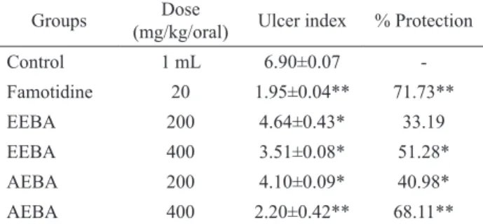
Table 2. Percentage (%) protection of plant extracts of Basella alba var. alba leaf extracts in ethanol induced ulcer model.
Groups Dose
(mg/kg/oral) Ulcer index % Protection
Control 1 mL 6.90±0.07
-Famotidine 20 1.95±0.04** 71.73**
EEBA 200 4.64±0.43* 33.19
EEBA 400 3.51±0.08* 51.28*
AEBA 200 4.10±0.09* 40.98*
AEBA 400 2.20±0.42** 68.11**
All values are the mean±SEM (n=6 rats/treatment) *p<0.05 and **p<0.01.
Histopathological examination of ulcerated stomach of experimental rats
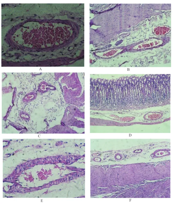
The cytoprotective effect was conirmed by histological examination. In the rats treated with absolute ethanol, there was markedly extensive damage to the gastric mucosa due to severe disruption of the surface epithelium, deep penetration of necrotic lesions into mucosa and edema of the submucosa layer with leukocyte iniltration of ulcerative tissues (Figure 1). However, rats treated with famotidine, AEBA and EEBA showed gastric mucosal protection as compared to control. Rats
treated with EEBA 200 or 400 mg/kg showed a marked reduction in ulcer areas, milder disruption of the surface epithelium and inhibition of edema and leucocyte iniltration of the submucosal layer. The rats treated with AEBA 200 or 400 mg/kg showed a mild disruption of the surface epithelium and edema of the submucosa layer with leukocyte iniltration which is comparatively similar to the effects of famotidine (20 mg/kg). Grossly, the leaf extracts of Basella alba has shown a substantial and signiicant protection against gastric ulcers in rats.
Discussion
Figure 1. Histological evaluation of gastric lesions of ulcerated rats stomach. A. Control (distilled water), (H&E stain, 10×); B. AEBA (200 mg/kg), (H&E stain, 10×); C. AEBA (400 mg/kg), (H&E stain, 10×); D. EEBA (200 mg/kg), (H&E stain, 10×); E. EEBA (400 mg/kg), (H&E stain, 10×); Standard (20mg/kg), (H&E stain, 10×).
(Middleton et al., 2000). Preliminary phytochemical tests on the aqueous as well as alcoholic extracts of Basella alba have shown the presence lavonoids as well as tannins and this could be responsible for its anti-ulcer activity. The presence of mucilage in the plant could also play a role in cytoprotection. The exact mechanisms underlying the protective action of the leaf extract against ethanol induced gastric lesions are unclear. Further studies are in progress in this lab to explore the compounds responsible for the protective effect and the mechanism of this activity.
Conclusion
The present study reveal that the leaf extracts of Basella alba var. alba possesses statistically signiicant antiulcerogenic activity. The activity may be due to enhancement of defensive mechanism through an improvement in gastric cytoprotection or inhibition of acid secretion or both.
A B
C D
References
Abdulla MA, Ali HM, Ahmed KA, Noor SM 2009. Evaluation of the anti-ulcer activities of Morus alba extract in experimentally induced gastric ulcer in rats. Biomed Res 20: 35-39.
Baggio CH, Freitas CS, Rieck L, Marques MCA 2003. Gastroprotective effects of a crude extract of Baccharis illinita DC in rats. Pharmacol Res 47: 93-98.
Bancroft JD, Stevens A 1990. Theory and practice of histological techniques. Churchill Livingstone Newyork, 3rd ed, p. 281-282.
Deshpal S, Shah GB, Deshpande I, Parmar NS 2003. Antiulcer activity of aqueous extract of Basella rubra in albino rats. J Nat Remedies 3: 212-214.
Deshpande SS, Shah GB, Parmar NS 2003. Antiulcer activity of Tephrosia purpurea in rats. Indian J Pharmacol 35: 168-172.
Ecobichon DJ 1997. The basis of toxicity testing. CRC Press, New York, 2nd ed. p. 43-49.
Gaskil DL, Serinek KL, Levine VA 1982. Effect of prostacyclin
on mucosal blood low. Surgery 92: 220-224.
Khandare RA, Gulecha VS, Mahajan MS, Mundada AS, Gangurde HH 2009. Evaluation of antiulcer activity of polyherbal formulation. Int J Pharm Res Develop 1: 1-6.
Khandelwal KR, Kokate CK 1995. Reaction of Cell Content, Practical Pharmacognosy. Nirali prakashan, N. Delhi, v. 2, p. 9-30.
Kumar V, Bhat ZA, Kumar D, Bohra P, Sheela S 2011. In-vitro
antiinlammatory activity of leaf extracts of Basella
alba var. alba L., Int J Drug Dev Res 3: 176-179. Maity S, Vedasiromoni J R, Ganguly D K 1986. Antiulcer effect
of hot water extract of black tea (Camellia sinensis). J Ethnopharmacol 46: 167-174.
Middleton E, Kandaswami C, Theoharides TC 2000. The effects of plant lavonoids on mammalian cells: implications
for inlammation, heart disease, and cancer. Pharmacol
Rev 52: 673-751.
Nandkarni KM 1976. Indian Materia Medica. Popular Prakashan Private Limited Publication, Mumbai: 3rd ed. p: 177-178.
Oates PJ, Hakkinen JP 1988. Studies on the mechanism of ethanol-induced gastric damage in rats,
Gastroenterology 94: 10-21.
Pareek V, Singh M, Bhat ZA, Singh P, Kumar D, Sheela S 2010. Studies on mucilage of Basella alba Linn. J Pharm Res 3: 1892-1894.
Sairam K, Priyambda S, Aryya NC, Goel RK 2003. Gastroduodenal ulcer protective activity of Asparagus racemosus; an experimental, biochemical and histological study. J Ethnopharmacol 86: 1-10. Shah AH, Khan ZA 1973. Gastro protective effect of
pretreatment with Zizyphus sativa fruits against toxic damage in rats. Fitoterapia 3: 226-234.
Shay H, Komarov SA, Feels SE, Meraze D, Grunenstein M, Siplet H 1945. A simple method of the uniform production of gastric ulceration in rat. Gastroenterology 5: 43-61.
Szabo S 1987. Mechanisms of mucosal injury in the stomach and duodenum: time-sequence analysis of morphologic, functional, biochemical and histochemical studies. Scan J Gastroent 22: 21-28.
Vyawahare NS, Deshmukh VV, Gadkari MR, Kagathara VG 2009. Plants with antiulcer activity. Phcog Rev 3: 118-125.
*Correspondence
Vijender Kumar
Department of Pharmaceutical Sciences, University of Kashmir, Srinagar (J & K)-190006