Neuroectodermal Differentiation by Silencing the
Pluripotency-Associated Oct-25 Gene
Dario Nicetto1, Matthias Hahn2, Julia Jung3, Tobias D. Schneider1, Tobias Straub1, Robert David3¤, Gunnar Schotta2, Ralph A. W. Rupp1*
1Adolf Butenandt Institut, Institut fu¨r Molekularbiologie, Ludwig Maximilians-Universita¨t, Mu¨nchen, Germany,2Center for Integrated Protein Science (Munich) at the Institut fu¨r Molekularbiologie, Adolf-Butenandt-Institut, LMU, Mu¨nchen, Germany,3Medizinische Klinik I am Klinikum der Universita¨t Mu¨nchen (LMU), Mu¨nchen, Germany
Abstract
Post-translational modifications (PTMs) of histones exert fundamental roles in regulating gene expression. During development, groups of PTMs are constrained by unknown mechanisms into combinatorial patterns, which facilitate transitions from uncommitted embryonic cells into differentiated somatic cell lineages. Repressive histone modifications such as H3K9me3 or H3K27me3 have been investigated in detail, but the role of H4K20me3 in development is currently unknown. Here we show that Xenopus laevis Suv4-20h1 and h2 histone methyltransferases (HMTases) are essential for induction and differentiation of the neuroectoderm. Morpholino-mediated knockdown of the two HMTases leads to a selective and specific downregulation of genes controlling neural induction, thereby effectively blocking differentiation of the neuroectoderm. Global transcriptome analysis supports the notion that these effects arise from the transcriptional deregulation of specific genes rather than widespread, pleiotropic effects. Interestingly, morphant embryos fail to repress the Oct4-related Xenopus gene Oct-25. We validate Oct-25 as a direct target of xSu4-20h enzyme mediated gene repression, showing by chromatin immunoprecipitaton that it is decorated with the H4K20me3 mark downstream of the promoter in normal, but not in double-morphant, embryos. Since knockdown of Oct-25 protein significantly rescues the neural differentiation defect in x20h double-morphant embryos, we conclude that the epistatic relationship between Suv4-20h enzymes and Oct-25 controls the transit from pluripotent to differentiation-competent neural cells. Consistent with these results in Xenopus, murine Suv4-20h1/h2 double-knockout embryonic stem (DKO ES) cells exhibit increased Oct4 protein levels before and during EB formation, and reveal a compromised and biased capacity forin vitrodifferentiation, when compared to normal ES cells. Together, these results suggest a regulatory mechanism, conserved between amphibians and mammals, in which H4K20me3-dependent restriction of specific POU-V genes directs cell fate decisions, when embryonic cells exit the pluripotent state.
Citation:Nicetto D, Hahn M, Jung J, Schneider TD, Straub T, et al. (2013) Suv4-20h Histone Methyltransferases Promote Neuroectodermal Differentiation by Silencing the Pluripotency-Associated Oct-25 Gene. PLoS Genet 9(1): e1003188. doi:10.1371/journal.pgen.1003188
Editor:Anne C. Ferguson-Smith, University of Cambridge, United Kingdom
ReceivedApril 3, 2012;AcceptedNovember 7, 2012;PublishedJanuary 31, 2013
Copyright:ß2013 Nicetto et al. This is an open-access article distributed under the terms of the Creative Commons Attribution License, which permits unrestricted use, distribution, and reproduction in any medium, provided the original author and source are credited.
Funding:RAWP was supported by SFB/TR5–DFG (http://www.dfg.de/foerderung/programme/listen/projektdetails/index.jsp?id = 5485239). GS was supported by SFB/TR5–DFG (http://www.dfg.de/foerderung/programme/listen/projektdetails/index.jsp?id = 5485239). This work was also supported by Episys–BMBF (http:// www.episys.org/EPISYS/Home.html). The funders had no role in study design, data collection and analysis, decision to publish, or preparation of the manuscript.
Competing Interests:The authors have declared that no competing interests exist. * E-mail: [email protected]
¤ Current address: Biomedical Research Center Rostock, Rostock University, Rostock, Germany
Introduction
Embryonic development is controlled by fine-tuned differen-tial gene expression. A succession of regulatory protein networks unfolds the zygotic gene expression program along a hierarchy of decisions, leading from the embryonic ground state to the epiblast and then to germ layers, which become patterned into cell type and organ precursor territories. The pluripotent trait, key feature of embryonic stem (ES) cells [1], is progressively restricted and finally lost as soon as embryonic cells become specified to germ layer fates. Recent studies have revealed that alterations in chromatin structure, dynamics and composition represent fundamental processes, which define the epigenetic landscape that directs cell type specification along this hierarchy [2,3].
Besides important contributions from ATP dependent chroma-tin remodelling factors [4,5] and histone variants [6] in modulating nucleosome dynamics, histone post-translational modifications (PTMs) have been linked to gene expression [3,7]. The transition from pluripotent to differentiated cells is charac-terized by a progressive increase in heterochromatin formation, in a process that changes the hyperdynamic open chromatin structure into a less accessible architecture [1,8]. At the same time transcriptional silencing of non-lineage specific genes is achieved via acquisition of repressive histone marks.In vivostudies have shown that dynamic alterations in the levels of histone modifications characterize early stages of development both in mammals [9–11] and other vertebrates [12–14].
linked both to transcriptional activation and repression [15,16]. In particular, repressive histone methyl marks are found on lysine residues at position 9 and 27 on histone H3 and in position 20 on histone H4. H3K27 trimethylation is catalyzed by polycomb repression complex (PRC) 2, which predominantly represses developmental regulatory genes [17–19]. Trimethyla-tion of H3K9 and H4K20 relies on Suv39h and Suv4-20h enzyme activities, respectively [20,21], and predominantly marks repetitive genomic DNA at pericentromeric and telo-meric heterochromatin [16,21]. While H3K9-specific HMTases have been characterized in significant depth [20,22,23], we know little about the functions of Suv4-20h1 and Suv4-20h2 enzymes with regard to gene regulation. In vivo analysis of H4K20 methylation states in mouse embryos reveals specific patterns both in cellular or subnuclear abundance [9,24]. Suv4-20h DKO pups die perinatally, indicating an essential function of the two enzymes during embryogenesis [24]. Moreover, quantitative analysis of histone PTMs in X. laevis revealed a progressive and significant accumulation of H4K20me3 levels during embryogenesis, suggesting developmental functions for these enzymes [13].
To characterize the functional role of H4K20me2/3 during vertebrate development we have investigated the consequences of both morpholino-mediated protein knockdown and mRNA-born overexpression of the Xenopus Suv4-20h1 and h2 homologs in frog embryos. Our data reveal a specific and selective requirement for Suv4-20h enzyme acitivities in neuroectodermal differentia-tion, in a process which involves transcriptional repression of pluripotency associated POU-V genes, both in Xenopus embryos and in murine ES cells.
Results
Characterization of Xenopus Suv4-20h1 and h2 enzymes We initially identified X. laevis Suv4-20h1 and h2 ESTs via database mining. Mouse and frog Suv4-20h1 and h2 protein
sequences are well conserved, particularly within the SET domains ($88% identity), even though the xSuv4-20h2 open reading frame is longer than its mouse homolog due to C-terminal insertions (supplementary data, Figure S1). XSuv4-20h1/h2 genes are both maternally and zygotically expressed in a ubiquitous manner, as shown by RNAin situ hybridisation and RT-PCR analysis (Figure S2A–S2D). XSuv4-20h1 mRNA abundance decreases during the initial stages of development and subsequently rises from mid-gastrula on, reflecting the switch from maternal-to-zygotic transcription at midblastula. In con-trast, the initially high xSuv4-20h2 mRNA level falls and stays low at late stages (Figure S2D).
To test the acivities of these Xenopus HMTases, we first analyzed their ability to rescue H4K20me3 levels in Suv4-20h1/ h2 DKO mouse embryonic fibroblasts (MEF Suv4-20h DKO; [24]), which lack this modification. Both frog cDNAs re-established a proper H4K20me3 pattern, which was strongly enriched at heterochromatic regions that were identified as DAPI-dense chromocenters within nuclei (Figure 1A). Thus, Xenopus laevis Suv4-20h homologs are biologically active and can direct H4K20 trimethylation. To test, whether they generate this histone mark in frog embryos, we designed antisense Morpholino oligonucleotides (MO) to reduce synthesis of xSuv4-20h1 and h2 proteins from endogenous mRNAs (Figure S3A, S3B). These MOs specifically inhibited translation of their cognate templates in vitro (Figure S3C). To avoid possible functional complementation between the xSuv4-20h enzymesin vivo, we decided to inject the two MOs simultaneously into both blastomeres of 2-cell stage embryos and performed western blots with nuclear protein extracts from these double-morphant embryos at the tadpole stage (NF30-33). Compared to uninjected controls or embryos injected with an unrelated control MO (control-morphants), the double morphants contained significant-ly less H4K20me2 (p = 0.0011) and H4K20me3 (p = 0.0164), which was coupled to an increase in H4K20me1 (p = 0.0034) (Figure 1B and 1C).
This result was confirmed by MALDI-TOF mass spectrom-etry (Figure S4A). As described in Schneider et al. [13], the relative abundance of histone modifications was calculated by quantifying the amount of a specific modification relative to the amount of all modification states determined for the same histone peptide. As reported before [13], the H4K20me3 mark could not be quantitated reproducibly for technical reasons. Compared to control embryos, however, xSuv4-20h double morphants contained approximately 2.5-fold less of H4K20me2 (p = 0.0153) and three-fold more H4K20me1 (p = 0.0185), while the abundance of the unmodified peptide state remained unaffected. Importantly, the levels of histone H3 methylation on two tryptic peptides, covering the K9, K27 and K36 positions, were indistinguishable between control and double-morphant embryos (Figure S4B and S4C). Western blot analysis with antibodies against trimethylated H3K9 or H3K27 also showed no difference in the abundance of these two marks between control embryos and xSuv4-20h double morphants (Figure S4D).
To further characterize the effects of xSuv4-20h enzyme depletion on the cellular level, we performed immunohistochem-istry on sections from tailbud stage embryos (NF30), which were injected with the xSuv4-20h MO-mix into one of two blastomeres at 2-cell stage together with fluorescently labelled dextranes as lineage tracer. While H3 staining was comparable between injected and uninjected sides under all conditions (Figure S5A), staining for H4K20me2 and –me3 was clearly reduced on the double-morphant side of the neural tube (Figure 1D). In
Author Summary
The quest of modern developmental biology is a detailed molecular description of the process that leads from the fertilized egg to the complex and highly differentiated adult organism. This process is controlled largely on the level of gene expression. While early embryonic cells are pluripotent and capable of transcribing most of their genome, older cells have become committed to the germ layer and differentiation programs during gastrulation. They express then a subset of genes compatible with their future physiological function. Young, pluripotent cells and post-gastrula, committed cells express different networks of transcription factors and contain chromatin of different structure and composition. How these two regulatory layers are interconnected during development is incom-pletely understood. We describe a novel and unexpected link between the pluripotency-associated POU-V gene Oct-25 and xSuv4-20h histone methyltransferases. XSuv4-20h enzymes are required to repress the Oct-25 gene, a homolog of mammalian Oct4, in the neuroectoderm of frog embryos as a prerequisite for neural differentiation. Consistently, murine Suv4-20h double-null ES cells show increased Oct4 protein levels before and during in vitro differentiation and display compromised differentiation in comparison to wild-type ES cells. Thus, Suv4-20h enzymes control specific POU-V genes and are involved in germ-layer specific differentiation.
agreement with our western blot and mass spec results, the reduction in the di- and tri-methyl mark was coupled to an increase in H4K20me1 staining. Altogether these results indicate that xSuv4-20h1 and h2 downregulation leads to a quantitative reduction of H4K20 di- and trimethyl marks in the frog embryo, without affecting the bulk abundance of other repressive histone marks such as H3 K9/K27 methylation.
RNA-based overexpression of Suv4-20h HMTases had the opposite effect. When injected singly, xSuv4-20h1 or h2 mRNAs increased both di- and trimethylated H4K20 in a dose-dependent manner (Figure S8A). A comparable result was achieved by injection of either mouse Suv4-20h1 or h2 mRNAs (Figure S9A). Together, these results identify the frog cDNAs as orthologs of mammalian Suv4-20h enzymes. Loss and gain of function experiments also indicate that the bulk abundance of di- and
trimethylated H4K20 can be manipulated over a wide range without compromising embryonic viability.
XSuv4-20h HMTases depletion inhibits eye and melanocyte formation
We next tested, whether depletion of xSuv4-20h HMTases affects embryonic development. We injected xSuv4-20h1/h2 MO-mix into one blastomere of two-cell stage embryos and scored phenotypic alterations by comparing injected with uninjected sides. No obvious differences were observed during early development, including gastrulation, axial extension and dorso-ventral patterning. From tailbud stages on, two main phenotypes became manifest. First, in the injected side of xSuv4-20h double morphants the eye formation was strongly compromised. The eye rudiments contained no or barely visible retinal pigment and
Figure 1. Functional analysis of xSuv4-20h HMTases.(A) Transiently transfected eGFP-tagged Suv4-20h1 and h2 enzymes from frog or mouse re-establish H4K20me3 marks in heterochromatic foci of Suv4-20h1/h2 DKO MEFs. (B–D) Bulk histones from tadpoles (NF30-33) injected with morpholinos targeting translation of endogenous xSuv4-20h1 and h2 mRNA show a strong reduction in H4K20me2 and H4K20me3 levels and a concomitant increase in the H4K20me1 mark. (B) Western Blot analysis of uninjected embryos, control morphants (ctrl-MO), and double morphants with antibodies against H4K20 mono-, di- and trimethylation. PanH3 antibody was used as loading control. (C) Western Blot quantification of three independent biological experiments; data represent mean values, error bars indicate SEM. (D) Immuno-histochemistry on xSuv4-20h double morphant tadpoles. Panels show details from neural tubes stained with antibodies against the histone epitopes indicated on the side. Whole sections shown in Figure S5A.
typically had no lens (Figure 2A). Secondly, melanophores that are found on the dorsal part of the head and the lateral portion of the trunk, were severely reduced in numbers or completely lost from the double-morphant side (Figure 2A). Both phenotypes had a penetrance between 80–90% in xSuv4–20h double morphants (p,0.0001, Fisher’s exact test) in several independent experiments (Figure 2B). Control-morphant embryos had normal eyes and melanocytes (Figure 2A) and were indistinguishable from unin-jected siblings in most cases (Figure 2B).
The distinct eye phenotype prompted us to investigate the underlying molecular changes. RNA in situ hybridization exper-iments revealed a clearly reduced expression of the homeobox
transcription factor Rx-1 (Figure 2C) and the paired box transcription factor Pax-6 (Figure S5D) in xSuv4-20h double morphants. The reduction of these two master regulators of eye differentiation explains the morphological eye phenotype, but we noticed that embryonic transcription was already misregulated upstream of these factors. The pan-neural markers Nrp1 (Figure 2C) and N-CAM (Figure S5E), which are induced during gastrula stages, were also strongly reduced in double morphants. However, several key markers of embryonic patterning were not perturbed, such as the organizer genes Chordin, Goosecoid and Xnr-3 at gastrula stages (Figure S5B). The anteroposterior patterning of the central nervous system (CNS) appeared also to
Figure 2. xSuv4-20h1/h2 double morphants lack eyes and melanophores.(A) Morphological phenotypes of representative tadpoles (NF30-33) from embryo cohorts injected into one of two blastomeres at two-cell stage with ctrl-MO, xSuv4-20h1/h2 MOs (double morphants), and double morphants coinjected with 3 ng mouse Suv4-20h1/h2 mRNAs (rescued). Injected body halves were identified by green fluorescence of the coinjected lineage tracer Fluor 488 Dextran. (B) Penetrance of the eye phenotype. Data from three to five independent experiments; n = total number of embryos scored. (C) RNAin situhybridization analysis for Rx-1 in tadpoles (NF30-33), and CNS markers Nrp1 at neural tube stage (NF19-20). For each condition, numbers refer to embryos showing the displayed morphology or staining, in comparison to the total number of analysed embryos. doi:10.1371/journal.pgen.1003188.g002
be normal given the wild-type-like expression patterns of Otx2 and Krox20 in fore- and hindbrain territories, respectively (Figure S5C). These results provide first evidence that H4K20 di- and trimethylation serves to regulate distinct developmental genes in a selective manner.
Xenopus Suv4-20h activity is required for normal development
The specificity of the developmental phenotypes arising from xSuv4-20h enzyme depletion was validated by rescue experiments, in which we coinjected increasing doses of murine Suv4-20h1/h2 mRNAs together with the xSuv4-20h MO-mix. Due to sequence divergence, transcripts of the murine orthologs escape inhibition by the MOs targeting the frog mRNAs. Already 2 ng of murine Suv4-20h transcripts were sufficient to rescue the eye defect in two thirds of the double morphant embryos (p,0.0001, Fisher’s exact test). In most cases, the retinal neuroepithelium regained its circular structure and near normal size, as well as a central lens (Figure 2A). The rescue efficiency did not increase with higher concentrations of mouse transcripts (Figure 2B, columns 4–6). The number of melanophores was also increased at their proper sites under rescue conditions (Figure 2A). Furthermore, the expression domains of Rx-1 and Nrp1 (Figure 2C), as well as Pax-6 and N-CAM (Figure S5D and S5E) were efficiently restored.
To test, whether this phenotypic rescue requires Suv4-20h proteins or their enzymatic activity, we generated catalytically inactive murine Suv4-20h protein variants (Figure S6A), based on structural predictions [25,26]. Unlike the wild-type proteins, neither variant restored the H4K20me3 mark at heterochromatic foci in Suv4-20h DKO MEFs (Figure S6B). When tested side by side with the wild-type enzymes, the mutants did neither increase the abundance of the H4K20me2 and -me3 marks in wild-type frog embryos (Figure S6C, compare lanes 1, 3 and 5), nor rescue H4K20 methylation levels in xSuv4-20h double morphants (Figure S6C compare lanes 1, 4 and 6), although being expressed at similar levels (Figure S6D). Consequently, the inactive variants also failed to rescue the eye and melanophore phenotype (Figure S7A–S7C and S7D, compare columns 2–4).
In the course of these experiments we noticed that overexpres-sion of either frog or mouse Suv4-20h1 and h2 proteins never caused any obvious morphological or molecular changes in the embryos (Figures S8B, S8C and S9B, S9C), despite strongly enhanced H4K20me3 levels in bulk chromatin (Figures S8A and S9A). In particular, morphological landmarks such as eyes and melanophores formed normal in size, number and location under overexpression conditions. Expression domains of marker genes such as Rx-1 and Pax-6 were unaffected (Figures S8D and S9D). Thus, H4K20 di- and trimethylation is required for normal development, but excess deposition of these marks has no apparent phenotypic consequences.
XSuv4-20h enzymes are required for ectoderm formation The apparent functional selectivity of the ubiquitously expressed enzymes encouraged us to test, whether xSuv4-20h HMTases control additional aspects of germ layer formation and patterning. Therefore, we compared the expression of key developmental regulatory genes in uni-laterally injected control-morphantsversus xSuv4-20h double morphants by RNAin situhybridisation (listed in Figure 3A).
Based on our previous results, we continued with genes involved in neurogenesis (Figure 3B). At the open plate stage, primary neurons are specified in three stripes next to the dorsal midline on each side. At this time, each stripe expresses the neural specific regulatory genes Neurogenin-related 1a (Ngnr-1a) and Delta-like
1, as well as the differentiation marker N-tubulin. The expression of these three genes was extinguished in almost all of the xSuv4-20h MO-injected sides, while being unaffected by control-MO (Figure 3B). In addition to these stripes, Delta-like 1 mRNA delineates the anterior border of the neural plate, and this domain was also extinguished (Figure 3B). In contrast, mesodermal expression of Delta-like 1 around the blastoporus remained unaffected in morphant condition (Figure 3B, arrow). Delta-like 1 and N-tubulin stripes were effectively rescued by coinjection of wild-type mSuv4-20h1/h2 mRNAs, while Ngnr-1a was restored in a broad, diffuse manner (Figure 3B, right column). Notably, inactive mouse Suv4-20h HMTases could not rescue N-tubulin expression (Figure S7E, middle column). At the same time, mesodermal control genes like MyoD were unaffected (Figure S7E, right column) Together, these results implicate xSuv4-20h enzymes in neuronal fate selection.
Next, we extented our analysis to marker genes expressed in other germlayers and territories (Figure 3C and Figure S5). The epidermal keratin gene XK81 demarcates non-neural ectoderm and was expressed normally on the surface of morphant epidermis; however, due to a slight retardation in neural tube closure on the injected side, its expression appears asymmetric in anterior views. This may indicate an involvement of xSuv4-20h enzymes in morphogenetic processes during neurulation and/or neural crest specification. This phenotype led to a mild broadening of the neural plate markers Sox2 (Figure 3C), Sox3 and Sox11 (Figure S5F) at apparently normal mRNA levels. Prior to these neural plate markers, a group of genes including FoxD5, Geminin, Zic1, Zic2, Zic3 and members of the Iroquois family are induced in the prospective neuroectoderm and stabilize the neural fate by their regulatory interactions (reviewed in ref [27]). At midgastrula (NF11), FoxD5 and Geminin did not respond to xSuv4-20h enzyme depletion (Figure S5F), but Xiro1, Zic1 (Figure 3C), Zic2 and Zic3 (Figure S5F) mRNAs were strongly reduced. In contrast, key mesodermal factors such as Xbra, MyoD (Figure 3C) and VegT (Figure S5F), as well as regulators of endodermal differentiation like Sox17 a and Endodermin (Figure 3C) were expressed normally in both morphants and in embryos overex-pressing frog xSuv4-20h proteins (Figure S8E). Taken together these results demonstrate that xSuv4-20h HMTases are critical for neural development, but apparently dispensable for mesoderm and endoderm formation inX. laevis.
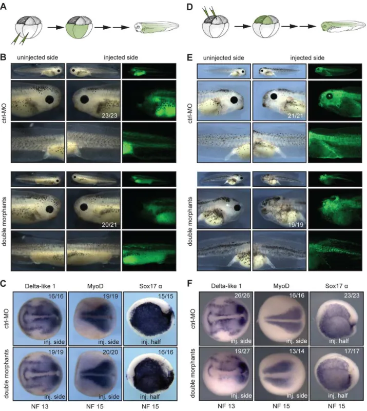
To further verify the specific role of Xenopus Suv4-20h enzymes in neural development, we considered two different approaches; in a first series of experiments we performed injections at 8-cell stage in the animal or vegetal pole blastomeres, selectively labelling cells predominantly belonging either to mesendoderm (vegetal injections, Figure 4A) or ectoderm (animal injections, Figure 4D). Vegetal pole blastomere injections led to no evident morphological and molecular phenotypes (Figure 4B and 4C). Conversely, animal injections reproduced the eye and melano-phore phenotypes from half-injected embryos, while mesodermal and endodermal structures developed normally (Figure 4E). Consistent with the morphological defects, Delta-like 1 expression in the neural plate was suppressed, while MyoD and Sox17 a genes were unaffected (Figure 4F). These results provide strong evidence that the neural and melanocyte phenotypes originate in the ectoderm.
(Figure 5A). They were also negative for Xbra, indicating absence of contaminating mesoderm. Noggin-mediated Nrp1 expression was clearly visible in wt caps, but strongly reduced upon co-injection of xSuv4-20h morpholinos, while XK81 expression was downregulated in both the samples (Figure 5A). Thus, double morphant caps are both refractory to neural induction and restrained in epidermal differentiation. However, they differentiate into mesoderm upon stimulation with Activin A just like control explants, as shown by immunostaining for muscle myosin heavy chain (Figure 5B). These results confirm the crucial role of xSuv4-20h enzymes in coordinating the formation of ectodermal tissues, and show that in the absence of the two enzymes neural induction is impaired.
XSuv4-20h enzymes are required for cell survival and proliferation
Loss of H4K20 di- and trimethylation is known to compromise DNA damage repair in mice and to partially block G1/S transition [24]. This prompted us to test, whether xSuv4-20h depletion affects apoptosis and cell proliferation in frog embryos. Immunostaining for activated Caspase3 revealed an increase in apoptotic cells on the injected side of double morphant embryos (Figure S10A). Coinjection of xBcl-2 mRNA, an anti-apoptotic factor, reduced the Caspase3 positive cells to levels of the uninjected control side, however, without re-establishing a proper Delta-like 1 and N-tubulin pattern in the double-morphant side. Overexpression of xBcl-2 mRNA alone had no effect on the expression of the tested markers (Figure S10A). Thus, although embryonic frog cells depleted for the H4K20me2/me3 marks become apoptotic at higher rate than wt cells, the absence of neurons in the double-morphant neural plate cannot be explained by selective cell death.
Double morphant embryos stained for the mitotic marker H3S10P, showed a two-fold reduction (p = 0.0058) in the number of proliferating cells at midneurula stage, compared to control morphant embryos (Figure S10B). This mild phenotype might be correlated with the observed increase in apoptosis. Since neural induction continues in frogs, even when cell proliferation is blocked from gastrulation onwards [28], it is unlikely that the nearly complete loss of N-tubulin positive neurons is brought about by this mild reduction in cell proliferation. Taken together, the main xSuv4-20h morphant phenotype represents not a selective loss of neuroblasts, but a block in neural differentiation.
XSuv4-20h double morphant frog embryos fail to silence Oct-25 transcription in sensorial ectoderm
So far, our analysis in xSuv4-20h morphant embryos has indicated a specific and selective loss of gene expression in ectodermally derived tissues. The earliest affected markers - Zic and Xiro genes - become induced at early gastrula stage and help establish the neural plate state [27]. At this time in frog development, embryonic cells in the animal hemisphere are still plastic and express members of the POU-V gene family – i.e. Oct-25, Oct-60 and Oct-91 - that encode paralogs of the mammalian
pluripotency regulator Oct4 [29,30]. Because Oct-25 and Oct-91 regulate germ layer differentiation in Xenopus [31–34], we investigated their expression (Figure 6A). Oct-25 is initially expressed throughout the animal hemisphere at early gastrula, but gets restricted to the presumptive floor plate (notoplate) by midneurula [31]. On the injected side of the vast majority of double morphants, however, Oct-25 expression was expanded from the notoplate down to the ventral midline. Interestingly, ectopic Oct-25 expression was restricted to the sensorial cell layer of the ectoderm, which contains neural and epidermal precursor cells, respectively (Figure 6A, sections). The Oct-60 gene, which is expressed during oogenesis, was not activated under these conditions. Oct-91 staining appeared normal in the majority of the embryos, although some showed a mild upregulation in double morphants as well (data not shown). The ectopic expression of Oct-25 is a specific consequence of xSuv4-20h enzyme depletion, because its normal pattern was re-established in morphants upon coinjection of mRNAs encoding wild-type, but not inactive, mouse Suv4-20h proteins (Figure S7E, left column). Notably, the selective derepression of the Oct-25 gene was also observed in double-morphant AC explants (Figure 6B), excluding indirect effects from non-ectodermal tissues.
We then performed qRT-PCR analysis to quantitate the relative changes in gene expression. It is frequently observed that embryo cohorts develop in slight asynchrony as a non-specific consequence of Morpholino injection, possibly obscuring tran-scriptional responses. To minimize this potential artifact, we analysed the RNAs of matching pairs of wt and xSuv4-20h depleted samples by dissecting embryos at early neurula stage (NF14) into uninjected and injected halves, based on the coinjected fluorescent lineage tracer (Figure S11A). As shown in Figure 6C, the Oct-25 mRNA is about three-fold higher in xSuv4-20h double-morphant halves (p = 0.0123), while being similar between control-morphant and uninjected halves. In the same sample, Oct-91 expression was unaffected (Figure 6C). We used this assay also to confirm the diminished expression of neural plate marker genes detected earlier by RNAin situhybridisation. With the exception of Ngnr 1a, Nrp1 and N-tubulin, mRNA levels were clearly reduced in the morphant halves (p = 0.0122 and 0.0163, respectively; Figure S11B).
To gain further information about the complexity of the underlying transcriptional misregulation, we performed transcrip-tome analysis in wild-type and double-morphant embryos, again dissecting embryos in corresponding pairs of injected and uninjected halves (Figure S12A). Six percent of the 11639 annotated probe sets present on the microarray were significantly altered in their expression as a consequence of xSuv4-20h enzyme depletion, about equally split into upregulated (n = 319) and downregulated (n = 404) probes (Figure S12B and S12C; for a complete list of the responding probesets see NCBI’s GEO Series accession number GSE41256). This result suggests that the observed phenotypes in the double morphants originate from transcriptional misregulation of distinct genes, rather than from global, pleiotropic effects. Indeed, Oct-25 mRNA is also specif-ically upregulated in the microarray data set, where it is among the
Figure 3. xSuv4-20h enzymes are required for differentiation of the neuroectoderm.(A) Schematic illustration of analysed markers of the different germ layers (germ layer colour code extended toin situpanels). Downregulated genes upon xSuv4-20h depletion are labelled in red. (B) Expression pattern of the neuroectodermal markers Ngnr 1a (NF12.5), Delta-like 1 (NF13), and N-tubulin (NF15). The pictures show dorsal views of the open neural plate with anterior to the left. (C) Expression patterns of XK81 (ectoderm), Sox2, Xiro1, Zic1 (neuroectoderm), Xbra, MyoD (mesoderm), Sox17a, Endodermin (endoderm) in ctrl-MO injected or double morphant embryos. XK81 - anterior views with dorsal side to the top. Sox2 and MyoD - dorsal views, anterior to the left. Xiro1 and Zic1 - dorsal views; injected halves are lineage-traced by coinjection of LacZ mRNA and subsequentb–Gal staining (light blue). Xbra - vegetal view. Sox-17a- internal stain from the injected side in bisected embryos, animal pole up. Endodermin – internal stain from the injected side in bisected embryos; anterior to the left.
ten most upregulated mRNAs in the double-morphant condition (Figure S12D).
The sustained expression of Oct-25 in xSuv4-20h morphant embryos fits the prediction of Oct-25 being a direct target of
H4K20me3 mediated transcriptional silencing. To test this assumption directly, we carried out chromatin-immunoprecipita-tion (ChIP) experiments with H4K20me3-specific antibodies at the neurula stage (NF15-16). For ChIP experiments we used X. Figure 4. xSuv4-20h1/h2 enzymatic activity is required in the ectodermal germ layer. (A, D) Schematic illustrations of targeting microinjections into mesendodermal or ectodermal territories at 8-cell stage. (B) Injecting xSuv4-20h MOs into the mesendoderm causes no apparent morphological phenotype in the embryo. (C) Neural, mesodermal and endodermal marker genes are expressed normally. (E) xSuv4-20h MOs reduce eyes, cranial and trunk melanophores, when injected into the ectoderm. (F) Expression of all tested markers in mesoderm and endoderm is normal, except for Delta-like 1, whose expression specifically in the open neural plate is strongly reduced on the injected side. Global morphology was assessed at hatching stage (NF36), molecular markers at indicated stages during neurulation. Top row images in (B) and (E) depict whole embryos for overview.
doi:10.1371/journal.pgen.1003188.g004
tropicalis embryos, since the available genome sequence of this closely related frog species [35] allowed us to design primer amplicons for non-exon derived DNA sequences. RNA in situ hybridization performed on neurula stage X. tropicalis embryos, confirmed that the expression patterns of Oct-25 and N-tubulin were up- and down-regulated, respectively, to the same extend as observed for X. laevis (Figure S13). We retrived the pericentro-meric major satellite repeat sequence (MSAT3) as positive control amplicon for the experiment. Genic regions, which are H4K20me3-free and, thus, could be used as negative controls, are difficult to predict, since genome-wide analysis in mammalian cells reported only enrichment of this modification on pericen-tromeric and subtelomeric heterochromatin [36,37]. As negative
controls we considered: GAPDH, a constitutively expressed housekeeping gene; thyroid hormone receptor a (thra), a gene whose expression can be detected at neurula; and thra-induced bzip protein (thibz) that is expressed from metamorphosis on (Figure S14A). Statistical analysis of qRT/PCR data indicates that expression of GAPDH and thra was not significantly altered under the double-morphant condition (Figure S14B). Therefore, the relative H4K20me3 levels at these genes were defined as background, and compared to the levels on other loci (Figure S14A). The modification strongly decorated the pericentromeric MSAT3 repeat region (Figure 6D), as expected from the analysis in murine cells [21]. At the 59UTR amplicon of the Oct-25 gene, H4K20me3 was significantly enriched compared to the control genes GAPDH (p = 0.0155), thra (p = 0.0103) and thibz (p = 0.0128) (Figure S14A and Figure 6D). In a second set of experiments, we compared the abundance of H4K20me3 between wild-type and xSuv4-20h double-morphant embryos (Figure 6E). In morphants, the modification was selectively reduced at the 59UTR amplicon of Oct-25 (p = 0.004). Together, these ChIP experiments validate the 59end of the Oct-25 gene as direct target of xSuv4-20h mediated transcriptional silencing.
Xenopus Oct-25 has been implicated in germ layer formation [32,34]. We wanted to know, whether the sustained expression of Oct-25 in xSuv4-20h morphants could cause the observed downregulation of early neural plate and neural differentiation markers. This question is difficult to address, since the role of Oct-25 in neural induction is ambiguous - both overexpression and morpholino knockdown inhibit neural differentiation [32,34]. Thus, Oct-25 acts in pleiotropic fashion, perhaps switching target genes or protein interaction partners. In a previous report [38], human Oct4 protein was shown by ChIP analysis to bind to promoters of early neural markers, including Zic and Sox genes. In order to link Xenopus Oct-25 mechanistically to these genes, we have mis-expressed constitutively activating and repressing Oct-25 fusion proteins in animal caps (Figure S15A). Zic1, Zic3 and Sox2 responded to the Oct-25 variants in a manner consistent with direct regulator/target gene interaction, i.e. they were hyperactivated by Oct-25-VP16 (p = 0.0143; 0.0456; 0.01622, respectively) and suppressed by Oct-25-EnR (p = 0.0236; 0.0167; 0.0231, respective-ly) compared to the uninjected sample. In line with this assumption, inspection of theX. tropicalisgene sequences detailed the presence of multiple Oct-25 DNA binding motifs within 2.0 Kb distance from the transcriptional start site for each of these genes (Figure S16). For the two Zic genes, which are misregulated in the forming neural plate of morphant embryos (Figure 3C and Figure S5F), we confirmed the misregulation by Oct-25 variants via RNAin situ hybridisation (Figure S15B).
Interestingly, Sox2 expression was affected only in AC explants, but not in the double morphant embryos. This can be explained by considering two points: First, in animal caps levels and activities of the injected Oct-25 protein variants most likely exceed endogenous Oct-25 protein activity and regulate Sox2 expression in a dominant fashion. Secondly, formation of neural tissue in the embryo requires inductive influences including FGF signalling [39],and Sox2 transcription is stimulated by FGF8 [27], which is normally expressed in the mesoderm. Thus, the stimulating influence of FGF signalling on Sox2 transcription in the embryo may neutralize the repressive influence from deregulated Oct-25 expression, while the repressive activity of the deregulated Oct-25 levels prevails in animal caps in the absence of FGF signalling.
The remaining genes either failed to respond to one of the two Oct-25 protein variants (Zic2, Xiro1), or did not respond (Ngnr 1a, N-tubulin). These observations suggest an indirect effect. While it is possible that additional factors that are misregulated in xSuv4-20h
Figure 5. In vitro induction of xSuv4-20h double-morphant animal cap explants. (A) Noggin-dependent neuralisation. XK81, Nrp1 and Xbra expression is monitored in uninjected control caps and double-morphant caps with or without Noggin mRNA. Note that explants coinjected with xSuv4-20h MOs together with Noggin mRNA show reduced Nrp1 expression, but normal downregulation of XK81 mRNA. (B) Muscle induction by Activin A in uninjected, ctrl-MO injected, and xSuv4-20h double morphant animal caps. Top row demonstrates comparable expression of myosin heavy chain (MHC-a) in non-dissected sibling embryos.
Figure 6. xSuv4-20h double morphants fail to silence Oct-25 expression in deep-layer ectoderm due to reduced H4K20me3 enrichment at Oct-25 promoter.RNAin situhybridization analysis for Oct-25, Oct-60 and Oct-91 in embryos (A) and animal caps (B) for ctrl-morphants or xSuv4-20h double-morphant embryos, injected unilaterally at two-cell stage and fixed at midneurula stage (NF15). Injected sides were defined by coinjected Alexa-fluorescence prior toin situhybridisation. (A) Dorsal views of stained embryos with anterior to the left. For Oct-25 Vibratome cross-sections, ctrl-MO and double-morphant embryos are shown. (B) Comparative expression analysis for Oct-25, Oct-60 and Oct-91 in animal caps from bilaterally injected embryos, fixed at midneurula stage (NF15). (C) qRT-PCR profiles for Oct-25 and Oct-91 in ctrl-MO and xSuv4-20h double-morphant embryos. Data represent normalized ratios of mRNA levels as means of four independent experiments, error bars indicate SEM. (D, E) Chromatin immunoprecipitation (ChIP) analysis on Oct-25, GAPDH, thra, thibz genes and major satellite repeat region 3 (MSAT3). (D) H4K20me3 levels on the indicated genes in normal embryos, and (E) in uninjectedversusdouble-morphant embryos. Fold enrichment was calculated as the ratio between H4K20me3 precipitated material over negative control (No antibody sample). Data represent mean values of three to five independent experiments, error bars indicate SEM.
doi:10.1371/journal.pgen.1003188.g006
morphants contribute to the neural phenotype, the combined results from ChIP experiments and Oct-25 variants define a pathway, in which xSuv4-20h enzyme dependent repression of Oct-25 is needed during gastrulation for proper neuroectoderm differentiation.
Deregulated Oct-25 expression in xSuv4-20h double morphants inhibits neural differentiation
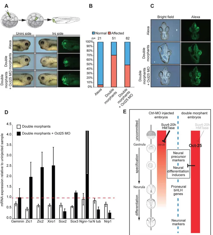
To further analyse the mechanistic interaction between xSuv4-20h enzymes and Oct-25, we performed rescue experiments with triple-morphant embryos, in which synthesis of Oct-25 and xSuv4-20h proteins was simultanously blocked (Figure 7). The Oct-25 morpholino that we used has been shown before to inhibit efficiently Oct-25 translation from both non-allelic gene copies [40]. Because global Oct-25 depletion inhibits the formation of anterior neural structures [40], we employed two different strategies for the triple-knockdown to circumvent this problem. In a first series of analysis we injected a single A1 blastomere of 32-cell stage embryos to target 32-cells that predominantly contribute to the retina and brain. Also in this experimental series, the morphology of double morphant eyes was strongly affected (Figure 7A). 71% of the injected embryos showed a clear reduction of retinal pigment, the remainders often restricted to the dorsal-most portion of the eyecup. The majority of the eyes contained no lens (Figure 7C). When the downregulation of xSuv4-20h enzymes was coupled to a concomitant knockdown of Oct-25 (triple morphants), the percentage of embryos showing this defect was reduced to 49% (p = 0.0188, Fisher’s exact test). The retinal pigment was rescued in the triple morphants, whose eyes also regained a properly structured lens (Figure 7C). To confirm the morphological phenotypes, we investigated the basal neural gene expression in AC explants. The expression of a subset of genes involved in the establishment of the neural plate state (Zic1, Zic2, Xiro1, Sox2 and Sox3) was strongly reduced upon downregulation of xSuv4-20h enzymes at early neurula (NF14-15), compared to uninjected animal caps (p = 0.0068; p = 0.0127; p = 0.0113; p = 0.0321; p = 0.0037, respectively). With the exception of Sox2, the simultaneous downregulation of xSuv4-20h enzymes and Oct-25, rescued neural gene expression. In fact, under the triple morphant condition most of these genes were expressed at higher levels than normal, suggesting that they are partly repressed by Oct-25 in unmanipulated explants (Figure 7D). Most impor-tantly, the combined results of the two triple-knockdown experiments indicate that both morphological and molecular features of the xSuv4-20h double morphant phenotype can be rescued to a significant extent by reducing Oct-25 protein levels. This result firmly establishes that the sustained and elevated expression of Oct-25 protein is responsible for the neural differentiation defect of xSuv4-20h double-morphant embryos.
Murine Suv4-20h1/h2 double-knockout ES cells have elevated Oct4 levels in undifferentiating and
differentiating conditions
Oct-25 plays multiple roles during early frog development, including interference with Activin/BMP-dependent mesendo-derm formation before gastrulation, and with neural induction during gastrulation [32,34]. A similar role is considered for its mammalian paralog Oct4, which is required for the pluripotent state of ES cells, but antagonizes ectodermal differentiation as soon as these cells exit pluripotency [30,41,42]. Although previous genome-wide studies of histone modifications in mammalian cells have not detected H4K20me3 on the Oct4 gene [36,37], this apparent similarity made us investigate Oct4 protein expression in wild-type and Suv4-20h1/h2 DKO murine ES cells. We tested
two independently derived DKO cell lines (B4-2 and B7-1), and compared them with two wild-type controls, i.e. wt26, an isogenic ES cell line, and the well-characterized GSES-1 cell line [43]. All four cell lines formed comparable ES cell colonies in LIF-containing medium (Figure 8A and Figure S17B), although the two DKO lines grew slightly slower. Upon aggregation they formed embryoid bodies, which were clearly smaller than those of the wild-type lines, both at day 2 and day 6 of differentiation (Figure 8A and Figure S17). After replating the differentiated cells for one day, the two DKO lines frequently formed again colonies resembling undifferentiated ES-cells (day 7 in Figure 8A and Figure S17B). To obtain a quantitative measure of Oct4 gene expression, we fixed and stained the four cell lines before (day 0) and during (day 6) differentiation for Oct4 protein and subjected equal cell numbers to FACS-analysis. The Oct4 signals were quite similar between wt26 and GSES-1 cells, as they were between the two DKO lines. In contrast to the wild-type cell lines, however, the signals of the DKO lines were reproducibly shifted to the right (Figure 8B and Figure S17C). Based on normalized median fluorescence intensity, the two DKO lines contained approxi-mately three-fold higher Oct4 protein amounts than the wild-type lines at day 0 (p = 0.00604), and still two-fold more at day 6 (p = 0.01266) (n = 3; Figure 8C and Figure S17B). We conclude that Oct4 expression is being reduced during differentiation in Suv4-20h1/h2 DKO cells. However, these cells have higher Oct4 levels in the undifferentiated state, and maintain higher levels during differentiation in comparison to wild-type cells.
Oct4 protein levels are known to be tightly regulated [1] and to influence lineage decisions during ES cell differentiation [41,42]. We therefore investigated the biological significance of the elevated Oct4 protein levels in Suv4-20h DKO ES cell lines. Unfortunately, the applied EB differentiation protocol promotes predominantly mesendodermal differentiation, which prevented the analysis of neural markers. Nevertheless, we performed FACS analysis on wt and Suv4-20h DKO cell lines stained for the chemokine receptor 4 (CXCR4) protein, whose expression indicates mesendoderm induction in embryoid bodies. At day 6 of differentiation, wt cell lines showed a robust increase in CXCR4 positive cells compared to day 0 (Figure 8D and data not shown). In contrast, both Suv4-20h DKO cell lines contained a significantly lower percentage of CXCR4 positive cells at day 6 when compared to the wild type cell lines (p = 0.03255; Figure 8). We also noted that replated wt cells frequently formed autono-mously beating areas at differentiation day 14 (see Video S1), indicating functional cardiomyocyte formation, while contracting areas were never observed in the Suv4-20h DKO cells (Video S2; n = 4 experiments). Finally, qRT-PCR analysis indicated a reproducible and statistically significant shift in mesendoderm gene expression in the DKO ES cells, which show enhanced induction of FoxA2 (p = 0.00706) and reduced levels of Gata4 (p = 0.00037), compared to the wt ES cell lines (Figure S17D). Together, these results reveal a compromised and biased differentiation capacity for Suv4-20h DKO ES cell lines, and provide an entrypoint for further experimentation in the murine system.
Discussion
Figure 7. Oct-25 knockdown in double-morphant embryos rescues xSuv4-20h phenotypes. (A) Schematic illustration of targeting microinjections of tadpoles injected into the A1 blastomere at 32-cell stage, and morphological phenotypes of representative embryos (NF35-37) from cohorts injected with Alexa, xSuv4-20h MOs (double morphants) and double morphants plus Oct-25 MO. (B) Penetrance of the eye phenotype. Data from three independent experiments; n = total number of embryos scored. (C) Vibratome cross-sections of representative embryos injected as in panel (A). (D) qRT-PCR profiles for the indicated genes in double morphants and double morphants plus Oct-25 MO animal cap explants at NF 14–15. Data represent normalized mRNA levels as mean of three to four independent experiments; error bars indicate SEM. (E) Model for Xenopus Suv4-20h1/h2 enzyme function during neuroectoderm differentiation. A global increase in H4K20me3 reduces widespread Oct-25 expression in the animal emisphere during gastrulation as a prerequisite for neural induction. In H4K20me3 depleted morphant embryos, Oct-25 expression persists in the ectodermal stem cell compartment (sensorial cell layer), interfering with the transcriptional activation or activities of key regulators of the neural plate state and neurogenesis. Additional genes that are deregulated like Oct-25 in xSuv4-20h morphant embryos, may also contribute to impaired ectoderm differentiation.
doi:10.1371/journal.pgen.1003188.g007
transcriptional regulation [44,45]. Our experiments identify a specific and selective role of xSuv4-20h HMTases in the formation of the ectodermal germlayer through control of mRNA expression of key regulators of the neural plate state and neuronal differentiation circuits. Indeed, our results indicate for the first time that H4K20me3 controls transcription in a rather gene-specific manner. The mRNA profile of double morphant embryos shows appr. 6% of the annotated probesets to be misregulated, when H4K20me3 levels have been reduced to appr. 25%. About half of the responding mRNAs are transcriptionally upregulated and, thus, their genes may qualify as being directly controlled by H4K20me3 deposition. Surprisingly, our molecular analysis revealed that xSuv4-20h enzymes are required to restrict the expression of the pluripotency-associated Oct-25 gene during gastrula and neurula stages. In the absence of proper H4K20me3 deposition, the Oct-25 gene becomes transcriptionally derepressed and interferes with neural differentiation. The successful rescue of key morphological and molecular aspects of the neural defect in double-morphant embryos by the simultanous inhibition of Oct-25 translation establishes this pathway formally. At least in Xenopus, the regulatory interaction between xSuv4-20h enzymes and Oct-25 is needed for embryonic cells to exit the pluripotent state and differentiate as neuroectoderm.
The genetic interaction between Suv4-20h enzymes and POU-V genes appears also to be conserved in mouse ES cells, although the H4K20me3 mark has not yet been detected on the Oct4 gene locus. To this point, we have shown that Suv4-20h DKO ES cells contain significantly elevated Oct4 protein levels, compared to wt ES cells. During ES cell differentiation the mammalian Oct4 gene is known to become repressed by a battery of epigenetic mechanisms including DNA methylation, incorporation of somatic linker histones and repressive histone modifications (H3K9me3/ H3K27me3), which cooperate to achieve chromatin compaction of the Oct4 gene locus [46]. Our finding that Oct4 protein levels are increased in the DKO ES cells both before and during differentiation actually suggests that Suv4-20h enzymes regulate mammalian Oct4 transcription in a way that is at least partly independent from the other repressive mechanisms targetting this locus.
Our results in Xenopus rest predominantly on loss of function analysis, achieved by morpholino-mediated knockdown of endog-enous xSuv4-20h protein translation. Specifically, we have shown that our antisense oligonucleotides block translation of xSuv4-20h1 and h2 isoforms in vitro, and significantly decrease H4K20me2 and –me3 levels in vivo, without altering the bulk
Figure 8. Suv4-20h double-null ES cells have elevated Oct4 and lower CXCR4 protein levels before and during differentiation.
Wild-type and Suv4-20h DKO ES cells were grown undifferentiated in
LIF-containing medium, or differentiatedin vitro by embryoid body formation. Wt26 - isogenic wild-type ES cell line; B4-2, B7-1 – independent Suv4-20h DKO ES cell lines. (A) Morphological appearance (scale bar 100mm). Top row: undifferentiated ES cells (day 0); middle:
embryoid bodies at day 6; bottom: cells from embryoid bodies, replated for 24h. (B) Before (day 0) and during (day 6) differentiation, cell lines were stained for Oct4 protein and 26104cells from each condition were
subjected to FACS analysis. Red graph: fluorescence of non-specific isotype control; black and green graphs represent the Oct4 protein levels in wild-type and Suv4-20h DKO ES cell lines, respectively. (C) Suv4-20h DKO cells have higher Oct4 protein levels compared to wild-type ES cells and maintain these during differentiation. Median fluorescence intensity was calculated from data in panel (B), error bars indicate SEM. (D) Suv4-20h DKO cells show a reduction in the percentage of CXCR4+cells at differentiation day 6. Data represent
normalized values of percentage of CXCR4+cells as means of three
abundance of other repressive histone marks such as H3K9me3 and H3K27me3. The morpholinos produced specific phenotypes, which were rescued on the morphological and molecular level by RNA-born co-expression of heterologous xSuv4-20h enzymes and, thus, originate from deficient H4K20me2/me3 states.
While xSuv4-20h double morphant embryos showed consistent phenotypes at high penetrance, we were surprised to see that H4K20me2 and –me3 states could be quantitatively increased in frog embryos without any obvious morphological or molecular consequences (Figures S8 and S9). This result can be explained considering first of all the higher stability of the knockdown by non-degradable morpholinos compared to the transient protein upregulation by RNA injection; secondly, demethylation of higher-methylated states may occur rather rapidly through H4K20me2 and me3 demethylases at specific sites, where H4K20me1 is required, e.g. Wnt/b-Catenin inducible genes [47]. However, we did not observe evidence for compromised transcription of Wnt target genes under overexpression (Figures S8 and S9) or morphant condition (Figure S5). Since mono- and dimethylated H4K20 states are quite abundant modifications in Xenopus embryos (30–40% each; see ref. [13]), it is most likely the loss of H4K20 trimethylation, which interferes with normal development.
XSuv4-20h double-morphant embryos were frequently defec-tive for eye and melanocyte differentiation, indicating a prominent impairment of neuroectodermal differentiation. This selectivity is surprising, given that the two HMTases are expressed throughout the entire embryo (Figure S2). As a matter of fact, the phenotypes originate in the neuroectoderm, as shown by targeted injection into animal or vegetal blastomeres of 8-cell stage embryos (Figure 4). A large panel of marker genes that were investigated by RNA in situ hybridisation indicates that mesodermal and endodermal gene expression patterns are not perturbed by xSuv4-20h enzyme depletion (Figure 3A). This includes markers, which are required for specification of embryonic axes and formation and patterning of the mesendodermal germlayers (Figure S5). We also note that morphant animal cap explants were refractory to Noggin-dependent neural induction, but could be induced to differentiated skeletal muscle by a mesoderm inducing signal (Figure 5). We therefore assume that a major function of xSuv4-20h enzymes lies in the transcriptional control of genes that coordinate and execute neuroectodermal differentiation. Consis-tent with this hypothesis, many of the genes that we found downregulated in xSuv4-20h morphants, are key regulators of eye development (Rx-1, Pax-6), neuronal differentiation (Ngnr 1a, Delta-like 1) or regulators of neural competence and neural plate state (Zic-1, -2, -3, Xiro-1, Nrp1, N-CAM; [27]).
While these molecular results explain the overt morphological phenotypes in a consistent manner, it should be noted that these HMTases are clearly involved in additional cellular aspects. The mild reduction in mitotic cells and the increased apoptotic rate of morphant embryos (Figure S10) is reminiscent of findings in Suv4-20h1/h2 DKO MEFs, which are less resistant to DNA damage and compromised at the G1/S checkpoint [24]. The data reported here indicates a need for deeper analysis of the regulatory impact of Suv4-20h enzymes on transcription in both mammals and non-mammalian vertebrates.
According to current models, xSuv4-20h enzymes mediate transcriptional repression, based on the enrichment of the H4K20me3 mark on heterochromatic foci. Genes that are regulated by these enzymes should therefore become derepressed under loss of function condition. Following this logic, many of the genes, which are misregulated in morphant frog embryos, would be classified as indirect targets, since they were downregulated.
One very notable exception, which we have validated as direct target, is Oct-25 (Figure 6). Oct-25 is induced broadly in the animal hemisphere at the blastula/gastrula transition, before it becomes restricted to the notoplate at neurula stages [31]. Oct-25 plays multiple roles during early frog development, including interference with Activin/BMP-dependent mesendoderm forma-tion before gastrulaforma-tion, and with neural inducforma-tion during gastrulation [31,32,34]. Our study reveals now a new function for Oct-25, namely to control the transit from a pluripotent cell to a neural cell that differentiates, when Oct-25 expression has faded. As depicted in our model (Figure 7E), this function depends on the precise dose and duration of Oct-25 transcription, which is controlled by the level of H4K20me3 deposition on the first exon of the Oct-25 gene through xSuv4-20h enzymes. As we have shown here, deregulated transcription of Oct-25 in double-morphant embryos elicits massive consequences on the differen-tiation of neuroectodermal organs and cell types. We have traced back the origin of the malformations to the gastrula stage, when a gene network, defining the neural state, become perturbed by Oct-25. Some members of this network are good candidates for direct regulation through Oct-25 (e.g. Zic and Sox genes). However, since Oct-25 transcription persists ectopically at least until the mid-neural fold stage in the ectoderm, subsequent gene cascades involved in regional differentiation of the neuroectoderm could also be directly misregulated by Oct-25.
The specific and selective deregulation of Oct-25 transcription in a precise spatial domain, i.e. the sensorial cell layer of the ectoderm, implies a very intriguing role for xSuv4-20h enzymes. This domain contains not only the uncommitted precursors of neuronal and epidermal cell types, but – with regard to the involuting marginal zone – includes also mesodermal and endodermal precursor cells. The observed derepression of Oct-25 in this domain may thus reflect a conserved mechanism, by which Suv4-20h enzymes control pluripotency in the embryo. As discussed above, we have found Oct4 protein to be increased in two independent Suv4-20h double knockout ES cell lines under LIF-maintained self-renewal conditions, when compared to wt ES cells (Figure 8 and Figure S17). The DKO cell lines also maintain higher Oct4 levels during differentiation than wt ES cells, although their Oct4 levels get diminished in the course of 6 days. Recent data from several labs suggest that the pluripotency regulators Sox2 and Oct4 guide ES cells towards specific germ layer differentiation programs, when they exit the pluripotent state [41,42]. Indeed, our findings are in agreement with Thomson and colleagues, who describe Oct4 to antagonize ectodermal specifi-cation and to direct mesendodermal cell fate decisions. The conserved Suv4-20h-dependent restriction of Oct4 expression may thus contribute to the germ-layer specification of embryonic cells, when they exit the pluripotent state.
Materials and Methods
Ethics statement
Animal work has been conducted in accordance with Deutsches Tierschutzgesetz; experimental use of Xenopus embryos has been licensed by the Government of Oberbayern (Projekt/AK ROB: 55.2.1.54-2532.6-3-11).
Expression constructs andin vitrotranscription
Full length X. laevis 20h1a (NM_001092308) and Suv4-20h2a (NM_001097050) cDNAs in pCMV-SPORT6 were provided by ImaGenes. Capped mRNAs were synthesizedin vitro with SP6 RNA-Polymerase after HpaI linearization. Both cDNAs were subcloned via XhoI/EcoRI sites into pBluescript II KS to
generate digoxygenin-labelled antisense probes with T3 RNA-Polymerase. Xenopus Bcl-2, Oct-25-VP16 and –EnR constructs were transcribed with SP6 RNA-Polymerase from NotI- (Bcl-2 and Oct-25-VP16) and SacII- (Oct-25-EnR) linearized pCS2+
plasmids, respectively. Mouse Suv4-20h1 and h2 enzymes were transcribed with SP6 from PvuI-linearized pCMVmyc-constructs [24]. Enzymatically inactive mouse Suv4-20h HMTases were generated via PCR-mutagenesis (see Text S1, Table S1 for primers). Synthetic mRNAs were injected in the animal pole of two-cell stage embryos at 2, 3 or 4ng per embryo. Rescue experiments with wt and mutated mRNAs were performed with 3ng of a 1:1 mix of wt or mutated Suv4-20h1 and h2 mRNAs, injected into the animal pole of a single blastomere at two-cell stage. Xenopus Bcl-2 mRNA was injected unilaterally in the animal pole of two-cell stage embryos at 800 pg per embryo. Xenopus Oct-25-VP16, -EnR mRNAs were injected in the animal pole of two-cell stage embryos at 100 pg per embryo.
Cell culture, microscopy, and FACS analysis
Mouse embryonic fibroblasts (MEF) wild type and Suv4-20h DKO cells [24] were cultivated in High Glucose DMEM with L-Glutamine and sodium pyruvate, complemented with 10% FCS, b-mercaptoethanol, non essential amino acids and penicillin/ streptomycin in a 37uC incubator at 5% CO2. Lipofectamine 2000
(Invitrogen) was used for the transfection of plasmid DNAs. Immunofluorescence analysis was performed as described in the Text S1.
Mouse ES cells were cultivated on gelatinized plates in High Glucose DMEM with L-Glutamine and sodium pyruvate, complemented with 15% FCS, 0.1 mM ß-mercaptoethanol, non essential amino acids, penicillin/streptomycin and LIF. Cells were maintained at 37uC in a humidified atmosphere of 5% CO2. ESin
vitro differentiation and FACS analysis were carried out as described [43] The incubation steps with the primary Oct4 (1:250, Abcam) or CxCR4 (1:50, BD Pharmingen) antibody and subsequently a FITC-conjugated secondary antibody (1:250, Invitrogen) were performed at RT for 45 min with two washing steps after each antibody incubation. For the isotype controls purified, IgG was used instead of the Oct4-antibody. All FACS analyses were performed with an Epics XL (Beckman-Coulter) using the analysis software FlowJo.
Morpholino oligonucleotides
Translation-blocking Morpholino oligonucleotides targeting Xenopus Suv4-20h1 (X.laevisandX.tropicalis: 59 -GGATTCGCC-CAACCACTTCATGCCA-39), Xenopus Suv4-20h2 (X.laevis: 59 -TTGCCGTCAACCGATTTGAACCCAT-39: X.tropicalis: 59 -CCGTCAAGCGATTTGAACCCATAGT-39) and Xenopus Oct-25 (X.laevis: 59 -TTGGGAAGGGCTGTTGGCTGTACAT-39) mRNAs were supplied by Gene Tools LLC. Each Morpholinos recognizes the two non-allelic isoforms of each gene inX.laevis(see Figure S3A, S3B). GeneTools’ standard control Morpholino was used to monitor non-specific effects. Morpholino activity was tested by in vitro translation (SP6-TNT Kit, Promega), adding 2 pg of control Morpholino or 1 pg of Suv4-20h1 and/or h2 Morpholinos per TNT reaction. Unless stated otherwise, embryos were injected at a dose of 60–80 ng per embryo (30–40 ng each of Suv4-20h1 and h2 Morpholinos, or 60–80 ng control Morpholino per embryo). For 8-cell stage experiments, morpholinos were injected in two neighbouring, animal or vegetal blastomeres on one side of the embryos, at half the dose (i.e. 40 ng total). For morphogical epistasis experiments, Xenopus Suv4-20h1 and h2 Morpholinos (5 ng each per embryo) and Oct-25 Morpholino (1 ng per embryo) were injected into A1 blastomere at 32-cell stage.
Embryo handling
Xenopus laeviseggs were collected, fertilizedin vitro, and handled following standard procedures; embryos were staged according to Nieuwkoop and Faber (1967). The embryos were injected with maximally 10 nl volume. When required, they were sorted into left side or right side injected cohorts before fixation, based on the coinjected lineage tracer Alexa Fluor-488 Dextran (Invitrogen). Alkaline-phosphatase stained and refixed embryos were either sectioned after embedding in paraffin (10mm), or in gelatine/ albumin mixture supplemented with 25% glutaraldehyde before sectioning (30–50mm) with a Vibratome 1000 (Technical Products International, INC.) as described [48]. Animal caps were manually dissected at NF9 and transferred singly into wells of a 96-well plate, coated with 1% agarose and filled with 1X Steinberg’s solution, 0.1% BSA with or without Activin A (1:10 diluted conditioned cell culture supernatant). For neural induction, embryos were injected into the animal pole with Noggin mRNA (60 pg per embryo) alone or together with xSuv4-20h1 and h2 morpholinos (40 ng each per embryo) at two- to four-cell stage. For mesoderm induction, embryos were injected animally 4 times with 2.5 nl of control morpholino (80 ng per embryo) or a mix of xSuv4-20h1 and h2 morpholinos (40 ng each) at two or four cell stage. For Oct-25-VP16 and –EnR overexpression experiments, embryos were injected animally 4 times with 2.5 nl of each mRNAs (100 pg per embryo). For epistasis experiments on animal caps, embryos were injected 4 times with 2.5 nl of xSuv4-20h1 and h2 Morpholinos (40 ng each per embryo) and Oct-25 Morpholino (30 ng per embryo) at two or four cell stage.
Analysis of histone modifications in Xenopus embryos Nuclei extraction from Xenopus embryos and mass spectrom-etry analysis of histone modifications were performed as described [13]. Histone marks were quantitated as relative abundances of a specific modification state as a fraction of the amount of all modifications found for this peptide (for details see ref 13).
RNAin situhybridization and immunocytochemistry Whole-mount RNA in situ hybridizations were performed as described (Sive et al. 2000). Embryos were photographed under bright light with a Leica M205FA stereomicroscope. The following antibodies were used for immunocytochemistry: H3S10P antibody (1:300, Upstate Biotechnology), active Caspase3 antibody (1:500, Promega), and myosin heavy chain antibody MF20 (1:100 hybridoma cell culture supernatant), anti-mouse or anti-rabbit alkaline phosphatase-conjugated secondary antibodies (1:1000, Chemicon).
Western blots and immunostaining
detected with ECL plus western blotting detection reagents (GE Healthcare). Histological sections were stained with pan H3 (1:2000, Abcam), H4K20me1 (1:5000), H4K20me2 (1:2000), H4K20me3 (1:5000) antibodies [24].
Quantitative RNA analysis
Total cellular RNA was isolated with TRizol (Qiagen) and phenol/chloroform extraction. On-column RNA clean-up, in-cluding a DNAse digestion step, was performed using RNeasy-Mini-Kit (Qiagen). Samples for qRT-PCR and microarray profiling were collected as described in the Text S1.
Microarray expression analysis
Microarray data were processed using R/Bioconductor (www. bioconductor.org). If not indicated otherwise, we used standard parameters in all functions calls. Expression values were calculated using ‘gcrma’. Probe sets were kept for differential expression analysis if there were more ‘present’ calls (calculated using ‘mas5calls’) in one of the treatment groups than non-‘present’ calls, if their expression level variance was higher than 0 across all arrays and if the probe set had an Entrez identifier annotation according to the Entrez database with a date stamp of 2011-Mar16. One gene to many probe set relationships were resolved by retaining only the probe set with the highest variance across all arrays. Differential expression statistics were obtained using a linear model (library ‘limma’). A significant response was defined if the local false discovery (‘locfdr’ package) rate calculated on the moderated t statistic was smaller than 0.2. The data discussed in this publication have been deposited in NCBI’s Gene Expression Omnibus and are accessible through GEO Series accession number GSE41256 (http://www.ncbi.nlm.nih.gov/geo/query/ acc.cgi?acc=GSE41256).
ChIP experiments
ChIP experiments were performed using Xenopus tropicalis as described [49], with minor changes (see Text S1 for details).
Identification of Oct-25 binding sites
A published weight matrix (PMID:17567999) was used to scan 2 kb upstream regions of selected X. tropicalis genes (Xenbase version 7.1) for binding site occurrence. Scanning was performed using RSA matrix-scan (PMID:18802439) with default parame-ters.
Supporting Information
Figure S1 Xenopus laevis versusMus musculusSuv4-20h proteins sequence alignment. Amino acid sequence alignment for Mus musculus(Refseq. NM_001167885.1)versus Xenopus laevisSuv4-20h1 (Refseq. NM_001092308) (A) and Mus musculus (Refseq. NM_146177.2) [24]versus Xenopus laevis Suv4-20h2 (Refseq NM_001097050) proteins (B). (C) Aminoacid sequence conserva-tion (% identity) of the SET domain between mouse and Xenopus proteins.
(PDF)
Figure S2 Xenopus laevis Suv4-20h expression during early development. XSuv4-20h1 (A) and xSuv4-20h2 (B) mRNA expression was detected by RNA in situ hybridization at the indicated developmental stages. (C) Total RNA was extracted from animal cap (AC), marginal zone (MZ) and vegetal pole (VP) explants of NF9 embryos; semiquantitative PCR shows relative levels of xSuv4-20h1 and xSuv4-20h2 transcripts in the three explants. ODC was used as loading control, -RT as negative
control. (D) qRT-PCR profiles of xSuv4-20h enzymes. The chart shows the expression of the two enzymes relative to ODC at the indicated developmental stages.
(PDF)
Figure S3 Morpholino specificity. 25-mer xSuv4-20h1 mor-pholino (A) and xSuv4-20h2 mormor-pholino (B) oligonucleotides perfectly target to the start codon of the respective two non-allelic isoforms. Sequence differences between the two morpho-linos confer specificity of each oligonucleotide for either xSuv4-20h1 or xSuv4-20h2 mRNA. (C)In vitro TNT assay performed as described in Materials and Method section. xSuv4-20h1 and h2 MOs specifically inhibited translation of their cognate templates.
(PDF)
Figure S4 Quantification of histone methylation states in xSuv4-20h morphants by MALDI-TOF mass spectrometry. Bulk histones from NF30-33 embryos were isolated and analysed as described in Materials and Methods. (A) H4 peptide 20–23, (B) H3 peptide 9– 17 and (C) H3 peptide 27–40 in uninjected, ctrl-MO and double morphant embryos. The values represent mean of three independent experiments; error bars indicate SEM. Star - for technical reasons H4K20me3 mark was quantitated only in some samples (star). (D) Western Blot analysis of uninjected, control morpholino (ctrl-MO) and xSuv4-20h1, h2 morpholino injected embryos using antibodies against H3K27me3 and H3K9me3. PanH3 antibody was used as loading control.
(PDF)
Figure S5 Germ layer marker gene expression in xSuv4-20h double morphants. (A) Immuno-histochemistry on ctrl-MO and xSuv4-20h double-morphant tadpoles. Panels show representa-tive cross-sections of neural tubes stained with antibodies against the histone epitopes indicated on top. Inj – injected side. Squares on double-morphant sections represent the croped pictures shown in Figure 1D. (B-F) RNAin situhybridization analysis of ctrl-MO injected and double morphant embryos at the indicated stages using probes against Chordin, Xnr-3 and Gooscoid (B), Krox20 and Otx2 (C), Pax-6 (D), N-CAM (E), FoxD5, Geminin, Zic2, Zic3, Sox3, Sox11 and VegT (F). Chordin, Xnr-3 and Gooscoid – dorsal side views; animal pole is on the top. Krox20 -dorsal views, anterior on the left. Otx2 - anterior views, -dorsal on the top. Pax-6 – head region; rescued embryos included. N-CAM - dorsal views of stained embryos with the anterior on the left; rescued embryos included. FoxD5, Geminin, Zic2, Zic3 - dorsal views; injected halves are lineage-traced by coinjection of LacZ mRNA and subsequentb–Gal staining (light blue). Sox2, Sox3 -dorsal views, anterior to the left. VegT - internal stain in bisected embryos.
(PDF)
Figure S6 Functional SET domains are required for proper Suv4-20h activity. (A) Schematic ofMus musculusSuv4-20h1 and h2 SET domain mutations. (B) Immunofluorescence of wild-type and Suv4-20h DKO MEFs transfected with the indicated constructs. (C) Western Blot analysis of NF11.5 uninjected embryos (lane 1), xSuv4-20h1, h2 double morphants (lane 2), active and inactive mouse Suv4-20h1, h2 mRNAs injected embryos (lane 3, 5 respectively) and double morphant embryos cojnected with active or inactive mSuv4-20h1, h2 mRNAs (lane 2, 4 respectively), using antibodies against H4K20 mono-, di- and trimethylation. PanH3 antibody was used as loading control. (D) Anti-myc western blot with the same samples used in B. Asterisks indicate unspecific bands.
(PDF)