Cop
yright
© ABE&M t
odos os dir
eit
os r
eser
vados
.
Hyperinsulinism/hyperammonemia
(HI/HA) syndrome due to a
mutation in the glutamate
dehydrogenase gene
Síndrome de hiperinsulinemia/hiperamonemia devido a uma mutação no gene da glutamato desidrogenase
Maria Lúcia Corrêa-Giannella1, Daniel Soares Freire2,
Ana Mercedes Cavaleiro1, Maria Angela Zanella Fortes1,
Ricardo Rodrigues Giorgi1, Maria Adelaide Albergaria Pereira2
SUMMARY
The hyperinsulinism/hyperammonemia (HI/HA) syndrome is a rare autosomal dominant disea se manifested by hypoglycemic symptoms triggered by fasting or highprotein meals, and by elevated serum ammonia. HI/HA is the second most common cause of hyperinsulinemic hypo glycemia of infancy, and it is caused by activating mutations in GLUD1, the gene that encodes
mitochondrial enzyme glutamate dehydrogenase (GDH). Biochemical evaluation, as well as direct sequencing of exons and exonintron boundary regions of the GLUD1 gene, were per
formed in a 6year old female patient presenting fasting hypoglycemia and hyperammonemia. The patient was found to be heterozygous for one de novo missense mutation (c.1491A>G;
p.Il497Met) previously reported in a Japanese patient. Treatment with diazoxide 100 mg/day promoted complete resolution of the hypoglycemic episodes. Arq Bras Endocrinol Metab. 2012;56(8):485-9
SUMÁRIO
A síndrome de hiperinsulinemia/hiperamonemia (HI/HA) é uma condição rara, de herança autos sômica dominante, que se manifesta por sintomas de hipoglicemia desencadeada por je jum ou refeições de alto conteúdo proteico, juntamente com elevação da concentração de amô nia sérica. HI/HA é a segunda causa de hipoglicemia hiperinsulinêmica da infância e é causa da por mutações ativadoras no GLUD1, o gene que codiica a enzima mitocondrial glutamato
desidrogenase (GDH). A avaliação bioquímica, bem como o sequenciamento direto dos éxons e junções éxoníntron do gene GLUD1, foi realizada em uma paciente de 6 anos de idade com hi
poglicemia de jejum e hiperamonemia. A paciente apresentava uma mutação de novo missense
(c.1491A>G; p.Il497Met) em heterozigose, que havia sido previamente relatada em um paciente japonês. O tratamento com diazóxido 100 mg/dia promoveu resolução completa dos episódios hipoglicêmicos. Arq Bras Endocrinol Metab. 2012;56(8):485-9
1 Laboratório de Endocrinologia Celular e Molecular (LIM-25), Faculdade de Medicina da Universidade de São Paulo (FMUSP), São Paulo, SP, Brazil 2 Serviço de Endocrinologia e Metabologia do Hospital das Clínicas, FMUSP, São Paulo, SP, Brazil
Correspondence to:
Maria Lúcia Corrêa-Giannella Av. Dr. Arnaldo, 455, 4o andar, sala
4.305
01246-903 – São Paulo, SP, Brazil [email protected]
Received on May/30/2012 Accepted on Sept/10/2012
INTRODUCTION
T
he hyperinsulinism/hyperammonemia syndrome(HI/HA) is the second most common cause of hyperinsulinemic hypoglycemia of infancy. It is a rare genetic disease caused by activating mutations in GLUD1, a gene located on chromosome 10q23.3., composed of 13 exons that encode the
hypo-Cop
yright
© ABE&M t
odos os dir
eit
os r
eser
vados
.
+1.89]; weight, 32 kg [Z-score, +4.1]). Laboratory test results, including liver and renal function tests and concentrations of GH, IGF-I, cortisol and ACTH were normal (data not shown). The child presented hypo-glycemic episodes after overnight fasting, as well as in the postprandial period. During an episode suggestive of fasting hypoglycemia, the following results were ob-served: plasma glucose, 42 mg/dL (2.3 mM); serum insulin, 7.7 µU/mL; C-peptide, 3.4 ng/dL; blood ketone bodies (strip test), negative. Administration of glucagon elevated plasma glucose to 113 mg/dL (6.3 mM).
Following demonstration of hyperinsulinemic hy-poglycemia, an abdominal CT scan was carried out to exclude pancreatic neuroendocrine tumor (NET), and blood was collected to determine ammonia concentra-tions. CT scan was normal and ammonia concentrations in two occasions were 97 and 166 μmol/L (reference range, 11-32 μmol/L). Both parents presented normal serum ammonia concentrations. With the diagnosis of HI/HA syndrome, the patient’s mother was instructed to avoid offering high protein meals and the patient was started on diazoxide 100 mg/day, with complete resolution of the hypoglycemic episodes.
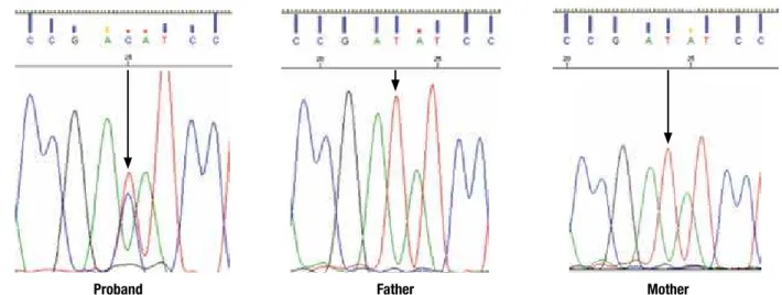
Direct sequencing of the coding region of the GLUD1 gene revealed that the affected child was heterozygous for a missense mutation in exon 11 (c.1491A>G; p.Ile497Met; based on NCBI Accession Number NM_005271.3). Neither parents carried this variant (Figure 1), suggesting a “de novo” mutation, which could not be deinitively conirmed because a pa-ternity test was not performed.
Figure 1. Reverse strand sequencing electropherogram showing the substitution of T by C at nucleotide 1491 (c.1491>G), resulting in the replacement of isoleucine by methionine at codon 497 (p.I497M) of the GLUD1 gene in the proband. The mutation was absent in the parents.
Proband Father Mother
glycemia is variable, and it is generally corrected by the administration of diazoxide (2).
In the present report, we describe the case of a Bra-zilian patient with HI/HA syndrome carrying an acti-vating mutation in the GLUD1 gene.
CASE REPORT
Cop
yright
© ABE&M t
odos os dir
eit os r eser vados .
DISCUSSION
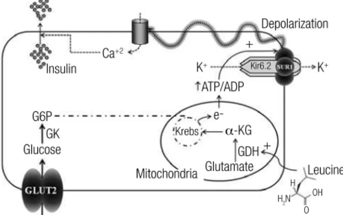
Hyperinsulinism is one of the most common causes of neonatal and childhood hypoglycemia (3). Insulin se-cretion by pancreatic beta cells is triggered by increased intracellular calcium concentrations. On the surface of these cells, potassium channels composed of Kir6.2 and SUR1 proteins control the polarization of the cell membrane by opening and closing its channels in re-sponse to increased or decreased concentration of in-tracellular adenosine triphosphate (ATP), respectively. When these channels are closed, the cell depolarizes, enabling the opening of calcium channels, increased in-tracellular concentrations of this ion, and consequent release of insulin (4) (Figure 2).
causes opening of potassium channels (which is nor-mal), and corrects hypoglycemia (Figure 2) (5).
The major regulator of insulin secretion is glucose which, in its metabolism, generates ATP and guano-sine triphosphate (GTP). In addition to glucose, other substrates may also generate ATP and stimulate insulin secretion, such as fatty acids and the amino acids gluta-mate and leucine. Glutagluta-mate is the substrate of GDH, an enzyme that catalyzes its oxidative deamination to alpha-ketoglutarate (α-KG) and ammonia. Within pan-creatic beta cells, α-KG enters the Krebs’ cycle, leading to an increased generation of ATP. Leucine, which is present in almost all proteins ingested, is a direct stimu-lator of GDH (Figure 3) (2,4). Under hyperglycemic conditions, however, the amino acids do not stimulate insulin release, as ATP, and mostly GTP, both gene-rated during glucose metabolization, inhibit intracel-lular GDH (6). Thus, GDH is allosterically activated by leucine and inhibited by GTP (7).
G6P GK Glucose Glucose b-cell Diazoxide Mitochondria ATP/ADP K+ K+ K+ + Ca+2 Ca+2 ATP:ADP GTP ADP GDH Glutamate -+ + Insulin Insulin Liver N-acetylglutamate Glutamate
NH3CPS Urea + + GDH α-KG Krebs e-α-KG α-KG Glutamate GDH+ Leucine Leucine Depolarization Depolarization OH OH Kir6.2 Kir6.2 O O H2N
H2N H
H
Figure 2. Mechanisms of insulin secretion in pancreatic b-cells. α-KG,
α-ketoglutarate; ADP, adenosine diphosphate; ATP, adenosine triphosphate; Ca+2, calcium ion; e-, electron; G6P, glucose 6-phosphate; GDH, glutamate
dehydrogenase; GLUT2, glucose transporter type 2; GK, glucokinase; K+,
potassium ion; Kir6.2, inward-rectiier potassium ion channel subunit 6.2; SUR1, sulfonylurea receptor 1. Adapted from Giurgea and cols. (5).
There are two basic mechanisms associated with ab-normal increase of insulin secretion by the beta cells: (1) defects in potassium channels (channelopathies) due to inactivating mutations in ABCC8 and KCNJ11 genes, which encode, respectively, the proteins SUR1 and Kir6.2. These mutations lead to constitutive clo-sure of potassium channels, so that beta cell membranes remain continuously depolarized, allowing constant insulin secretion irrespective of intracellular concen-trations of ATP. These mutations are usually inherited in an autosomal recessive manner, and result in severe hypoglycemia during the neonatal period. In such situ-ations, diazoxide, a drug that acts on potassium chan-nels, is ineffective; and (2) increased generation of mi-tochondrial ATP (metabolopathies), with consequent closure of potassium channels and increased insulin secretion. In this case, the administration of diazoxide
Figure 3. Mechanisms of hyperinsulinism and hyperammonemia in HI/HA syndrome. α-KG, α-ketoglutarate; ADP, adenosine diphosphate; ATP, adenosine triphosphate; Ca+2, calcium ion; CPS, carbamoyl phosphate
synthetase; GDH, glutamate dehydrogenase; GTP, guanosine triphosphate; K+, potassium ion; Kir6.2, inward-rectiier potassium ion channel subunit
6.2; SUR1, sulfonylurea receptor 1. Adapted from Stanley and cols. (11).
In the presence of activating mutations in the gene encoding GDH, there is a reduction in the sensitivity of the enzyme to allosteric inhibition by GTP and ATP, followed by increased response of GDH to leucine, increased deamination of glutamate, and consequent rise in ATP production, which causes excessive insu-lin secretion from beta cells in presence of glutamate and leucine. This sequence of events explains hyperin-sulinemic hypoglycemia that occurs during fasting, and particularly in the postprandial period after protein in-gestion (5,6,8).
Cop
yright
© ABE&M t
odos os dir
eit
os r
eser
vados
.
diagnosis of the disease – and in impaired urea pro-duction. In the liver, increased GDH activity converts all glutamate into ammonia and α-KG. Urea synthesis from ammonia is carried out by the action of carbamoyl phosphate synthetase (CPS), an enzyme activated by N-acetylglutamate (NAG), which is decreased as a re-sult of GDH overactivity (2,9).
HI-HA syndrome was irst described in 1977 by Weinzimer and cols. (10) in two boys with severe hy-perinsulinemic hypoglycemia together with hyperam-monemia. In 1998, Stanley and cols. studied eight chil-dren with the syndrome, identiied the gene GLUD1 and unraveled the pathophysiological mechanisms in-volved in hyperinsulinemia and hyperammonemia (11). In 2002, in a multicenter series of 175 patients, hyper-ammonaemia was found in 12 out of 69 tested patients with hyperinsulinemic hypoglycemia (12). Although the disease is rare, several case reports and some reviews have been published (2,9,13-18). An interesting clini-cal aspect of HI-HA syndrome is that epilepsy is a fre-quent inding; in a cohort of 16 patients, 15 presented seizures and 43% of them developed epilepsy. This is probably explained not only by recurrent hypoglyce-mia, but also by chronic hyperammonaemia and by decreased brain concentrations of the neurotransmitter GABA due to increased GDH activity (15).
To date, GLUD1 mutations described were located in exons 6, 7, 10, 11 and 12, encoding the catalytic and allosteric domains of GDH (15). The mutation c.1491A>G; p.Ile497Met found at exon 11 of the Brazilian patient was also described in a Japanese pa-tient (the mutation was previously named c.1504A>G; p.Ile444Met), and the functional study revealed that the inhibitory effects of GTP on GDH activity were decreased in presence of this mutation (18).
In conclusion, HI/HA syndrome is a serious condi-tion with harmful consequences related to permanent brain damage that occurs when diagnosis is delayed. Diagnosis should be considered in all infants with hy-perinsulinemic hypoglycemia, and should motivate the determination of ammonia concentrations. It is worthy commenting that the accuracy of ammonia measure-ment is extremely dependent on sample collection. Blood samples should be collected from a stasis-free vein into an EDTA evacuated tube, which must be immediately placed on ice and delivered to the lab as quickly as possible; plasma should be separated from the sample without delay, and ammonia analysis per-formed within 30 minutes. Some rare patients may ex-hibit serum ammonia within the normal range. If HI/
HA is highly suspected in presence of normal serum ammonia, the sensitivity to leucine can be assessed by an oral leucine tolerance test, with administration of 0.15 g/kg of leucine after a 4-hour fasting and deter-mination of plasma glucose and serum insulin at times -30, 0, 30, 60, 90 and 120 minutes. Patients with HI/ HA syndrome develop hypoglycemia induced by leu-cine (15). For deinitive diagnosis, direct sequencing of the GLUD1 gene should be performed.
Disclosure: no potential conlict of interest relevant to this article was reported.
REFERENCES
1. Miki Y, Taki T, Ohura T, Kato H, Yanagisawa M, Hayashi Y. Novel missense mutations in the glutamate dehydrogenase gene in the congenital hyperinsulinismhyperammonemia syndrome. J Pedi atr. 2000;136(1):6972.
2. Palladino AA, Stanley CA. The hyperinsulinism/hyperammone mia syndrome. Rev Endocr Metab Disord. 2010;11(3):1718. 3. Daly LP, Osterhoudt KC, Weinzimer SA. Presenting features of id
iopathic ketotic hypoglycemia. J Emerg Med. 2003;25(1):3943. 4. Palladino AA, Bennett MJ, Stanley CA. Hyperinsulinism in infancy
and childhood: when an insulin level is not always enough. Clin Chem. 2008;54(2):25663.
5. Giurgea I, BellanneChantelot C, Ribeiro M, Hubert L, Sempoux C, Robert JJ, et al. Molecular mechanisms of neonatal hyperinsulin ism. Horm Res. 2006;66(6):28996.
6. Kelly A, Ng D, Ferry RJ Jr, Grimberg A, KooMcCoy S, Thornton PS, et al. Acute insulin responses to leucine in children with the hyperinsulinism/hyperammonemia syndrome. J Clin Endocrinol Metab. 2001;86(8):37248.
7. Fahien LA, MacDonald MJ, Kmiotek EH, Mertz RJ, Fahien CM. Regulation of insulin release by factors that also modify gluta mate dehydrogenase. J Biol Chem. 1988;263(27):136104. 8. Hsu BY, Kelly A, Thornton PS, Greenberg CR, Dilling LA, Stanley
CA. Proteinsensitive and fasting hypoglycemia in children with the hyperinsulinism/hyperammonemia syndrome. J Pediatr. 2001;138(3):3839.
9. Stanley CA. Hyperinsulinism/hyperammonemia syndrome: in sights into the regulatory role of glutamate dehydrogenase in ammonia metabolism. Mol Genet Metab. 2004;81 Suppl 1:S4551. 10. Weinzimer SA, Stanley CA, Berry GT, Yudkoff M, Tuchman M,
Thornton PS. A syndrome of congenital hyperinsulinism and hy perammonemia. J Pediatr. 1997;130(4):6614.
11. Stanley CA, Lieu YK, Hsu BY, Burlina AB, Greenberg CR, Hopwood NJ, et al. Hyperinsulinism and hyperammonemia in infants with regulatory mutations of the glutamate dehydrogenase gene. N Engl J Med. 1998;338(19):13527.
12. de Lonlay P, Fournet JC, Touati G, Groos MS, Martin D, Sevin C, et al. Heterogeneity of persistent hyperinsulinaemic hypoglycae mia. A series of 175 cases. Eur J Pediatr. 2002;161(1):3748. 13. ElGharbawy AH. Hyperinsulinism/hyperammonemia syndrome:
a synopsis. Mol Genet Metab. 2005;84(2):1013.
Cop
yright
© ABE&M t
odos os dir
eit
os r
eser
vados
.
15. Kapoor RR, Flanagan SE, Fulton P, Chakrapani A, Chadefaux B, BenOmran T, et al. Hyperinsulinismhyperammonaemia syn drome: novel mutations in the GLUD1 gene and genotypephe notype correlations. Eur J Endocrinol. 2009;161(5):7315. 16. Diao C, Chen S, Xiao X, Wang T, Sun X, Wang O, et al. Two un
related Chinese patients with hyperinsulinism/hyperammonemia (HI/HA) syndrome due to mutations in glutamate dehydrogenase gene. J Pediatr Endocrinol Metab. 2010;23(7):7338.
17. Stanley CA. Two genetic forms of hyperinsulinemic hypoglyce mia caused by dysregulation of glutamate dehydrogenase. Neu rochem Int. 2011;59(4):46572.