Chronic Endotoxemia in Subjects with Type-1
Diabetes Is Seen Much before the Onset of
Microvascular Complications
Vivekanandhan Aravindhan1,2*, Viswanathan Mohan3, Namasivayam Arunkumar4, Sreedharan Sandhya1, Subash Babu5
1AU-KBC Research Centre, MIT Campus of Anna University, Chennai, India,2Dept of Genetics, Dr ALM PG IBMS, University of Madras, Taramani, Chennai, 600113, India,3Madras Diabetes Research Foundation & Dr. Mohan’s Diabetes Specialties Centre, WHO Collaborating Centre for Non-Communicable Diseases Prevention and NGT, International Diabetes Federation (IDF) Centre for Education, Chennai, India,
4Biostatistics & Programming, Algorithm Inc, Bengaluru, India,5National Institutes of Health-International Center for Excellence in Research, National Institute for Research in Tuberculosis, Chennai, India
*cvaravindhan@gmail.com
Abstract
Background
Lipopolysaccharide (LPS)/Endotoxin is hypothesized to play an important role in chronic inflammation associated with Type-1 diabetes (T1DM) and its complications. Endotoxin core antibodies (EndoCAb), LPS binding protein (LBP) and soluble CD14 (sCD14) act as modulators of LPS induced activation of innate immune systemin vivo. For the present
study we estimated the levels of LPS and its translocation markers in T1DM subjects with and without microvascular complications (MVC) and correlate them with clinical parameters of T1DM and serum inflammatory cytokine levels (TNF-α, IL-6, IL-1βand GM-CSF).
Methods
A total of 197 subjects (64 normal glucose tolerance (NGT) subjects, 97 T1DM subjects without MVC and 36 with MVC) were included in this study and the levels of serum LPS, its translocation markers and cytokines measured by immunoassays.
Results
Compared to NGT, T1DM subjects (both with and without MVC) had significantly higher lev-els of LPS, reduced levlev-els of LBP and EndoCAb along with significant increase in the levlev-els of IL-1β, IL-6, TNF-αand GM-CSF (p<0.05). No significant change was seen in the levels of these biomarkers between T1DM subjects with and without MVC.
Conclusions
Decreased levels of EndoCAb and LBP suggest sustained endotoxin activity in T1DM sub-jects even before the onset of microvascular complications.
a11111
OPEN ACCESS
Citation:Aravindhan V, Mohan V, Arunkumar N, Sandhya S, Babu S (2015) Chronic Endotoxemia in Subjects with Type-1 Diabetes Is Seen Much before the Onset of Microvascular Complications. PLoS ONE 10(9): e0137618. doi:10.1371/journal. pone.0137618
Editor:Alan Stitt, Queen's University Belfast, UNITED KINGDOM
Received:May 9, 2015
Accepted:August 20, 2015
Published:September 14, 2015
Copyright:This is an open access article, free of all copyright, and may be freely reproduced, distributed, transmitted, modified, built upon, or otherwise used by anyone for any lawful purpose. The work is made available under theCreative Commons CC0public domain dedication.
Data Availability Statement:All relevant data are within the paper and its Supporting Information files.
Background
Type-1 diabetes mellitus (T1DM) is a bonafide autoimmune disorder characterized by chronic inflammation with rapid loss of pancreatic beta cells leading to insulin deficiency [1]. However, the exact cause of autoimmunity is not known, even though, both genetic [2] and environmen-tal factors have been implicated [3]. The prevalence of T1DM and other autoimmune diseases has increased dramatically over the past few decades [4]. Although genetic factors may play an important role in the susceptibility to T1DM, the dramatic worldwide increase in prevalence is likely due to changes in the environmental factors [5]. One environmental factor which has gained prominence in recent years is the alterations in gut microbiota which leads to increased gut permeability and metabolic endotoxemia [6]. Bacterial Lipopolysaccaride (LPS)/ endo-toxin, a unique glycolipid located at the outer membrane of gram negative bacteria can induce inflammation [7]. More than the actual endotoxin levels, the levels of endogenous anti-endo-toxin core antibodies (EndoCAb), LPS binding protein (LBP) and soluble CD14 (sCD14) were found to be more important in determining the activity of endotoxin underin vivoconditions
[8]. Studies reporting levels of EndoCAb, LBP and sCD14 in T1DM subjects are not available. While EndoCAb antibodies can neutralize LPS, sCD14 and LPB can augment/antagonize LPS activity in a dose dependent manner. Recent studies have demonstrated chronic inflammation in T1DM subjects with microvascular complications (MVC) with systemic increase in the lev-els of pro-inflammatory cytokines [9]. We hypothesized that a systemic increase in endotoxe-mia could aggravate inflammation and promote MVC. The objective of the present study was to estimate the levels of LPS and its translocation markers in T1DM subjects with/without MVC (DN and/or DR) and correlate them with clinical parameters for T1DM and serum inflammatory cytokines (TNF-α, IL-6, IL-1βand GM-CSF).
Materials and Methods
Study participants
Patients with T1DM (n = 133; 97 T1DM without MVC and 36 T1DM with MVC) were recruited from Dr. Mohan’s Diabetes Specialties Centre, Chennai, India. T1DM was diagnosed by the absence of insulin reserve shown by C-peptide assay (C-peptide values<0.3 pg/ml) and requiring insulin from the time of diagnosis. Subjects with serum glutamic acid decarboxylase (GAD)–specific autoantibody levels10 IU/ml were classified as GAD+. Only fasting blood samples were used for all analysis. Institutional Ethical Committee approval was obtained from the Madras Diabetes Research Foundation Ethics Committee (Ref. No.MDRF-EC/SOC/2009/ 05), and written informed consent was obtained from all the study participants. The study was conducted as per principles of the declaration of Helsinki as revised in 2008.
Study Design and sample size calculation
It is a cross-sectional observational study. Initially, 20 normal glucose tolerant (NGT) and 20 age and gender matched T1DM subjects with and without MVC were used for analysis. On the basis of the preliminary results, with a confidence interval of 95%, an estimated p value<0.05, and a power of 80%, the sample size was estimated to be 150 ie, 60 NGT subjects, 60 T1DM subjects without MVC and 30 T1DM with MVC. Few more samples were included to account for the large variation seen in serum biomarker levels.
Estimation of biochemical parameters
Blood parameters were measured using a Hitachi-912 Autoanalyser (Hitachi, Mannheim, Ger-many). Glycated hemoglobin (HbA1c) was estimated by high pressure liquid chromatography
and analysis, decision to publish, or preparation of the manuscript.
Competing Interests:The authors have declared that no competing interests exist.
Abbreviations:T1DM, Type-1 diabetes mellitus; MVC, microvascular complication; NGT, Normal glucose tolerance; LPS, Lipopolysaccharide; LBP, Lipopolysaccharide binding protein; sCD14, Soluble cluster differentiation 14; EndoCAb, Endotoxin core antibody; TNF-α, Tumor necrosis factor- alpha; IL-1β, Interleukin-1 beta; GM-CSF, Granulocyte
(Bio-Rad, Hercules, CA). Urine samples were collected in the early morning after an overnight fast. Urine creatinine was measured using Jaffe’s method. Urine microalbumin concentration was measured using commercially available immunoturbidometric assay kits from Randox (Randox, UK) on Opera Technicon Auto Analyser (Bayer Diagnostics, USA). The urine sam-ple was added to a buffer containing anti-albumin antibody. The turbidity of the resulting solu-tion was measured and the albumin concentrasolu-tion was determined by constructing a standard curve with known concentrations of albumin. The mean inter-assay and intra-assay coefficient of variation were 3.4% and 2.4% respectively. Microalbuminuria was diagnosed if the albumin excretion was between 30 and 299μg/mg of albumin [10]. The expected protein excretion
(EPE) was calculated as the urinary protein to creatinine ratio [11]. The intra- and inter assay coefficient of variation for the biochemical assays ranged between 3.1% and 5.6%.
Screening for microvascular and macrovascular complications
All T1DM subjects were screened for both microvascular (diabetic retinopathy/DR, diabetic nephropathy/DN and diabetic neuropathy) and macrovascular complications (diabetic coro-nary artery disease/ DM-CAD and perivascular diseases/DM-PVD).
Doppler screening. Doppler screening for PVD was performed by recording of pressure tracings while in the supine position by doppler probe using the KODY Vaslab Machine (Kody Labs, Chennai, India). The ankle–brachial index (ABI) ratio was calculated in every subject as previously described [12].
Retinal photography. Screening for retinopathy was done using four-field stereo colour retinal photography (Zeiss FF 450 plus camera) which were graded by an ophthalmologist using the Early Treatment Diabetic Retinopathy Study (ETDRS) grading system as previously described [13].
Biothesiometry studies. Biothesiometer (Biomedical Instrument Co., Newbury, OH, USA) was used to assess vibratory perception threshold (VPT) of the great toes in a standard-ized fashion as previously described [12].
Electrocardiogram. Resting 12-lead electrocardiogram (ECG) was performed using Myo-card R electroMyo-cardiograph (Marks Electronics, Chennai, India) to asses CAD. Carotid Intimal Medial Thickness (IMT) was measured as previously described [13].
Arterial stiffness measurement. Arterial stiffness was measured using the Sphygmocor apparatus (Sphygmocor BPAS-1; PWV Medical, Sydney, Australia). Augmentation index was defined as the difference between the first and second peaks of the central arterial waveform, expressed as a percentage of the pulse pressure as previously described [13].
Definitions and cutoffs
Hypertension. It was diagnosed in subjects who were on antihypertensive medication or had systolic BP140 mmHg or diastolic BP90 mmHg as previously described [12,13].
Diabetic Perivascular disease. An ABI of0.9 was the criterion used for the diagnosis of PVD as per American College of Cardiology Foundation/American Heart Association Task Force (ACCF/AHA) 2011 Guidelines as previously described [12,13].
Diabetic Coronary Artery Disease. This was diagnosed based on a past history of docu-mented myocardial infarction and/or drug treatment for CAD (aspirin or nitrates) and/or elec-trocardiographic changes suggestive of ST segment depression and/or Q-wave changes and/or T-wave changes using appropriate Minnesota codes as previously described [12,13].
Diabetic Neuropathy. Diagnosed if VPT of the great toe exceeded mean + 2 SD of a healthy nondiabetic population aged 20–45 years (cut point20 V) as previously described [12,13].
Diabetic Nephropathy. Microalbuminuria was diagnosed if the albumin excretion was between 30 and 299μg/mg of creatinine and macroalbuminuria/overt nephropathy was
diag-nosed if albumin excretion was300μg/mg of creatinine as previously described [12,13].
Inclusion and exclusion criteria
Only adult type-1 diabetic subjects (age 30–50) were included in the study. The exclusion crite-ria were patients with type-2 diabetes and T1DM subjects with macrovascular complications including cardiovascular disease and perivascular disease. Patients with a previous diagnosis of urolithiasis or any known renal disease, liver cirrhosis, congestive heart failure, chronic lung diseases, chronic infections and viral hepatitis were also excluded.
Detection of serum LPS and translocation markers
Serum lipopolysaccaride (LPS) activity was determined with a Limulus amoebocyte lysate assay (Hycult Biotechnology). Serum LBP (Hycult Biotechnology), IgG antibody to LPS core (EndoCAb) (Hycult Biotechnology) and sCD14 (R&D Systems, Abingdon, United Kingdom) levels were measured by ELISA. The detection limit for LPS, LBP, sCD14 and EndoCAb were 0.04 Endotoxin Units (EU) /ml, 4.4 ng/ml, 0.125ng/ml and 0.125 EndoCAb Median Unit (EMU)/ml respectively. One EU equals approximately 0.1 to 0.2ng endotoxin/mL.
Estimation of serum pro-inflammatory cytokines
Serum levels of IL-1beta, IL-6, TNF-alpha and GM-CSF were determined by ELISA following kit protocol (Invitrogen CytoSetTMfor IL-1β, IL-6 and TNF-α; R&D DuoSet Antibody pair for GM-CSF). The lowest detection limits were: TNF-α-1.0 pg/ml, IL-6- 0.1 pg/ml, IL-1β-0.01 pg/ ml and GM-CSF-0.01pg/ml.
Statistical analysis
Student t-test was used to compare groups for continuous variables, whereasχ2 test or Fisher exact test (as appropriate) was used to compare proportions. Mann–Whitney U test was used for multiple pair-wise comparisons that did not show normal distribution. Multiple compari-sons were corrected using the Holm’s correction. Spearman’s Correlation analysis was per-formed between serum inflammatory markers and clinical parameters with the diabetic group. Since there was no statistical difference in the levels of inflammatory markers between the T1DM and T1DM-MVC groups, both groups were pooled for the correlation analysis. All the analyses were done using GraphPad Prism version 5.0 (GraphPad Software, CA, USA) and SPSS statistical package (Version 20.0; SPSS, Chicago, IL). p value less than 0.05 was considered significant.
Results and Discussion
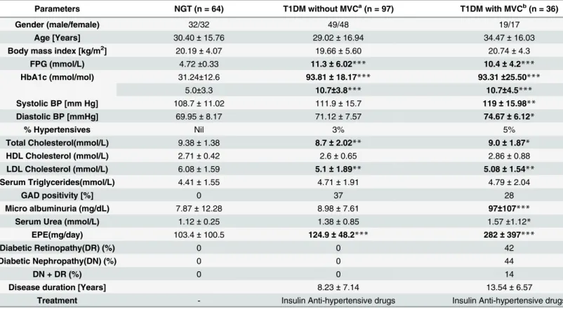
Table 1shows the clinical and biochemical characteristics of the study subjects. Compared
phenotype. Systolic BP, diastolic BP, serum urea and microalbuminuria were significantly ele-vated only in the T1DM-MVC group. 3% of T1DM and 5% (T1DM-MVC) had hypertension and were under hypertensive drugs. 37% of T1DM and 28% T1DM-MVC were GAD antibody positive. All the T1DM subjects had well established disease with minimal pancreatic beta cell reserve (as determined by C-peptide assay) and were under insulin. Among subjects with MVC, 42% had diabetic retinopathy (DR), 44% had diabetic nephropathy (DN) and 14% had both DN and DR.
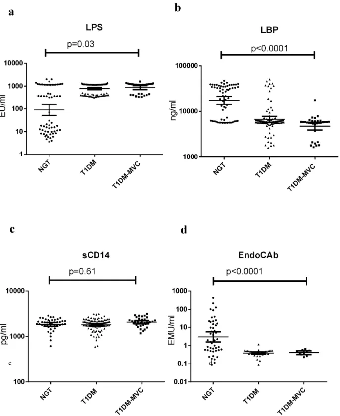
Fig 1shows the serum levels of LPS, LBP, sCD14 and EndoCAb in NGT and T1DM subjects
(with and without MVC). Compared to NGT, T1DM subjects (both with and without MVC) had significantly higher levels of LPS, reduced levels of LBP and EndoCAb with no major dif-ference in the sCD14 levels. T1DM subjects had undetectable levels of EndoCAb. No signifi-cant difference was seen in these biomarkers between T1DM subjects with and without MVC. At least among the NGT subjects (Fig 1a), two distinct sub-sects could be identified: one with high levels (MEAN 915 EU/ml) and another with low levels (MEAN 12 EU/ml) of LPS. On fur-ther analysis it was found that subjects who had high levels of endotoxemia also had high levels of IL-6, TNF-αand GM-CSF and low levels of LBP and EndoCAb. Interestingly this group also
Table 1. Clinical characteristics of the study population.
Parameters NGT (n = 64) T1DM without MVCa(n = 97) T1DM with MVCb(n = 36)
Gender (male/female) 32/32 49/48 19/17
Age [Years] 30.40±15.76 29.02±16.94 34.47±16.03
Body mass index [kg/m2] 20.19±4.07 19.66±5.60 20.74±4.3
FPG (mmol/L) 4.72±0.33 11.3±6.02*** 10.4±4.2***
HbA1c (mmol/mol) 31.24±12.6 93.81±18.17*** 93.31±25.50***
5.0±3.3 10.7±3.8*** 10.7±4.5***
Systolic BP [mm Hg] 108.7±11.02 111.9±15.7 119±15.98**
Diastolic BP [mmHg] 69.95±8.17 71.12±7.57 74.67±6.12*
% Hypertensives Nil 3% 5%
Total Cholesterol(mmol/L) 9.38±1.38 8.7±2.02** 9.0±1.87*
HDL Cholesterol (mmol/L) 2.71±0.42 2.6±0.65 2.86±0.88
LDL Cholesterol (mmol/L) 6.08±1.59 5.1±1.89** 5.08±1.54**
Serum Triglycerides(mmol/L) 4.41±1.55 4.71±1.91 4.79±2.04
GAD positivity [%] 0 37 28
Micro albuminuria (mg/dL) 7.87±12.28 8.98±7.61 97±107***
Serum Urea (mmol/L) 1.12±0.25 1.38±0.85 1.57±1.12*
EPE(mg/day) 103.4±100.5 124.9±48.2*** 282±397***
Diabetic Retinopathy(DR) (%) 0 0 42
Diabetic Nephropathy(DN) (%) 0 0 44
DN + DR (%) 0 0 14
Disease duration [Years] 8.23±7.14 13.54±6.57
Treatment - Insulin Anti-hypertensive drugs Insulin Anti-hypertensive drugs
a- Comparison between NGT and T1DM; b- Comparison between NGT and T1DM-MVC.
Data are given as mean±SD for continuos variables and as % for proportions. Statistical significance was determined by One-way ANOVA. *p<0.05;
**p<0.01; ***p<0.0001.
FPG-fasting plasma glucose; HbA1C-glycated hemoglobin. Letters in bold highlight values which are significant.
Fig 1. T1DM is characterized by systemic endotoxemia with significantly decreased levels of LBP and EndoCAb.Serum levels of LPS (a), LBP (b), sCD14 (c) and EndoCAb (d) were determined in NGT (n = 64), T1DM without MVC (n = 97) and T1DM subjects with MVC (n = 36) by immune-assays. Each dot represents individual values with the horizontal line representing the geo mean. 36 T1DM subjects without MVC and 26 with MVC had undetectable levels of EndoCAb. Significance was calculated by non-parametric Mann–Whitney U test and p<0.05 was considered significant. The indicated p values are
for pair-wise comparisons.
had significantly elevated levels of triglyceridemia (data not shown). Within the T1DM-MVC group, LPS, LBP, sCD14 and EndoCAb levels were not significantly different between those who had retinopathy from those who had microalbuminuria (LPS: DR(Mean ± SD)-1035±354 EU/ml, DN (Mean ± SD)- 943.8±441 EU/ml, p = 0.4; LBP:DR(Mean ± SD)-5± 1.8μg/ml, DN
(Mean ± SD)-6±3.9μg/ml, p = 0.5; sCD14: DR(Mean ± SD)- 2.2±0.48μg/ml and DN
(Mean ± SD)-2.0±0.46μg/ml, p = 0.2 and EndoCAb: DR(Mean ± SD)- 0.38±0.16 EMU/ml and
DN(Mean ± SD)- 0.43±0.09 EMU/ml, p = 0.7).
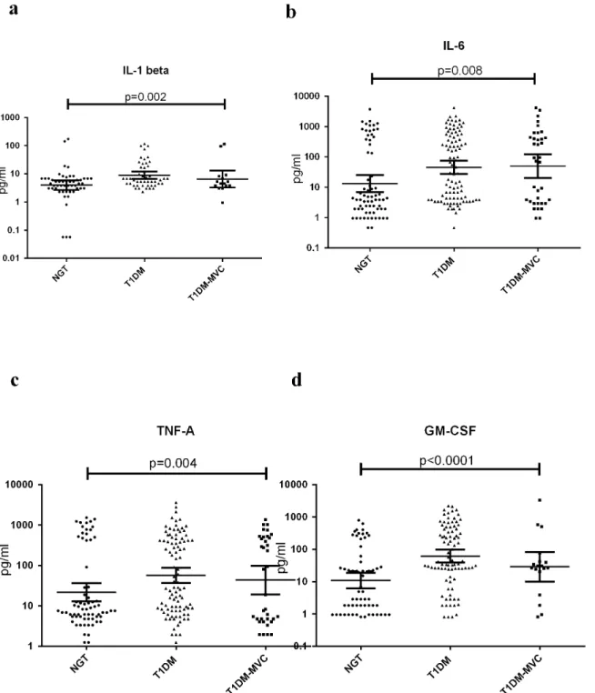
Fig 2shows the serum levels of TNF-α, IL-6, IL-1βand GM-CSF in NGT and T1DM
sub-jects (with and without MVC). Compared to NGT, TIDM subsub-jects (both with and without MVC) had significantly elevated levels of TNF-α, IL-6, IL-1βand GM-CSF in the serum. No significant difference was seen in cytokine levels between T1DM subjects with and without MVC. Spearman’s correlation analysis within the diabetic group (S1 Table) showed positive correlation of LPS levels with FGP, EPE, IL-1β, IL-6, TNF-αand GM-CSF. LBP levels showed a negative correlation with BMI, FPG, SBP, DBP, TGL, EPE, IL-1βand IL-6 levels. EndoCAb showed negative correlation with FPG, EPE TNF-αand GM-CSF levels.
In the present study, we attempted to investigate LPS and its translocation markers in T1DM subjects with/without MVC by measuring serum LPS, EndoCAb, sCD14 and LBP along with serum cytokines and other clinical parameters. The major findings of the study are: 1. T1DM subjects with/without MVC had significantly higher levels of LPS and lower levels of LBP and EndoCAb with no significant change in the sCD14 levels compared to NGT. 2. How-ever, even for these markers, there was no significant difference between T1DM subjects with and without MVC and 3. While LPS showed a positive correlation FPG and pro-inflammatory cytokines (IL-1β, IL-6 and TNF-α), LBP and EndoCAb showed negative association with these markers.
Changes in gut microbiota leading to leaky gut have now been identified as a major etiologi-cal factor for chronic inflammation as seen in T1DM [14]. Previously, increased levels of serum Zonulin, a gap-junction protein which serves as a marker for gut permeability was found to be significantly elevated in T1DM subjects indicating leaky gut [15]. However, com-paratively very few studies have looked at the end result of leaky gut which is the metabolic endotoxemia and associated inflammation. Recently, increased levels of serum LPS in T1DM subjects with metabolic syndrome and/or kidney disease was reported [16,17]. However, the levels of sCD14, LBP and EndoCAb which are the major regulators of LPS activity underin vivoconditions were not reported in these subjects. LBP is an acute-phase protein that
[28]. But the significance of EndoCAb levels under conditions of autoimmunity is less well explored. Our study for the first time showed low levels of EndoCAb in T1DM subjects.
The moderate endotoxemia with decreased EndoCAb and LBP together suggests sustained immune activation resulting in chronic inflammation. The end result is a systemic increase in
Fig 2. T1DM is characterized by systemic inflammation with high levels of serum pro-inflammatory cytokines.Serum levels of IL-1β(a), IL-6 (b), TNF-α(c), and GM-CSF (d) were determined in NGT (n = 64), T1DM without MVC (n = 97) and T1DM subjects with MVC (n = 36) by ELISA. Each dot represents individual values with the horizontal line representing the geo mean. Significance was calculated by non-parametric Mann–Whitney U test and p<0.05 was considered significant. The indicated p values are for pair-wise comparisons.
the levels of IL-1β, TNF-α, IL-6 and GM-CSF, as seen in this study. Several immune and non-immune cells express LPS receptor (TLR4) and respond to LPS stimulation by secreting pro-inflammatory cytokines [29]. The end result is a systemic increase in the levels of IL-1β, TNF-α, IL-6 and GM-CSF in the T1DM subjects as seen in this study. Our results are in agreement with previous reports were in elevated levels of IL-1βand IL-6 were reported in T1DM subjects [30]. Recently we reported increased levels of serum GM-CSF levels in T2DM subjects which were associated with the activated phenotype of dendritic cells [31]. Like T2DM, GM-CSF lev-els were found to be elevated even in T1DM subjects as can be seen in this study.
Conclusion
To summarize, chronic low-grade endotoxemia, as seen in the case of T1DM, was associated with decreased levels of LBP and EndoCAb and with elevated levels of TNF-α, IL-6, IL-1βand GM-CSF, indicating chronic inflammation. The most important finding of this study is that, this phenomenon was seen even during the early stages of the disease and seems to persist till the late stages of the disease were in MVC sets in, indicating that, yet to be identified factors may play a role in the onset of MVC in T1DM subjects. One of the limitations of this study is that, being cross-sectional in nature, no conclusions regarding a causal relationship between autoimmunity, endotoxemia and inflammation can be made. Further, since the prevalence of T1DM is less than 1% [32] this study was performed in a limited sample size with known dia-betic subjects. However, the strength of this study is that, it systematically reviews all the com-ponents of microbial translocation in age and gender matched T1DM subjects in a non-Caucasians population were in the incidence of T1DM has started increasing. In summary, this study suggests that T1DM in Asian Indians is characterized by high levels of LPS and pro-inflammatory cytokines and low levels of EndoCAb and LBP.
Supporting Information
S1 Table. Spearman’s Correlation analysis for LPS and translocation markers with clinical parameters and log cytokine concentration within the diabetic group (n = 133).
(DOC)
Acknowledgments
The project was partially funded by DST FAST TRACK (http://www.serb.gov.in/home.php) grant obtained by VA (SR/FT/LS-105/2009). This study was also supported by the Intramural Research Program of the Division of Intramural Research, NIAID, NIH, US. The Dept of Genetics, University of Madras has received funds for infrastructural support from DST-FIST and UGC-SAP programs. The funders had no role in study design, data collection and analysis, decision to publish, or preparation of the manuscript.
Author Contributions
Conceived and designed the experiments: VA VM SB. Performed the experiments: VA SS. Analyzed the data: VA NA. Contributed reagents/materials/analysis tools: VA VM SB. Wrote the paper: VA VM NA SS SB.
References
2. Notkins AL. Immunologic and genetic factors in type 1 diabetes. J Biol Chem 2002; 277: 43545–8. PMID:12270944
3. Peng H, Hagopian W. Environmental factors in the development of Type 1 diabetes. Rev Endocr Metab Disord 2006; 7: 149–62. PMID:17203405
4. Cooper GS, Bynum ML, Somers EC. Recent insights in the epidemiology of autoimmune diseases: improved prevalence estimates and understanding of clustering of diseases. J Autoimmun 2009; 33: 197–207. doi:10.1016/j.jaut.2009.09.008PMID:19819109
5. Nistico L, Iafusco D, Galderisi A, Fagnani C, Cotichini R, Toccaceli V, et al. Emerging effects of early environmental factors over genetic background for type 1 diabetes susceptibility: evidence from a Nationwide Italian Twin Study. J Clin Endocrinol Metab 2012; 97: E1483–91. PMID:22569240
6. Vaarala O. Leaking gut in type 1 diabetes. Curr Opin Gastroenterol 2008; 24: 701–6. doi:10.1097/ MOG.0b013e32830e6d98PMID:19122519
7. Bosshart H, Heinzelmann M. Targeting bacterial endotoxin: two sides of a coin. Ann N Y Acad Sci 2007; 1096: 1–17. PMID:17405910
8. Mussap M, Noto A, Fravega M, Fanos V. Soluble CD14 subtype presepsin (sCD14-ST) and lipopoly-saccharide binding protein (LBP) in neonatal sepsis: new clinical and analytical perspectives for two old biomarkers. J Matern Fetal Neonatal Med 24 Suppl 2: 12–4.
9. Navarro-Gonzalez JF, Mora-Fernandez C. The role of inflammatory cytokines in diabetic nephropathy. J Am Soc Nephrol 2008; 19: 433–42. doi:10.1681/ASN.2007091048PMID:18256353
10. Unnikrishnan RI, Rema M, Pradeepa R, Deepa M, Shanthirani CS, Deepa R, et al. Prevalence and risk factors of diabetic nephropathy in an urban South Indian population: the Chennai Urban Rural Epidemi-ology Study (CURES 45). Diabetes Care 2007; 30: 2019–24. PMID:17488949
11. Mohan V, Shah S, Saboo B. Current glycemic status and diabetes related complications among type 2 diabetes patients in India: data from the A1chieve study. J Assoc Physicians India 2013; 61: 12–5.
12. Eshcol J, Jebarani S, Anjana RM, Mohan V, Pradeepa R. Prevalence, incidence and progression of peripheral arterial disease in Asian Indian type 2 diabetic patients. J Diabetes Complications 2014; 28: 627–31. doi:10.1016/j.jdiacomp.2014.04.013PMID:24930714
13. Pradeepa R, Surendar J, Indulekha K, Chella S, Anjana RM, Mohan V. Relationship of diabetic retinop-athy with coronary artery disease in Asian Indians with type 2 diabetes: the Chennai Urban Rural Epide-miology Study (CURES) Eye Study—3. Diabetes Technol Ther 2015; 17: 112–8. doi:10.1089/dia. 2014.0141PMID:25375662
14. Vaarala O, Atkinson MA, Neu J. The "perfect storm" for type 1 diabetes: the complex interplay between intestinal microbiota, gut permeability, and mucosal immunity. Diabetes 2008; 57: 2555–62. doi:10. 2337/db08-0331PMID:18820210
15. Sapone A, de Magistris L, Pietzak M, Clemente MG, Tripathi A, Cucca F, et al. Zonulin upregulation is associated with increased gut permeability in subjects with type 1 diabetes and their relatives. Diabetes 2006; 55: 1443–9. PMID:16644703
16. Nymark M, Pussinen PJ, Tuomainen AM, Forsblom C, Groop PH, Lehto M. Serum lipopolysaccharide activity is associated with the progression of kidney disease in finnish patients with type 1 diabetes. Dia-betes Care 2009; 32: 1689–93. doi:10.2337/dc09-0467PMID:19502539
17. Lassenius MI, Pietilainen KH, Kaartinen K, Pussinen PJ, Syrjanen J, Forsblom C, et al. Bacterial endo-toxin activity in human serum is associated with dyslipidemia, insulin resistance, obesity, and chronic inflammation. Diabetes Care 34: 1809–15. doi:10.2337/dc10-2197PMID:21636801
18. Vreugdenhil AC, Rousseau CH, Hartung T, Greve JW, van 't Veer C, Buurman WA. Lipopolysaccharide (LPS)-binding protein mediates LPS detoxification by chylomicrons. J Immunol 2003; 170: 1399–405. PMID:12538700
19. Aravindhan V, Madhumitha H, Valarmathi S, Babu S. Increased Levels of Endotoxin Core Antibodies and Decreased Levels of sCD 14 Indicate Chronic Endotoxemia in Coronary Artery Disease (CURES-127). Journal of Clinical & Experimental Cardiology 2013; 4: 1–6.
20. Gonzalez-Quintela A, Alonso M, Campos J, Vizcaino L, Loidi L, Gude F. Determinants of serum con-centrations of lipopolysaccharide-binding protein (LBP) in the adult population: the role of obesity. PLoS One 2013; 8: e54600. doi:10.1371/journal.pone.0054600PMID:23349936
21. Monte SV, Caruana JA, Ghanim H, Sia CL, Korzeniewski K, Schentag JJ, et al. Reduction in endotoxe-mia, oxidative and inflammatory stress, and insulin resistance after Roux-en-Y gastric bypass surgery in patients with morbid obesity and type 2 diabetes mellitus. Surgery 2012; 151: 587–93. doi:10.1016/j. surg.2011.09.038PMID:22088821
role of endotoxin in the pathogenesis of non-alcoholic steatohepatitis. Obes Surg 2007; 17: 1374–80. PMID:18000721
23. Gutsmann T, Muller M, Carroll SF, MacKenzie RC, Wiese A, Seydel U. Dual role of lipopolysaccharide (LPS)-binding protein in neutralization of LPS and enhancement of LPS-induced activation of mononu-clear cells. Infect Immun 2001; 69: 6942–50. PMID:11598069
24. Zweigner J, Gramm HJ, Singer OC, Wegscheider K, Schumann RR. High concentrations of saccharide-binding protein in serum of patients with severe sepsis or septic shock inhibit the lipopoly-saccharide response in human monocytes. Blood 2001; 98: 3800–8. PMID:11739189
25. Lamping N, Dettmer R, Schroder NW, Pfeil D, Hallatschek W, Burger R, et al. LPS-binding protein pro-tects mice from septic shock caused by LPS or gram-negative bacteria. J Clin Invest 1998; 101: 2065– 71. PMID:9593762
26. Pavan Kumar N, Anuradha R, Andrade BB, Suresh N, Ganesh R, Shankar J, et al. Circulating biomark-ers of pulmonary and extrapulmonary tuberculosis in children. Clin Vaccine Immunol 2013; 20: 704–11. doi:10.1128/CVI.00038-13PMID:23486418
27. Anuradha R, George PJ, Pavan Kumar N, Fay MP, Kumaraswami V, Nutman TB, et al. Circulating microbial products and acute phase proteins as markers of pathogenesis in lymphatic filarial disease. PLoS Pathog 2012; 8: e1002749. doi:10.1371/journal.ppat.1002749PMID:22685406
28. Moretti EW, Newman MF, Muhlbaier LH, Whellan D, Petersen RP, Rossignol D, et al. Effects of decreased preoperative endotoxin core antibody levels on long-term mortality after coronary artery bypass graft surgery. Arch Surg 2006; 141: 637–41; discussion 642. PMID:16847232
29. Alkanani AK, Rewers M, Dong F, Waugh K, Gottlieb PA, Zipris D. Dysregulated Toll-like receptor-induced interleukin-1beta and interleukin-6 responses in subjects at risk for the development of type 1 diabetes. Diabetes 2012; 61: 2525–33. PMID:22751696
30. Cavallo MG, Pozzilli P, Bird C, Wadhwa M, Meager A, Visalli N, et al. Cytokines in sera from insulin-dependent diabetic patients at diagnosis. Clin Exp Immunol 1991; 86: 256–9. PMID:1934594
31. Surendar J, Mohan V, Pavankumar N, Babu S, Aravindhan V. Increased levels of serum granulocyte-macrophage colony-stimulating factor is associated with activated peripheral dendritic cells in type 2 diabetes subjects (CURES-99). Diabetes Technol Ther 2012; 14: 344–9. doi:10.1089/dia.2011.0182 PMID:22149626