MAFB mediates the therapeutic effect of sleeve
gastrectomy for obese diabetes mellitus by
activation of
FXR
expression
Jian Xu
1, Yong Wang
1, Jiajun Yin
2, Min Yin
2, Mofei Wang
1and Jingang Liu
1 1Department of General Surgery, the Fourth Affiliated Hospital of China Medical University, Shenyang City, Liaoning Province, P.R. China. 2
Department of General Surgery, Affiliated Zhongshan Hospital of Dalian University, Dalian City, Liaoning Province, P.R. China.
Abstract
Farnesoid X receptor (FXR) and related pathways are involved in the therapeutic effect of sleeve gastrectomy for overweight or obese patients with diabetes mellitus. This study aimed to investigate the mechanism ofFXRexpression regulation during the surgical treatment of obese diabetes mellitus by sleeve gastrectomy. Diabetic rats were established by combined streptozotocin and high-fat diet induction. Data collection included body weight, chemical indexes of glucose and lipid metabolism, liver function, and the expression levels of musculoaponeuroticfibrosarcoma oncogene family B (MAFB),FXR, and related genes induced by sleeve gastrectomy. Chang liver cells overexpressingMAFBgene were established to confirm the expression of related genes. The binding and activation ofFXRgene by MAFB were tested by Chip and luciferase reporter gene assays. Vertical sleeve gastrectomy induced significant weight loss and decreased blood glucose and lipids in diabetic rat livers, as well as decreased lipid deposition and recovered lipid function. The expression ofMAFB, FXR, and FXR-regulated genes in diabetic rat livers were also restored by sleeve gastrectomy. Overexpression ofMAFBin Chang liver cells led toFXRgene expression activation and the alteration of multiple FXR-regulated genes. Chip assay showed thatMAFBcould directly bind withFXRpromoter, and the activation ofFXRexpression was confirmed by luciferase reporter gene analysis. The therapeutic effect of sleeve gastrectomy for overweight or obese patients with diabetes mellitus was mediated by activation ofFXRexpression through the binding of MAFBtranscription factor.
Key words: Diabetes mellitus; Obesity; Sleeve gastrectomy;MAFB;FXR
Introduction
Diabetes mellitus (DM), also referred to as diabetes, is a common metabolic disorder characterized by aberrantly increased blood sugar level leading to severe complica-tions such as cardiovascular disease, kidney disease, stroke, foot ulcers, and eye disease if left untreated (1). Type 2 DM, previously known as non-insulin-dependent DM, is the most common type of DM caused by insulin resistance, the condition in which human cells fail to properly respond to insulin (1). DM patients are frequently overweight or obese, and recent progress in an etio-logical study suggested that obesity and lack of physical exercise are the main causes of type 2 DM (2). Moreover, a current systematic review and meta-analysis confirmed that weight control by a healthy eating pattern, energy intake reduction, and regular physical activity should be encouraged as the primary prevention and treatment strategies for obese patients with type 2 DM (3). How-ever, the specific mechanisms underlying the causative
effect of obesity in diabetes development and the thera-peutic efficacy of weight control for diabetics are not well known.
Sleeve gastrectomy (SG) has been applied as a prom-ising surgical way of weight control for obese diabetics. A large-scale meta-analysis including 27 independent studies and 673 DM patients demonstrated that sleeve gastrectomy produced significant DM resolution and improvement of DM markers in most diabetics (4). Similar results were also observed by a mice model investigation showing that vertical sleeve gastrectomy (VSG) could lead to sustainable weight loss and relieve fatty liver and insulin resistance (5). In an animal model study using the University of California Davis-type 2 diabetes mellitus rat, VSG was reported to be effective not only for weight loss and DM resolution, but also for preventing the onset of type 2 DM (6). However, little is known about the molecular mechanisms mediating the roles of sleeve
Correspondence: Jingang Liu:<[email protected]>
gastrectomy in causing weight loss and diabetes mellitus remission.
Farnesoid X receptor (FXR) acts as the sensor and nuclear receptor of bile acids, which activates FXR activity as nutrient signaling molecules (7). It has been well established that bile acids, the amphipathic deter-gent-like molecules as the end-products of cholesterol catabolism, could promote the solubilization of cholesterol and dietary lipids and are critically involved in lipid, choles-terol, and glucose metabolism (8,9). Bile acids binding withFXRinduce the expression offibroblast growth factor 15/19 to regulate bile acid synthesis, glycogen metabo-lism, and gallbladder filling (10). Consistent with the key roles ofFXRin metabolism,FXRhas been demonstrated to be associated with obesity-linked DM. For instance, FXR activity enhancement through agonist treatment or FXR gene overexpression leads to significantly decreased blood glucose levels in normal and diabetic mice, showing the critical function ofFXRin glucose metabolism regula-tion (11). It is also worth menregula-tioning that FXR agonists have been successfully applied as promising therapeutic agents for DM and other non-alcoholic fatty liver diseases (12). More importantly, the therapeutic value of VSG was revealed to be mediated by FXR signaling, thus leading to reduced body weight and improved glucose tolerance in DM mice (13). However, the mechanisms by which FXR was regulated during the substantial resolution of DM by sleeve gastrectomy deserve further investigation.
In this study, the effect of VSG on body weight, blood glucose, and lipid content, as well as on liver functions, was analyzed using a rat model of obese diabetes. To address the molecular mechanisms underlying the function of VSG in effectively inducing weight loss and diabetes symptom resolution, we predicted the musculo-aponeuroticfibrosarcoma oncogene family B (MAFB) as one of the candidate transcription factors that might bindFXRpromoter through bioinformatics analysis using JASPAR. A previous investigation showed that MAFB functions as a key regulator of isleta-cell activity andbcell maturation (14). Here, we investigated the influence of SG onMAFBexpression, the regulation ofFXRexpression by MAFB, and also the downstream regulatory mechanisms, which provided novel insights into the mechanisms under-lying the therapeutic effect of sleeve gastrectomy for obese patients with DM.
Material and Methods
Diet and animal models
Male Sprague-Dawley 8-week-old rats were housed individually in wire cages in the Animal Feeding Center of the Affiliated Zhongshan Hospital at the Dalian University and maintained on a 14-h light and 10-h dark cycle. The obese diabetic rats were established by the combination of high-fat diet (Guangdong Medical Laboratory Animal Center, China) and administration of streptozotocin (STZ)
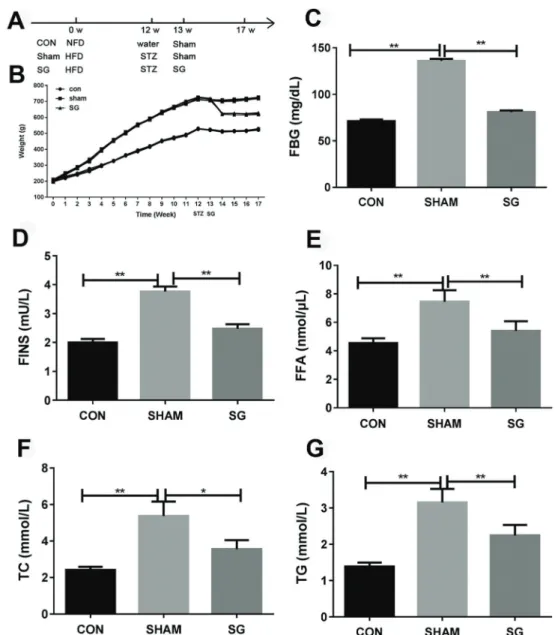
as previously described with minor modifications (6,15). Briefly, rats werefirst fed with a high-fat diet for 12 weeks, and then given a single intraperitoneal injection of 65 mg/ kg of STZ. Three days after the STZ injection, the glucose content in the venous blood from the tail of STZ-treated rats was analyzed using a blood glucose meter (iChem-540, iCubio Company, China). Obese diabetic rats were defined by blood glucose level over 16.7 mmol/L and a weight of more than 395 g. Rats fed with normal-fat diet (Guangdong Medical Laboratory Animal Center) and given a single intraperitoneal injection of water were used as the control. One week later, the VSG or sham surgery was carried out separately on the obese diabetic rats. The experimental protocols of this study were approved by the Ethics Committee of the Affiliated Zhongshan Hospital of the Dalian University. Rats in this study were classified into three groups as shown in Figure 1A: the control group (Con) of normal rats fed with normal-fat diet in combina-tion with injeccombina-tion of water and sham surgery, the Sham group (Sham) of diabetic rats that underwent sham surgery, and the sleeve gastrectomy (SG) group of diabetic rats that underwent VSG.
Vertical sleeve gastrectomy and the sham surgery VSG was performed on diabetic rats as previously described (6). Briefly, rats were fed with a liquid diet for consecutive 3 days before the VSG and sham surgery. Approximately 3–5% isoflurane was used to induce and maintain anesthesia of operated rats. A midline abdominal incision of approximately 3 cm was first made, followed by the transection of connective tissue attachments to rat liver and spleen, which allowed isolating the stomach. Approximately 70% of the stomach containing the entire fundus was removed and the remnant part in the tubular shape was left to connect the esophagus and pylorus. After being lavaged properly, the remnant of stomach was replaced into abdominal cavity. Finally, the abdominal cavity was closed. A combination of enrofloxacin (20 mg/kg) and meloxicam (2 mg/kg) was given to the operated rats for 14 consecutive days after surgery. The Sham group underwent a similar VSG as previously described and nothing was removed before suturing the abdominal incision of approximately 3 cm (6).
High Tech, China) by an automated RIA-immunity analyzer (Xi’an Nuclear Instrument Factory, China).
Lipid deposition analysis by oil red-O staining Histological visualization of lipid deposition in liver sections of VSG or sham surgery-operated diabetic rats and control group was carried out using the oil red-O (Lipid Stain) kit (#ab150678; Abcam, UK) according to the manufacturer’s instructions. Briefly, the rat liver sections were first incubated in propylene glycol for 2 min, in oil red-O solution for 6 min, and then in 85% propylene glycol
for 1 min. After being rinsed 2 min with distilled water, the slides were incubated at 37°C with hematoxylin for 2 min, rinsed again with tap water and distilled water, andfinally mounted using aqueous mounting medium under a cover-slip. The lipid deposited in rat liver tissues was stained red and observed under microscopy.
Quantitative RT-PCR and western blotting
Briefly, total RNA samples from rat liver tissues or Chang liver cells were extracted with Trizol solution (Sigma-Aldrich, USA) following the manufacturer’s instructions. The cDNA synthesis was carried out using approximately 2 mg RNA with EasyScript First-Strand cDNA Synthesis SuperMix kit (TransGen Biotech, China). The quantitative RT-PCR was performed by mixing 2 mL cDNA and 1mL specific primers with the TransStarttSYBR Green qPCR Supermix (TransGen Biotech) following the manufacturer’s instructions. GAPDH was used as the internal control for the quantitation of gene expression. The sequences of primers used in this analysis are listed in Table 1. The relative protein abundance of MAFB and FXR in rat liver tissues and Chang liver cells was determined by western blotting using the anti-MAFB (#ab56242, Abcam, UK) and FXR (#AB10304, Merck Millipore, Germany) anti-bodies according to the manufacturer’s instructions.
Cell culture and transfection
Human hepatocyte Chang liver cells were purchased from the Cell Bank of the Chinese Academy of Sciences (China) and cultured in DMEM medium containing 10% fetal bovine serum with 100mg/mL streptomycin and 100 U/mL penicillin at 37°C in an incubator under 95% humidity and 5% CO2. The establishment of Chang liver cells over-expressing MAFB gene was finished according to the previously described protocol with minor modifications (17). Briefly, the humanMAFBgene ORF was cloned into the pcDNA3.0 expression vector as described by the reference. For overexpression ofMAFBgene, Chang liver cells were seeded in a culture dish and transfected with the MAFB-pcDNA3.0 construct using the Lipofectamine
2000 reagent (Invitrogen, USA) following the manufac-turer’s instructions. The pcDNA3.0 plasmid was used as the control plasmid DNA transfected with Lipofectamine 2000 reagent following the manufacturer’s recommenda-tions. The expression levels werefinally checked by real-time PCR and western blotting 48 h after transfection.
Chromatin immunoprecipitation (ChIP)
The binding of MAFB withFXRpromoter was tested by chromatin immunoprecipitation (ChIP) using the ChIP Kit (#ab500, Abcam) following the manufacturer’s instruc-tions. The anti-MafA antibody ChIP Grade (#ab17976, Abcam) was used for immunoprecipitation. Finally, the binding was determined by PCR amplification of promoter sequence. The antibody against IgG was used as negative control.
Transcriptional activity by luciferase reporter assays The activation of FXR expression by MAFB was confirmed by luciferase-based reporter assay using the pGL3 promoter vector (#E1761, Promega, USA) following the manufacturer’s instructions with reference to pre-viously described protocol (18). Briefly, the wild-typeFXR promoter as well as the mutant version ofFXRpromoter with mutation of the predicted binding sites were sepa-rately cloned into the pGL3 promoter vector. The FXR promoter was amplified using primers namedFXR promo-ter forward and reverse as in Table 1. HEK293 cells purchased from the Cell Bank of the Chinese Academy of Sciences were cultured in DMEM containing 10% fetal calf serum at 37°C under 95% humidity and 5% CO2. The transfection of HEK293 cells with the pGL3 promoter Table 1. qPCR primers used in the study.
Gene Primer
FXR forward GGGCAACTGCGTGATGGA
FXR reverse AGGAGGGTCTGCTGGTCTACA
MAFB forward GCAGCAACGGTAGTGTGGA
MAFB reverse TGACCTTGTAGGCGTCTCTCT
SHP-1 forward GCCCTCTCTTCCTGCTTGG
SHP-1 reverse GGTTGTGGTGGGTCTGGTG
SREBP-1 forward CCAGCCTTTGAGGATAACCA
SREBP-1 reverse CCGAAGCATCAGAGGGAGT
CYP7A1: forward TGCCTTCTGTTACCGAGTGATGTT
CYP7A1 reverse ACCGGCAGGTCATTCTCTACC
PEPCK forward GCTGACAGACTCGCCCTATG
PEPCK reverse CACCGTATCCGCTTCCG
G6Pase forward CTCAGGAACGCCTTCTATGT
G6Pase reverse GTGACGGGGAACTGTTTTATC
PPARa forward AGAATCCACGAAGCCTACCT
PPARa reverse AGAATCGGACCTCTGCCTC
vectors containing the wild-type or mutant FXRpromoter was carried out using the Fugene HD transfection reagent (Roche, USA) following the manufacturer’s instructions. The cells were then lysed using passive lysis buffer (Promega) 48 h after transfection. The luciferase enzyme activity was presented as fold-change relative to the vehicle control.
Statistical analysis
Statistical analysis was performed using the SPSS software package (version 18.0, SPSS). The significance of differences was statistically tested by the Student’s t-test using data from at least three biological replicates. Significant differences were defined by a P valueo0.05.
Results
VSG induced weight loss and decreased blood glucose and lipids in diabetic rats
The body weights of rats were measured each week from the beginning of this study, showing that the high-fat diet induced a significant weight increase in the Sham and SG groups before surgery, compared to the control group. The sleeve gastrectomy significantly decreased body weights of diabetic rats in the SG group (Figure 1A). Consistent with the change of weight, blood glucose, and lipid analysis also demonstrated that FBG, FINS, FFA, TC, and TG of the SG group were greatly lowered compared to the Sham group (Figure 1B–G), showing the effective function of VSG in causing weight loss and diabetes mellitus resolution via improved glucose and lipid metabolism.
VSG improved liver function of diabetic rats
The levels of two common liver function markers, AST and ALT, were remarkably elevated in the sham group, compared with the control group, but VSG effectively recovered the AST and ALT levels (Figure 2A and B). Results of the oil red-O dyed-tissues showed that the fat deposition in the STZ-induced diabetic rats was greatly enhanced compared with the control group, but sleeve gastrectomy significantly repressed lipid deposition in dia-betic rat liver tissues (Figure 2C). These results indicated that sleeve gastrectomy could effectively recover the liver functions of diabetic rats, showing the important mediating role of the liver organ during DM therapy by surgery.
RecoveredMAFB and FXR expression by VSG To explore the molecular mechanisms underlying the physiological effects of VSG, the MAFB protein, which regulates islet a-cell activity and b cell maturation (14), was predicted as a possible regulator ofFXRexpression (data not shown). Our results showed that the mRNA levels of bothMAFBandFXRwere greatly decreased in the liver tissues of sham-operated diabetic rats, but VSG recovered theMAFBandFXRmRNA levels to the degrees comparable to those of the control group (Figure 3A and B). Consistently, the decreased protein abundance
of MAFB and FXR in diabetic rat livers was also restored by VSG (Figure 3C). The changes of MAFB and FXR expression in diabetic rats, which are strictly correlated with glucose metabolism and liver function, indicated that these two proteins might be key players mediating DM resolution by sleeve gastrectomy.
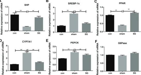
Modulation of FXR-regulated signaling components by sleeve gastrectomy
To further investigate the role of FXR in VSG-regulated glucose metabolism and diabetes mellitus progression, the expression levels of six key signaling components regulated by FXR in VSG-operated diabetic rat livers were also measured by quantitative RT-PCR method. The mRNA level of small heterodimer partner 1 (SHP-1) was significantly decreased in diabetic rats but recovered by VSG (Figure 4A). On the contrary, the expression of sterol regulatory element binding protein-1 (SREBP-1) was remarkably increased in diabetic rat livers compared with the control group, but markedly repressed by the VSG (Figure 4B). The expression level of peroxisome-proliferator-activated receptor a (PPARa) showed the same alteration in these groups as SHP-1 (Figure 4C). Also, the expression of cholesterol 7a-hydroxylase gene (CYP7A1) exhibited correlative alteration in the sham and SG-operated diabetic liver rats compared with the control group (Figure 4D). Moreover, our results showed that the phosphoenolpyruvate carboxykinase (PEPCK) expression was contrarily influenced by the sham surgery and VSG in diabetic rats (Figure 4E). No significant change of the glucose-6-phosphatase (G6Pase) was detected in this assay (Figure 4F). The significant and correlated expres-sion changes of these FXR-regulated genes provided multiple lines of evidence suggesting the strong regulatory functions of FXR in resolution of DM by VSG.
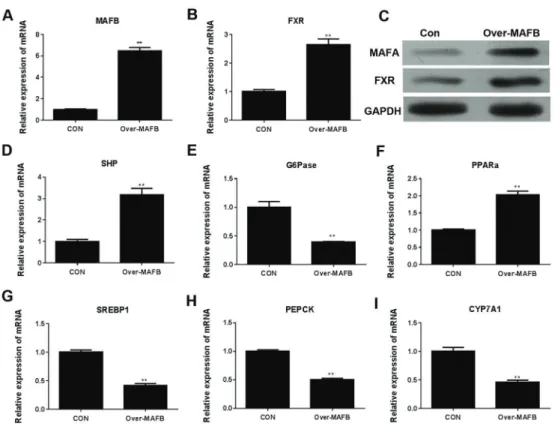
Overexpression ofMAFB regulated FXR-associated signaling cascades
showed the potent ability of MAFB in regulating FXR and related molecular processes in human liver cells.
MAFB binded withFXR promoter to induce FXR expression
The RT-PCR amplification following Chip assay demon-strated that MAFB protein directly associated with FXR promoter in HEK293 cells (Figure 6A). For the investiga-tion of activating ability of MAFB binding forFXR expres-sion, luciferase reporter assay was also performed in HEK293 cells, and results showed that the luciferase gene was significantly activated by the binding of MAFB with FXR promoter, while the mutant version showed no significant activation of reporter gene expression.
These results clearly demonstrated that MAFB was a key transcription factor responsible for the activation of FXR expression by directly binding theFXRgene promoter.
Discussion
Figure 3.A, MAFB andB, FXR mRNA levels in sleeve gastrectomy (SG), sham, and control (CON) rats measured by quantitative RT-PCR. C: MAFB and FXR protein levels determined by western blotting. GAPDH was applied as the internal control. MAFB: musculoaponeuroticfibrosarcoma oncogene family A; FXR: farnesoid X receptor; GAPDH: glyceraldehyde-3-phosphate dehydrogenase. Data are reported as means±SD. *Po0.05 and **Po0.01 (t-test).
Figure 5. mRNA levels of MAFB (A) and FXR (B) in Chang liver cells overexpressing MAFB gene (over-MAFB) were analyzed by quantitative RT-PCR and by western blotting (C). The mRNA levels of SHP (D), G6Pase (E), PPARa(F), SREBP (G), PEPCK (H), and CYP7A1 (I) were analyzed by quantitative RT-PCR. GAPDH was applied as the internal control in qRT-PCR analysis. Con: control group; FXR: farnesoid X receptor; SHP: small heterodimer partner; SREBP-1: sterol regulatory element binding protein-1; CYP7A1: cholesterol 7a-hydroxylase gene (CYP7A1); PEPCK: phosphoenolpyruvate carboxykinase; G6Pase: glucose-6-phosphatase; PPARa: peroxisome-proliferator-activated receptora. Data are reported as means±SD. **Po0.01 (t-test).
mediator of DM development by regulating glucose and lipid metabolism and was revealed to be involved in the therapeutic effect of VSG (12,13). However, the regulation ofFXRgene expression during DM progression is not well investigated, especially the transcription factors respon-sible for the activation ofFXRexpression associated with DM. Using bioinformatics analysis,MAFBgene was found to be a potential transcription factor regulatingFXRgene expression. In this study, we reported that the expression of MAFB was strictly correlated with FXR and multiple FXR-regulated genes in diabetic rats that underwent VSG. The enhancement of FXRexpression by MAFB was further confirmed by the overexpression ofMAFBgene in Chang liver cells. This regulation was supported by the expression of downstream genes controlled by FXR, including SHP-1 as the nuclear receptor induced by FXR and involved in bile acid biosynthesis (19), SREBP-1, which is associated with lipid metabolism and negatively regulated by FXR (20), PPARa, which controls SREBP activity and lipid synthesis (21), CYP7A1, which acts as another important regulator of bile acid metabolism inhibited by both FXR and SHP (19,22), and also PEPCK, which regulates the rate-limiting step of hepatic gluconeo-genesis and is activated by FXR (23). G6Pase acts as regulator of hepatic gluconeogenesis (24), and we noticed that its expression in rat livers exhibited no significant alteration as shown in Figure 4F, which might be due to the functional abundance of multiple metabolism regula-tors (25) or the specific expression and functioning of G6Pase isozymes (26). Moreover, by Chip assay and luciferase-based reporter gene analysis, we found that the activation ofFXRexpression was mediated by the direct binding of MAFB withFXRpromoter. These results revealed a novel mechanism of FXR-mediated DM development and treatment by sleeve gastrectomy, through the trans-cription factor activity of MAFB. Combined with roles of FXR in bile acid, glycogen, and lipid metabolism (10,27), these findings indicated that surgery resulted in FXR expression change, downstream signaling activation, and metabolic adaptions. This discovery provided important insight into the molecular pathogenesis of DM, as well as the therapeutic effect of sleeve gastrectomy.
MAFB protein, as a basic leucine-zipper-containing transcription factor, was first identified as the interacting partner of the DNA-bound Ets-1 protein and involved in erythroid differentiation (28). Further investigation showed that MAFB plays roles in multiple biological and patholog-ical process such as monocytic differentiation, osteoclast differentiation, self-renewal of differentiated functional macrophages, myeloid commitment divisions of hemato-poietic stem cells, respiratory rhythmogenesis, and fatal central apnea (29). More importantly, MAFB was later found to be a potent regulator of pancreatica-cell activity and b cell maturation (14,30). For instance, MAFB was shown to regulate cell type-specific glucagon gene expression and associated with islet cell development
and obesity (31). One recent study using a mice model reported that MafB deficiency in hematopoietic cells could lead to a greatly increased percentage of body fat, thus accelerating the development of obesity (31). Considering the close association of islet beta cell failure in obesity-associated diabetes (32), it is reasonable to predict the involvement of MAFB in DM development because of the beta cell compensation for insulin resistance. Not surpris-ingly, MAFB was found to possess a protective role for diabetic nephropathy by regulation of multiple pathways including antioxidative enzymes and Notch pathways (33). In this study, we discovered the direct link between MAFB-regulated target gene expression and DM development and treatment, which could lay down key research basis for the understanding of diabetes pathogenesis and novel therapy development. It should be noted that MAFB, as a transcription factor, might regulate the expression of multiple genes besidesFXR, suggesting that further study of its downstream regulatory pathways could broaden our understanding of DM development. In consideration of the several functions of MAFB and FXR in metabolism, the possible involvement of MAFB-regulatedFXRexpression in other metabolic disease deserves further investigation. Moreover, the application of diabetic rat or mouse models induced by high-fat diet and STZ, an antibiotic causing the destruction of pancreatic islet b-cells, has been accepted as a feasible way to test clinical thera-peutic effects and underlying mechanism of VSG for diabetic patients (34). Both the mice and rats are sensitive to the pancreatic b-cell cytotoxic effects induced by STZ treatment (34), but rats are more suitable for surgical operation due to body shape and thus were applied in this study. By strictly following the widely proven protocol, we successfully established the obese DM rat model, which was confirmed by a change of the body weight and a number of physiological indexes associated with glucose and lipid metabolism and liver function after VSG. This establishment provided the basis for the mechanism investigation of DM combined with obesity and confirmed the applicability of this model for obese DM studies.
In summary, we reported the characterization of MAFB as a novel transcription factor responsible for the activation of FXR expression and downstream pathway during DM resolution induced by sleeve gastrectomy. The transcription factor activity of MAFB for FXRexpression was confirmed by expression analysis in diabetic rat livers and Chang liver cells, combined with the binding and luciferase reporter gene assays. The discovery provides meaningful insight into the molecular processes under-lying DM development, as well as the therapeutic effects of sleeve gastrectomy.
Acknowledgments
References
1. Taylor R. Pathogenesis of Type 2 diabetes: Tracing the reverse route from cure to cause.Diabetologia2008; 51: 1781–1789, doi: 10.1007/s00125-008-1116-7.
2. Carnethon MR, Rasmussen-Torvik LJ, Palaniappan L. The obesity paradox in diabetes.Curr Cardiol Rep2014; 16: 446, doi: 10.1007/s11886-013-0446-3.
3. Franz MJ, Boucher JL, Rutten-Ramos S, Vanwormer JJ. Lifestyle weight-loss intervention outcomes in over-weight and obese adults with type 2 diabetes: a systematic review and meta-analysis of randomized clinical trials. J Acad Nutr Diet 2015; 115:1447–1463, doi: 10.1016/ j.jand.2015.02.031.
4. Gill RS, Birch DW, Shi X, Sharma AM, Karmali S. Sleeve gastrectomy and type 2 diabetes mellitus: a systematic review.Surg Obes Relat Dis2010; 6: 707–713, doi: 10.1016/ j.soard.2010.07.011.
5. Ding L, Sousa KM, Jin L, Dong B, Kim BW, Ramirez R, et al. Vertical sleeve gastrectomy activates GPBAR-1/TGR5 to sustain weight loss, improve fatty liver, and remit insulin resistance in mice. Hepatology 2016; 64:760–773, doi: 10.1002/hep.28689.
6. Cummings BP, Bettaieb A, Graham JL, Stanhope KL, Kowala M, Haj FG, et al. Vertical sleeve gastrectomy improves glucose and lipid metabolism and delays diabetes onset in UCD-T2DM rats.Endocrinology2012; 153: 3620–3632, doi: 10.1210/en.2012-1131.
7. Li T, Chiang JY. Bile acid signaling in metabolic disease and drug therapy. Pharmacol Rev 2014; 66: 948–983, doi: 10.1124/pr.113.008201.
8. Li T, Chiang JY. Bile Acid signaling in liver metabolism and diseases.J Lipids2012; 2012: 754067.
9. Manley S, Ding W. Role of farnesoid X receptor and bile acids in alcoholic liver disease.Acta Pharm Sin B2015; 5:158–167, doi: 10.1016/j.apsb.2014.12.011.
10. Kliewer SA, Mangelsdorf DJ. Bile acids as hormones: The FXR-FGF15/19 pathway.Dig Dis2015; 33: 327–331, doi: 10.1159/000371670.
11. Zhang Y, Lee FY, Barrera G, Lee H, Vales C, Gonzalez FJ et al. Activation of the nuclear receptor FXR improves hyper-glycemia and hyperlipidemia in diabetic mice. Proc Natl Acad Sci USA2006; 103: 1006–1011, doi: 10.1073/pnas. 0506982103.
12. Carr RM, Reid AE. FXR agonists as therapeutic agents for non-alcoholic fatty liver disease.Curr Atheroscler Rep 2015;17: 500, doi: 10.1007/s11883-015-0500-2.
13. Ryan KK, Tremaroli V, Clemmensen C, Kovatcheva-Datchary P, Myronovych A, Karns R, et al. FXR is a molecular target for the effects of vertical sleeve gastrectomy.Nature2014; 509: 183–188, doi: 10.1038/nature13135.
14. Conrad E, Dai C, Spaeth J, Guo M, Cyphert HA, Scoville D, et al. The MAFB transcription factor impacts islet b-cell function in rodents and represents a unique sig-nature of primate islet b-cells.Am J Physiol Endocrinol Metab2016; 310:E91–E102, doi: 10.1152/ajpendo.00285. 2015.
15. Degenhardt TP, Alderson NL, Arrington DD, Beattie RJ, Basgen JM, Steffes MW, et al. Pyridoxamine inhibits early
renal disease and dyslipidemia in the streptozotocin-diabetic rat.Kidney Int2002; 61: 939–950, doi: 10.1046/j.1523-1755. 2002.00207.x.
16. Zhang W, Li C, Liu B, Wu R, Zou N, Xu YZ, et al. Pioglitazone upregulates hepatic angiotensin converting enzyme 2 expression in rats with steatohepatitis.Ann Hepatol 2013; 12: 892–900.
17. Ong CK, Leong C, Tan PH, Van T, Huynh H. The role of 50untranslated region in translational suppression of OKL38
mRNA in hepatocellular carcinoma. Oncogene 2007; 26: 1155–1165, doi: 10.1038/sj.onc.1209896.
18. Gage MC, Pourcet B, Pineda-Torra I. Luciferase Reporter Assays to Assess Liver X Receptor Transcriptional Activity. Methods in Molecular Biology 2016; 1376: 77–85, doi: 10.1007/978-1-4939-3170-5.
19. Goodwin B, Jones SA, Price RR, Watson MA, McKee DD, Moore LB, et al. A regulatory cascade of the nuclear receptors FXR, SHP-1, and LRH-1 represses bile acid biosynthesis. Mol Cell 2000; 6: 517–526, doi: 10.1016/ S1097-2765(00)00051-4.
20. Ting TC, Miyazaki-Anzai S, Masuda M, Levi M, Demer LL, Tintut Y, et al. Increased Lipogenesis and Stearate Accel-erate Vascular Calcification in Calcifying Vascular Cells. J Biol Chem 2011; 286: 23938–23949, doi: 10.1074/jbc. M111.237065.
21. Knight BL, Hebbachi A, Hauton D, Brown AM, Wiggins D, Patel DD, et al. A role for PPARalpha in the control of SREBP activity and lipid synthesis in the liver.Biochem J 2005; 389: 413–421, doi: 10.1042/BJ20041896.
22. Chiang JY, Kimmel R, Weinberger C, Stroup D. Farnesoid X receptor responds to bile acids and represses cholesterol 7alpha-hydroxylase gene (CYP7A1) transcription. J Biol Chem 2000; 275: 10918–10924, doi: 10.1074/jbc.275.15. 10918.
23. Stayrook KR, Bramlett KS, Savkur RS, Ficorilli J, Cook T, Christe ME, et al. Regulation of carbohydrate metabolism by the farnesoid X receptor. Endocrinology 2005; 146: 984–991, doi: 10.1210/en.2004-0965.
24. Huang X, Yang C, Luo Y, Jin C, Wang F, McKeehan WL, et al. FGFR4 prevents hyperlipidemia and insulin resistance but underlies high-fat diet induced fatty liver.Diabetes2007; 56: 2501–2510, doi: 10.2337/db07-0648.
25. Ahn SW, Gang GT, Tadi S, Nedumaran B, Kim YD, Park JH, et al. PEPCK and G6Pase are required for Steroidogenesis in Testicular Leydig Cells.J Biol Chem2012; 287: 41875– 41887, doi: 10.1074/jbc.M112.421552.
26. Yu W, Hui W, Li M, Zhan G, Zhang S. Identification, expression and regulation of amphioxus G6Pase gene with an emphasis on origin of liver.Gen Comp Endocrinol2015; 214: 9–16, doi: 10.1016/j.ygcen.2014.12.021.
27. Li G, Guo GL. Farnesoid X receptor,the bile acid sensing nuclear receptor, in liver regeneration.Acta Pharma Sin B 2015; 5:93–98, doi: 10.1016/j.apsb.2015.01.005.
29. Sarrazin S, Mossadegh-Keller N, Fukao T, Aziz A, Mourcin F, Vanhille L, et al. MafB restricts M-CSF-dependent myeloid commitment divisions of hematopoietic stem cells.Cell2009; 138: 300–313, doi: 10.1016/j.cell.2009.04.057.
30. Hang Y, Stein R. MafA and MafB activity in pancreatic b-cells. Trends Endocrinol Metab2011; 22: 364–373, doi: 10.1016/ j.tem.2011.05.003.
31. Tran MT, Hamada M, Nakamura M, Jeon H, Kamei R, Tsunakawa Y, et al. MafB deficiency accelerates the development of obesity in mice.FEBS Open Bio2016; 6: 540–547, doi: 10.1002/2211-5463.12058.
32. Prentki M, Nolan CJ. Islet beta cell failure in type 2 dia-betes.J Clin Investi 2006; 116: 1802–1812, doi: 10.1172/ JCI29103.
33. Morito N, Yoh K, Ojima M, Okamura M, Nakamura M, Hamada M, et al. () Overexpression of Mafb in podo-cytes protects against diabetic nephropathy. J Am Soc Nephrol 2014; 25: 2546–2557, doi: 10.1681/ASN.2013 090993.