Critical for Its Full Pro-Apoptotic Function
Nadia Godin-Heymann
¤☯, Yihua Wang
☯, Elizabeth Slee, Xin Lu
*Ludwig Institute for Cancer Research Ltd., Nuffield Department of Clinical Medicine, University of Oxford, Oxford, United Kingdom,
Abstract
We reported recently that apoptosis-stimulating protein of p5γ (ASPP) β, an activator of p5γ, co-operates with oncogenic RAS to enhance the transcription and apoptotic function of p5γ. However, the detailed mechanism remains unknown. Here we show that ASPPβ is a novel substrate of mitogen-activated protein kinase (MAPK). Phosphorylation of ASPPβ by MAPK is required for RAS-induced increased binding to p5γ and increased transactivation of pro-apoptotic genes. In contrast, an ASPPβ phosphorylation mutant exhibits reduced p5γ binding and fails to enhance transactivation and apoptosis. Thus phosphorylation of ASPPβ by RAS/MAPK pathway provides a novel link between RAS and p5γ in regulating apoptosis.
Citation: Godin-Heymann N, Wang Y, Slee E, Lu X (β01γ) Phosphorylation of ASPPβ by RAS/MAPK Pathway Is Critical for Its Full Pro-Apoptotic Function. PLoS ONE 8(1β): e8β0ββ. doi:10.1γ71/journal.pone.008β0ββ
Editor: Wei-Guo Zhu, Peking University Health Science Center, China
Received August 19, β01γ; Accepted October β5, β01γ; Published December β, β01γ
Copyright: © β01γ Godin-Heymann et al. This is an open-access article distributed under the terms of the Creative Commons Attribution License, which permits unrestricted use, distribution, and reproduction in any medium, provided the original author and source are credited.
Funding: This work is supported by the Ludwig Institute for Cancer Research Ltd. The funders had no role in study design, data collection and analysis, decision to publish, or preparation of the manuscript.
Competing interests: The authors have declared that no competing interests exist. * E-mail: [email protected]
☯ These authors contributed equally to this work.
¤ Current address: Cancer Research UK, London, United Kingdom
Introduction
p5γ is the most commonly mutated tumour suppressor protein thus far identified. Although p5γ is mutated in more than 50% of tumours, its mutation rate varies significantly between different types of human cancer, with a particularly high incidence in colorectal and pancreatic tumours. Interestingly, the mediator of signal transduction RAS is also commonly mutated in these particular tumour types. It remains unclear why there exists such a tight association between the
p53 and RAS mutation status [1]. We reported recently that apoptosis-stimulating protein of p5γ (ASPP) β co-operates with oncogenic RAS to enhance the transcription and apoptotic function of p5γ in cancer cells [β]. This may be achieved via the ability of active RAS to induce ASPPβ, thereby promoting ASPPβ’s interaction with p5γ and enhancing the activity of p5γ. However, the detailed mechanism underlying this observation remains to be elucidated.
Activated RAS promotes the protein kinase activity of RAF, which phosphorylates and activates MEK (also known as MAPKK). MEK phosphorylates and activates a mitogen-activated protein kinase (MAPK/ERK), a serine/threonine-selective protein kinase. The MAPK enzymes require a specific phosphorylation sequence where a serine or threonine is followed by proline (S/TP) [γ]. It was shown that endogenous
RAS is necessary for the full apoptotic activity of ASPPβ, which suggests that RAS signalling may modify ASPPβ, potentially via a phosphorylation event. Phosphorylation by RAS/MAPK modulates the activation of most of their substrates and in some cases the phosphorylation mediates changes in subcellular localisation [4].
ASPPβ belongs to an evolutionarily conserved ASPP family of proteins, alongside ASPP1 and iASPP. All three contain signature sequences in their C-termini; ankyrin repeats, SHγ domain and proline rich sequences [5]. ASPPβ binds to RAS through its N-terminus [β,6]. The functions of ASPPβ are potentially controlled by its binding partners and localisation. When ASPPβ locates at the cell-cell junctions, it binds and co-localises with PARγ via its N-terminus to maintain the integrity of cell polarity and adherence junction [7,8], whereas in the cytosol/nucleus, ASPPβ enhances p5γ-induced apoptosis in cancer cells [9]. It also binds ATG5 and inhibits RAS-induced autophagy, independently of p5γ [10]. Thus it is important to find the molecular event that controls the localisation of ASPPβ.
cascade as ASPPβ phosphorylation mutant fails to do so. Thus phosphorylation of ASPPβ by RAS/MAPK pathway provides a novel link between RAS and p5γ in regulating apoptosis.
Results
ASPP2 is a novel substrate of MAPK
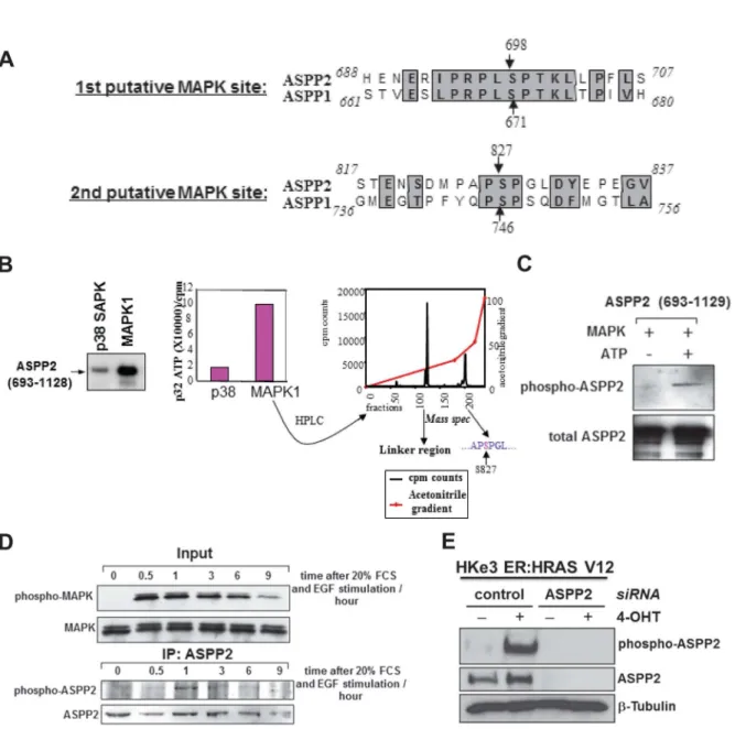
It has recently been shown that oncogenic RAS can enhance the apoptotic function of p5γ via ASPP1 and ASPPβ. Mechanistically ASPP1 and ASPPβ bind RAS-GTP and potentiates RAS signalling to enhance p5γ mediated apoptosis [β]. As RAS is upstream of several signalling cascades [1γ], we queried whether the activity of ASPPβ is regulated by the activation of a RAS-mediated signalling pathway. One of the most studied downstream pathways of RAS signalling is the Raf-MAPK pathway. Interestingly, we observed two conserved putative MAPK phosphorylation sites in ASPP1 and ASPPβ. The ASPP1 sites are at residues 671 and 746, and the ASPPβ sites are at residues 698 and 8β7 (Figure 1A). We thus tested whether RAS activation may regulate ASPPβ phosphorylation. An in vitro phophorylation assay was performed with a purified C-terminus fragment of ASPPβ (69γ-11β8) containing both MAPK putative phosphorylation sites. When compared to pγ8 SAPK, MAPK1 was clearly able to phosphorylate the ASPPβ fragment in vitro (Figure 1B, left and middle panels). As shown in Figure S1, histone βB phosphorylated by pγ8 SAPK had high levels of incorporated γβP, suggesting that pγ8 SAPK was active; while under the same conditions, ASPPβ (69γ-11β8) fragment phosphorylated by pγ8 SAPK had very low levels of incorporated γβP, indicating that pγ8 SAPK is not an efficient kinase for ASPPβ phosphorylation. The phosphorylated ASPPβ fragment by MAPK1 was digested by trypsin and fractioned on a high performance liquid chromatography (HPLC). Each eluted fraction was measured for its radioactivity content (Figure 1B, right panel). The fractions representing these radioactive peaks were analysed by mass spectrometry. Of the two radioactive peaks, one represented the linker region between the GST and our ASPPβ fragment and the other corresponded to a fragment of the same mass as that containing the second putative phosphorylation site, serine 8β7. Hence ASPPβ can be phosphorylated at serine 8β7 by MAPK1 in vitro.
A synthetic peptide encoding amino acids 8β4-8γβ, with a phosphoserine at residue 8β7, was used to raise antibodies. A polyclonal antibody NGH.S4 was purified by affinity column purification. To test the efficacy of the purified phospho-specific antibody, a non-radioactive in vitro phosphorylation assay was performed on the purified GST-ASPPβ fragment (69γ-11β8) with recombinant MAPK1. Figure 1C shows that the phospho-specific antibody is phospho-specific for the ASPPβ fragment phosphorylated in vitro by MAPK. To test whether endogenous ASPPβ could be phosphorylated in cells, Saosβ cells were grown in low serum for 50 hours to remove all background stimulation of RAS, after which the cells were stimulated with EGF and β0% fetal calf serum (FCS). Phosphorylated endogenous ASPPβ was detected by the phospho-specific antibody γ0 minutes after RAS stimulation (Figure 1D). ASPPβ phosphorylation was rapid and transient as γ hours after EGF
stimulation phosphorylated ASPPβ was barely detectable. Moreover, with another different phospho-ASPPβ antibody, ES1, ASPPβ phosphorylation was also observed in a human colon cancer cell line HKeγ ER:HRASV1β cells, in which RAS activation is induced upon the addition of 4-hydroxytamoxifen (4-OHT) [β,10,11] (Figure 1E). The phospho-specific antibody for ASPPβ is specific as knockdown of ASPPβ resulted in a lack of detection of phospho-ASPPβ. All these demonstrate that ASPPβ is a novel substrate of MAPK and Ser8β7 of ASPPβ can be phosphorylated by RAS/MAPK pathway.
Raf/MAPK Pathway Activates the Pro-Apoptotic Function of ASPP2
One of the most studied downstream pathways of RAS signalling is the Raf/MAPK pathway [1γ]. Knowing ASPPβ is a substrate of MAPK, we thus tested whether activation of Raf/ MAPK pathway is sufficient to regulate ASPPβ activity using a mutant form of Raf (Raf CAAX), which is constitutively present at the plasma membrane, so the Raf pathway is constitutively active [14]. The impact of co-expression of Raf CAAX with p5γ and ASPPβ was tested by analysing the transcriptional activity of p5γ on the pro-apoptotic Bax reporter. Raf CAAX increases Bax-luciferase levels by β.5 fold over the baseline of p5γ and ASPPβ alone (P=0.05). This effect is likely to be mediated by ASPPβ as Raf CAAX had little effect on p5γ in its absence (Figure βA).
The effect of Raf CAAX on ASPPβ and p5γ was compared to that of HRAS V1β or KRAS V1β. All three activate ASPPβ and p5γ transactivation activity to a similar extent, namely β.5 fold (Figure βB). This suggests that the effect of activated RAS on ASPPβ and p5γ is mediated via the downstream Raf /MAPK pathway.
ASPP2 phosphorylation by MAPK is necessary for ASPP2 full pro-apoptotic activity
To assess the effect of MAPK phosphorylation on ASPPβ activity, alanine substitution mutants of the two putative MAPK phosphorylation sites were constructed. In conditions where the cells were starved of serum, these mutants had identical activity to wild-type ASPPβ in their ability to enhance the transactivation function of p5γ (Figure γA). However, whereas activated Raf CAAX was able to stimulate wild-type ASPPβ and ASPPβ (S698A) by β.5 fold, it was unable to increase the activity of mutant ASPPβ (S8β7A) (Figure γB). These results suggest that MAPK phosphorylation of ASPPβ Ser8β7 is necessary for Raf CAAX to stimulate the full transcriptional activity of p5γ via ASPPβ.
Figure 1. MAPK phosphorylates ASPP2. (A) ASPP1 and ASPPβ have two conserved putative MAPKβ phosphorylation sites in their C-terminus. (B) The C-terminus fragment of ASPPβ is phosphorylated in vitro by MAPK1 (left panel). The intensity of phosphorylation is quantified (middle panel). The MAPK1 phosphorylated ASPPβ fragment was digested with trypsin and chromatographed and the radioactive peptides were measured by mass spectrometry (right panel). The first peak represents the GST linker region whereas the second presented a region of equal mass to the fragment containing serine 8β7. (C) An in vitro
phosphorylation assay was performed on the ASPPβ C-terminus fragment with recombinant MAPK1 and non-radioactive ATP. The phosphorylation status of ASPPβ was assessed using the purified NGH.S4 phospho-specific ASPPβ antibody (upper panel). Total ASPPβ is shown in the lower panel. (D) Saosβ cells were starved then stimulated with serum and EGF. At the indicated times the cells were harvested and either blotted for phospho/total MAPK (upper panel) or immunoprecipitated for total ASPPβ and blotted with NGH.S4 phospho-ASPPβ antibody. (E) Total cell lysates from HKeγ ER:HRASV1β cells treated with or without 4-OHT were transfected with control siRNA or siRNA against ASPPβ. ASPPβ phosphorylation was detected with ES1 phospho-ASPPβ antibody and total ASPPβ.
activation resulted in the increased ASPPβ expression (Figure 1E; Figure SγC). The mRNA level of ASPPβ was not affected upon RAS/MAPK pathway activation (Figure SγA), indicating that the regulation is not at the transcriptional level. Thus we investigated whether RAS/MAPK pathway activation could result in the increased protein stability of ASPPβ. The ASPPβ protein levels in the presence or absence of oncogenic RAS were measured after cycloheximide (CHX) addition (Figure SγB and C). ASPPβ was a bit more stable when oncogenic RAS is induced in HKeγ ER:HRASV1β cells (Figure SγB) or co-transfected in Saosβ cells (Figure SγC): ASPPβ levels were considerably decreased without oncogenic RAS, while in the presence of HRAS V1β, ASPPβ protein levels remained high after CHX treatment. These data indicate that ASPPβ phosphorylation by RAS/MAPK is necessary for ASPPβ full pro-apoptotic activity and this may be mediated by the stabilization of ASPPβ protein.
Ser827 phosphorylation is required for RAS-induced translocation of ASPP2
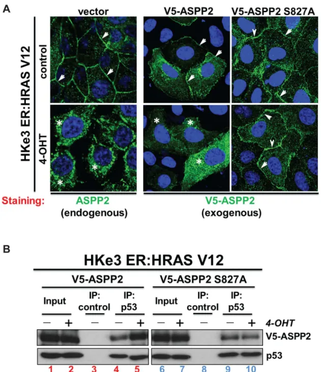
It has recently been shown that activation of RAS results in ASPPβ translocation from the plasma membrane to the cytosol and nucleus [β]. We therefore tested whether MAPK phosphorylation at Ser8β7 could affect the cellular localization of ASPPβ. Interestingly, we observed that in contrast to wild type ASPPβ, ASPPβ (S8β7A) remains at the plasma
membrane following RAS activation by 4-OHT in the HKeγ ER:HRASV1β system (Figure 4A). This suggests that ASPPβ binding to RAS at the plasma membrane occurs prior to MAPK phosphorylation of ASPPβ and that Ser 8β7 phosphorylation is required for RAS-induced translocation of ASPPβ to the cytosol.
As the translocation of wild-type ASPPβ from the plasma membrane to the cytosol and nucleus results in increased binding to p5γ [β], we tested whether the p5γ binding ability of ASPPβ phosphorylation mutant would be influenced by RAS activation. Indeed, upon activation of RAS by 4-OHT in HKeγ ER:HRASV1β cells, we observed an increase in the amount of p5γ co-immunoprecipitated with transfected V5-tagged wild-type ASPPβ (Figure 4B, compare lanes 4 and 5). Under the same conditions, the amount of p5γ in complex with transfected V5-ASPPβ (S8β7A) is not affected by RAS activation (Figure 4B, compare lanes 9 to 10). Together these data illustrate that phosphorylation of ASPPβ at Ser8β7 by MAPK is necessary for its ASPPβ to fully enhance p5γ’s pro-apoptotic activity.
Discussion
ASPPβ is a haploinsufficient tumor suppressor [15] [16] and it can cooperate with p5γ to suppress tumour growth in vivo
[15]. In human cancer, ASPPβ expression is down-regulated by Figure 2. Activated Raf enhances the transactivation activity of ASPP2 and p53 to the same extent as activated RAS. (A) Saosβ cells were transfected as indicated with a Bax-luciferase reporter and the luciferase activity shown. * P=0.05 (B) The value of ASPPβ+p5γ was taken as 1.0 to reflect the fold increase of ASPPβ and p5γ in the presence of activated Raf and mutant RAS. **
P=0.0055; **** P=0.0001. doi: 10.1γ71/journal.pone.008β0ββ.g00β
DNA methylation [9,17-19]. Importantly, in diffuse large B cell lymphomas, reduced ASPPβ expression associates with poor prognosis [β0]. ASPPβ expression is also down-regulated in both invasive and metastatic cells compared with normal breast epithelium [β1]. These findings established ASPPβ as a tumor suppressor and an activator of p5γ family. ASPPβ is involved in both senescence in fibroblasts and apoptosis in cancer cells [β,10]. ASPPβ acts at several steps in promoting senescence in fibroblasts. It can do so in a RAS-dependent, p5γ-independent manner, through its ability to bind ATG5 and to inhibit RAS-induced autophagy [10]. Additionally, it can bind
active RAS directly leading to enhanced activation of c-Raf/ MAPK-mediated senescence [6].
We and others have recently shown that ASPPβ can potentiate RAS signaling by binding directly via the ASPPβ N-terminus [β,6]. Moreover, the RAS-ASPP interaction enhances the transcription function of p5γ in cancer cells [β]. Until now, it has been unclear how RAS could affect ASPPβ to enhance p5γ function. We show here that ASPPβ is phosphorylated by the RAS/Raf/MAPK pathway and that this phosphorylation leads to its increased translocation to the cytosol/nucleus and increased binding to p5γ, providing an explanation of how RAS Figure 3. Phosphorylation of ASPP2 by the Raf/MAPK pathway enhances p53-mediated transactivation. (A) Saosβ cells were transfected with p5γ and either wild-type or mutant ASPPβ as indicated together with a Bax-luciferase reporter. Luciferase activity is shown following harvesting of cells. Values are Relative Light Units (RLU). (B) Saosβ cells were transfected with p5γ, constitutively active Raf CAAX and either wild-type or mutant ASPPβ as indicated together with a Bax-luciferase reporter. **
P=0.01γ; **** P=0.0001 (C) Saosβ cells were transfected with a Bax-luciferase reporter, ASPPβ and p5γ and treated with β0 µM UO1β6 or DMSO for β0 hours. Luciferase activity is shown in the left panel and protein expression was verified by Western Blot (right panel). **** P=0.0001.
Figure 4. Wild-type ASPP2, but not mutant ASPP2 (S827A), translocates to the cytosol and nucleus upon oncogenic RAS activation and this results in an increased interaction with p53. (A) RAS activation induces cytoplasmic and nuclear translocation of wild-type ASPPβ but not ASPPβ (S8β7A) in HKeγ ER:HRAS1β cells as detected by immunofluorescence. Arrows indicate cell membrane and stars indicate cytosol. (B) RAS activation enhances the binding of wild-type ASPPβ but not ASPPβ (S8β7A) to p5γ. Total cell lysates from HKeγ ER:HRASV1β cells treated with or without 4-OHT were immunoprecipitated with an anti-p5γ antibody or control IgG as indicated.
doi: 10.1γ71/journal.pone.008β0ββ.g004
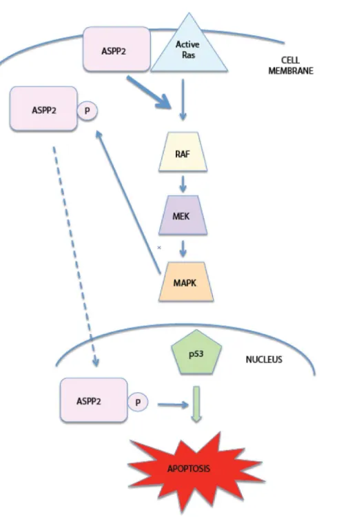
can activate p5γ pro-apoptotic functions (Figure 5).
Additionally, RAS/Raf/MAPK pathway activation stabilizes ASPPβ protein, although the underlying mechanism remains tobe investigated. Figure 5. Diagram summarizes the inter-regulation between ASPP2 and RAS. ASPPβ binds active RAS at the plasma membrane, thereby increasing RAS signaling to its downstream pathway effectors Raf/MAPK. Activated MAPK phosphorylates ASPPβ which can then relocate to the nucleus and activate p5γ pro-apoptotic signaling.
Thus ASPPβ is both upstream of RAS, by binding active RAS and enhancing its downstream pathway activity, as well as downstream of the RAS/RAF/MAPK pathway. It is likely that this possible feedback loop leads to an amplified pro-apoptotic signal. In a scenario where RAS is mutated and thereby constitutively active, it binds ASPPβ, resulting in increased RAS/MAPK signaling. This in turn activates ASPPβ via MAPK phosphorylation which will translocate and activate p5γ to promote apoptosis. ASPPβ will also continue to bind active RAS, thereby propagating the pro-apoptotic signal from RAS to p5γ. If RAS is only temporarily active, as happens in natural growth conditions, one could hypothesize that ASPPβ would bind active RAS, potentiate its downstream pathways, and MAPK-mediated phosphorylation of ASPPβ would lead to increased binding to p5γ. However, once RAS reverts to its inactive state, it would be unable to bind to ASPPβ and would therefore be unable to amplify the RAS signal to p5γ. Only in conditions where RAS is constitutively active would the ASPPβ feedback loop reach a threshold of signaling high enough to result in p5γ-dependent apoptosis. The ability of oncogenic RAS to stimulate apoptosis allows the cell to have a fail-proof mechanism: mutated RAS signals to p5γ via ASPPβ to induce apoptosis instead of uncontrolled proliferation. However, under normal conditions, growth factors or growth receptors would only activate RAS for short periods of time, preventing the amplification of ASPPβ signaling to p5γ via the feedback loop, thereby eliminating the apoptotic stimulus. The novel RAS/Raf/ MAPK/ASPPβ pathway is thus involved in an important feedback loop between RAS and p5γ, and is an effective way for mutant RAS to induce apoptosis in cancer cells with wild-type p5γ.
Materials and Methods
Cell culture, plasmids and siRNA
Saosβ cells were obtained from the American Type Culture Collection (Manassas, VA). HKeγ ER:HRASV1β cells were kindly provided by Dr Julian Downward [β,10,11]. Cells were cultured in DMEM supplemented with 10% fetal bovine serum (FBS) and penicillin streptomycin (Gibco-BRL, Invitrogen Ltd, Paisley, UK). For growth factor stimulation, cells were serum starved for indicated time, followed by stimulation with β0% serum plus epidermal growth factor (EGF), EGF alone or insulin (Sigma, Dorset, UK) as indicated.
Full-length ASPPβ and ASPPβ (69γ-11β8) was tagged with the V5-epitope. HRASV1β and KRASV1β were tagged with the haemagglutinin (HA) epitope. All expression plasmids used in this study were driven by the cytomegalovirus immediate-early promoter, with the exception of the RAS and Raf CAAX plasmids which were driven by the EF1a promoter. The ASPPβ mutants were constructed using the QuickChange Site-Directed Mutagenesis Kit (Stratagene).
siRNA oligos against ASPPβ were purchased from Dharmacon. Sequences are available from Dharmacon or upon request. We used siGENOME RISC-Free siRNA (Dharmacon, Lafayette, CO, United States) as a negative control. Cells were transfected with the indicated siRNA oligos at a final concentration of γ5nM using Dharmafect 1 reagent
(Dharmacon, Lafayette, CO, United States), according to the manufacturer's instructions.
Generation of Phosphor-ASPP2 Antibody
Using the peptide CPAPSpPGLDY (representing residues 8β4-8γβ) with the serine phosphorylated as an immunogen, a rabbit polyclonal antibody NGH.S4 which specifically recognizes phosphorylated ASPPβ at amino acid 8β7 was raised. An affinity column was made by cross-linking the phospho-peptide to epoxy-sepharose-6B (Amersham Pharmacia Biotech) according to the manufacturer’s instructions. Serum from the final bleed was clarified by centrifugation and filtration through a 0.45μm filter and was supplemented with 1X TTBS (0.5 M NaCl, β0 mM Tris [pH 8.0], 0.1% Tween-β0). This was passed over the affinity column and washed with TTBS until the flow-through had an ODβ80nm <0.01. The antibody was then eluted with 0.βM glycine (pH β.8) and neutralized with Tris-HCl (pH 8.0). A second phospho-specific antibody ES1 was raised against the peptide SDMPAPS[P]PGLDYE where S[P] is the phosphorylated serine 8β7, and was conjugated to KLH. The serum was double affinity purified against the non-phosphorylated peptide SDMPAPSPGLDYE.
Transactivation Assay
Saosβ cells (7 x 10⁵) were plated β4 hours prior to transfection in 6-cm-diameter dishes. All transactivation assays contained 1 μg of reporter plasmid. 50 ng of p5γ, 4 μg of ASPPβ, and 1.5 μg of HRASV1β or KRASV1β or Raf CAAX expression plasmids were used as indicated. Cells were lysed in reporter lysis buffer 16 to β4 hours after transfection and assayed using the luciferase assay kit (Promega). The fold increase of p5γ and ASPP by HRASV1β/KRASV1β/Raf CAAX was determined by the activity of p5γ and ASPP in combination with RASV1β/Raf CAAX divided by the activity of p5γ and ASPP alone.
Western blot analysis
and the haemagglutinin epitope is recognized by the anti-HA antibody (mAb) (Covance, Princeton, NJ USA). To detect RAS by immunoprecipitation the rat monoclonal antibody βγ8 was used (Santa Cruz, CA USA). The mouse and rabbit antibodies to ASPPβ were described previously [9]: rabbit anti-ASPPβ polyclonal antibody ASPPβ/77 and mouse monoclonal anti-ASPPβ antibody DX54.10. The mouse monoclonal DO-1 is specific to p5γ. Signals were detected using ECL detection system (GE Healthcare, Pollards Wood, Chalfont, Buckinghamshire, UK).
Immunofluorescence
Cells were fixed in 4% PBS-paraformaldeyde for 15 min, incubated in 0.β% Triton-X-100 for 5 min, then in 0.β% Fish Skin Gelatine in PBS for 10 min and stained for 1 hr with anti-V5 (Invitrogen, Paisley, UK) or with anti-ASPPβ with DX54.10. Antibodies were used at 1:100 dilution in 0.β% Fish Skin Gelatine-PBS, respectively. Staining with the secondary antibody and Hoechst was performed as described [β], followed by visualisation under a fluorescence microscope.
Immunoprecipitation
For immunoprecipitation, cells were lysed in IP buffer (β0 mM Trish-HCl [pH 7.5], 1 mM EDTA, 1 M KCl, 5 mM MgClβ , 10% v/v glycerol, 1% v/v Triton X-100, 0.05% v/v β-Mercaptoethanol and protease and phosphatase inhibitors). 1-4 mg of lysate was pre-cleared with protein G beads for γ0 min at 4°C and subsequently incubated with antibody pre-bound to protein G beads for β-16 hrs at 4°C. The beads were washed three times with NP40 buffer. The immunoprecipitation beads were mixed with 6x sample buffer and loaded on SDS-PAGE gels.
In vitro phosphorylation assay
β μg of GST-ASPPβ (69γ-11β8) expressed in E. coli, β μg of 1mg/ml recombinant Histone βB or β μl of water was incubated in γ0 μl of reaction mixture containing 50 mM Tris-HCl, pH 7.5, 0.1% β-mercaptoethanol, 1 μM microcystine and 100 μM [ -γβP]ATP (10 000 c.p.m./pmol) at γ0°C for γ0 min. 0.γ5 units MAPK1 or 0.1 units pγ8 SAPK were used. The reactions were terminated as described [1β] and analyzed by chromatography on a C18 column or resolved on SDS-PAGE gels and visualized by autoradiograph.
Quantitative reverse transcription–PCR
Real-time reverse transcriptase–PCR was performed using gene-specific primers (QuantiTect Primer Assays) for human ASPPβ or GAPDH with the QuantiTect SYBR Green RT-PCR Kit (Qiagen, Hilden, Germany). Relative transcript levels of ASPPβ were normalized to GAPDH mRNA levels.
Statistical analysis
SPSS for Windows (SPSS Inc.) was used to analyze the data. A two-tailed unpaired t-test was used to compare the
statistical significance of the differences in data from the two groups.
Supporting Information
Figure S1. p38 SAPK is not an efficient kinase for ASPP2 phosphorylation. ASPPβ (69γ-11β8) fragment was used as a substrate for an in vitro phosphorylation assay by the kinase pγ8 SAPK. As a negative control no substrate was used and Histone βB was the substrate for the positive control. γβ P-labelled ATP was added to the kinase assay and the P-labelled proteins were resolved on SDS-PAGE gels and visualized by autoradiograph.
(TIF)
Figure S2. Phosphorylation of ASPP2 by the Raf/MAPK pathway enhances p53-mediated transactivation. Saosβ cells were transfected with a Bax-luciferase reporter, ASPPβ and p5γ and treated with 100 μM PD 98059 or DMSO for β0 hours. The cells were harvested, luciferase activity shown. (TIF)
Figure S3. Oncogenic RAS stabilizes ASPP2. (A) Quantitative RT-PCR analysis of ASPPβ mRNA levels in HKeγ ER:HRASV1β cells with indicated treatment. (B) HKeγ ER:HRASV1β cells were transfected with ASPPβ wild-type (wt) expression plasmid in the presence or absence of 4-OHT. 16 hours after transfection, 10 μg/ml cycloheximide (CHX) was added to the cells for the time indicated. The protein levels of V5ASPPβ were determined by western blot analysis. -Tubulin was used as a loading control. (C) Saosβ cells were transfected with ASPPβ wt expression plasmid in the presence or absence of co-transfected HRAS V1β. 16 hours after transfection, 50 μg/ml CHX was added to the cells for the time indicated. The protein levels of V5-ASPPβ or HRAS V1β were determined by western blot analysis. -actin was used as a loading control.
(TIF)
Acknowledgements
We thank Kimberley Bryon-Dodd for critical reading of the manuscript. We thank Dario Alessi for his help with the in vitro
phosphorylation data. We are grateful to Dr Julian Downward for providing HKeγ ER:HRASV1β cells and Senji Shirasawa for HKeγ cells. We thank Richard Marais and Chris Marshall for providing us with the expression plasmids for HRAS, KRAS and HRAS V1β.
Author Contributions
References
1. Russo A, Bazan V, Agnese V, Rodolico V, Gebbia N (β005) Prognostic and predictive factors in colorectal cancer: Kirsten Ras in CRC (RASCAL) and TP5γCRC collaborative studies. Ann Oncol 16 Suppl 4: iv44-iv49. PubMed: 159βγ4β8.
β. Wang Y, Godin-Heymann N, Dan Wang X, Bergamaschi D, Llanos S et al. (β01γ) ASPP1 and ASPPβ bind active RAS, potentiate RAS signalling and enhance p5γ activity in cancer cells. Cell Death Differ β0: 5β5-5γ4. doi:10.10γ8/cdd.β01γ.γ. PubMed: βγγ9β1β5.
γ. Yoon S, Seger R (β006) The extracellular signal-regulated kinase: multiple substrates regulate diverse cellular functions. Growth Factors β4: β1-44. doi:10.1080/0β699050500β84β18. PubMed: 16γ9γ69β. 4. Cargnello M, Roux PP (β011) Activation and function of the MAPKs
and their substrates, the MAPK-activated protein kinases. Microbiol Mol Biol Rev 75: 50-8γ. doi:10.11β8/MMBR.000γ1-10. PubMed: β1γ7βγβ0. 5. Trigiante G, Lu X (β006) ASPP [corrected] and cancer. Nat Rev Cancer
6: β17-ββ6. doi:10.10γ8/nrc1818. PubMed: 16498444.
6. Wang Z, Liu Y, Takahashi M, Van Hook K, Kampa-Schittenhelm KM et al. (β01γ) N terminus of ASPPβ binds to Ras and enhances Ras/Raf/MEK/ERK activation to promote oncogene-induced senescence. Proc Natl Acad Sci U S A 110: γ1β-γ17. doi:10.107γ/ pnas.1β01514110. PubMed: βγβ48γ0γ.
7. Cong W, Hirose T, Harita Y, Yamashita A, Mizuno K et al. (β010) ASPPβ regulates epithelial cell polarity through the PAR complex. Curr Biol β0: 1408-1414. doi:10.1016/j.cub.β010.06.0β4. PubMed: β0619648.
8. Sottocornola R, Royer C, Vives V, Tordella L, Zhong S et al. (β010) ASPPβ binds Par-γ and controls the polarity and proliferation of neural progenitors during CNS development. Dev Cell 19: 1β6-1γ7. doi: 10.1016/j.devcel.β010.06.00γ. PubMed: β0619750.
9. Samuels-Lev Y, O'Connor DJ, Bergamaschi D, Trigiante G, Hsieh JK et al. (β001) ASPP proteins specifically stimulate the apoptotic function of p5γ. Mol Cell 8: 781-794. doi:10.1016/S1097-β765(01)00γ67-7. PubMed: 11684014.
10. Wang Y, Wang XD, Lapi E, Sullivan A, Jia W et al. (β01β) Autophagic activity dictates the cellular response to oncogenic RAS. Proc Natl Acad Sci U S A 109: 1γγβ5-1γγγ0. doi:10.107γ/pnas.11β019γ109. PubMed: ββ8474βγ.
11. Wang Y, Ngo VN, Marani M, Yang Y, Wright G et al. (β010) Critical role for transcriptional repressor Snailβ in transformation by oncogenic RAS in colorectal carcinoma cells. Oncogene β9: 4658-4670. doi:10.10γ8/ onc.β010.β18. PubMed: β056β906.
1β. Sapkota GP, Boudeau J, Deak M, Kieloch A, Morrice N et al. (β00β) Identification and characterization of four novel phosphorylation sites
(Serγ1, Serγβ5, Thrγγ6 and Thrγ66) on LKB1/STK11, the protein kinase mutated in Peutz-Jeghers cancer syndrome. Biochem J γ6β: 481-490. doi:10.104β/0β64-60β1:γ6β0481. PubMed: 1185γ558. 1γ. Downward J (β00γ) Targeting RAS signalling pathways in cancer
therapy. Nat Rev Cancer γ: 11-ββ. doi:10.10γ8/nri979. PubMed: 1β50976γ.
14. Sridhar SS, Hedley D, Siu LL (β005) Raf kinase as a target for anticancer therapeutics. Mol Cancer Ther 4: 677-685. doi: 10.1158/15γ5-716γ.MCT-04-0β97. PubMed: 158β7γ4β.
15. Vives V, Su J, Zhong S, Ratnayaka I, Slee E et al. (β006) ASPPβ is a haploinsufficient tumor suppressor that cooperates with p5γ to suppress tumor growth. Genes Dev β0: 1β6β-1β67. doi:10.1101/gad. γ74006. PubMed: 1670β401.
16. Kampa KM, Acoba JD, Chen D, Gay J, Lee H et al. (β009) Apoptosis-stimulating protein of p5γ (ASPPβ) heterozygous mice are tumor-prone and have attenuated cellular damage-response thresholds. Proc Natl Acad Sci U S A 106: 4γ90-4γ95. doi:10.107γ/pnas.0809080106. PubMed: 19β51665.
17. Zhao J, Wu G, Bu F, Lu B, Liang A et al. (β010) Epigenetic silence of ankyrin-repeat-containing, SHγ-domain-containing, and proline-rich-region- containing protein 1 (ASPP1) and ASPPβ genes promotes tumor growth in hepatitis B virus-positive hepatocellular carcinoma. Hepatology 51: 14β-15γ. doi:10.100β/hep.βγβ47. PubMed: β00γ40β5. 18. Liu ZJ, Lu X, Zhang Y, Zhong S, Gu SZ et al. (β005) Downregulated
mRNA expression of ASPP and the hypermethylation of the 5'-untranslated region in cancer cell lines retaining wild-type p5γ. FEBS Lett 579: 1587-1590. doi:10.1016/j.febslet.β005.01.069. PubMed: 15757645.
19. Sarraf SA, Stancheva I (β004) Methyl-CpG binding protein MBD1 couples histone Hγ methylation at lysine 9 by SETDB1 to DNA replication and chromatin assembly. Mol Cell 15: 595-605. doi:10.1016/ j.molcel.β004.06.04γ. PubMed: 15γβ7775.
β0. Lossos IS, Natkunam Y, Levy R, Lopez CD (β00β) Apoptosis stimulating protein of p5γ (ASPPβ) expression differs in diffuse large B-cell and follicular center lymphoma: correlation with clinical outcome. Leuk Lymphoma 4γ: βγ09-βγ17. doi:10.1080/104β8190β1000040017. PubMed: 1β61γ517.
β1. Sgroi DC, Teng S, Robinson G, LeVangie R, Hudson JR Jr. et al. (1999) In vivo gene expression profile analysis of human breast cancer progression. Cancer Res 59: 5656-5661. PubMed: 1058β678.