Uropathogenic
Escherichia coli
’s Ability to Exploit
Abundant Host Metabolites
Wentong Cai1, Yvonne Wannemuehler1, Giuseppe Dell’Anna2, Bryon Nicholson1, Nicolle L. Barbieri1,3, Subhashinie Kariyawasam4, Yaping Feng5, Catherine M. Logue1, Lisa K. Nolan1, Ganwu Li1*
1Department of Veterinary Microbiology and Preventive Medicine, College of Veterinary Medicine, Iowa State University, Ames, Iowa, United States of America, 2Laboratory Animal Resources, College of Veterinary Medicine, Iowa State University, Ames, Iowa, United States of America,3Departamento de Biofı´sica, Universidade Federal do Rio Grande do Sul, Porto Alegre, Rio Grande do Sul, Brasil,4Department of Veterinary and Biomedical Sciences, Pennsylvania State University, University Park, Pennsylvania, United States of America,5Laurence H. Baker Center for Bioinformatics and Biological Statistics, Iowa State University, Ames, Iowa, United States of America
Abstract
Two-component signaling systems (TCSs) are major mechanisms by which bacteria adapt to environmental conditions. It follows then that TCSs would play important roles in the adaptation of pathogenic bacteria to host environments. However, no pathogen-associated TCS has been identified in uropathogenicEscherichia coli(UPEC). Here, we identified a novel TCS, which we termed KguS/KguR (KguS:a-ketoglutarate utilization sensor; KguR:a-ketoglutarate utilization regulator) in UPEC CFT073, a strain isolated from human pyelonephritis.kguS/kguRwas strongly associated with UPEC but was found only rarely among otherE. coliincluding commensal and intestinal pathogenic strains. Anin vivocompetition assay in a mouse UTI model showed that deletion ofkguS/kguRin UPEC CFT073 resulted in a significant reduction in its colonization of the bladders and kidneys of mice, suggesting that KguS/KguR contributed to UPEC fitnessin vivo. Comparative proteomics identified the target gene products of KguS/KguR, and sequence analysis showed that TCS KguS/KguR and its targeted-genes,c5032toc5039, are encoded on a genomic island, which is not present in intestinal pathogenicE. coli. Expression of the target genes was induced bya-ketoglutarate (a-KG). These genes were further shown to be involved in utilization ofa -KG as a sole carbon source under anaerobic conditions. KguS/KguR contributed to the regulation of the target genes with the direct regulation by KguR verified using an electrophoretic mobility shift assay. In addition, oxygen deficiency positively modulated expression ofkguS/kguRand its target genes. Taken altogether, this study describes the first UPEC-associated TCS that functions in controlling the utilization ofa-ketoglutaratein vivothereby facilitating UPEC adaptation to life inside the urinary tract.
Citation:Cai W, Wannemuehler Y, Dell’Anna G, Nicholson B, Barbieri NL, et al. (2013) A Novel Two-Component Signaling System Facilitates Uropathogenic
Escherichia coli’s Ability to Exploit Abundant Host Metabolites. PLoS Pathog 9(6): e1003428. doi:10.1371/journal.ppat.1003428 Editor:Matthew A. Mulvey, University of Utah, United States of America
ReceivedDecember 1, 2012;AcceptedApril 25, 2013;PublishedJune 27, 2013
Copyright:ß2013 Cai et al. This is an open-access article distributed under the terms of the Creative Commons Attribution License, which permits unrestricted use, distribution, and reproduction in any medium, provided the original author and source are credited.
Funding:This work was partly supported by the USDA NRICGP Microbial Functional Genomics program (grant no. 2008-3560418805). The funders had no role in study design, data collection and analysis, decision to publish, or preparation of the manuscript.
Competing Interests:The authors have declared that no competing interests exist. * E-mail: liganwu@iastate.edu
Introduction
Urinary tract infection (UTI) is one of the most common bacterial infections in humans and is a significant clinical issue worldwide. Annually, UTIs are associated with 7 million office visits, 1 million emergency room visits, 100,000 hospitalizations,
and $1.6 billion in healthcare costs [1]. In fact, ,80–90% of
community-acquired UTIs are caused by uropathogenicEscherichia
coli(UPEC) [2]. Many virulence factors are required for UPEC to
cause UTIs. Typically, UTIs begin with urethral contamination with UPEC from the bowel [3]. Successful attachment to the uroepithelium requires specific adhesins including P, type 1 and other fimbriae (such as F1C, S, M, and Dr fimbriae) [4,5]. UPEC may then ascend the urethra to enter the bladder and kidneys, where several highly regulated virulence factors, including fimbriae, secreted toxins (hemolysin, Vat, Sat, and CNF) and polysaccharide capsule, may contribute to colonization and pathogenesis [6]. It is likely that UPEC’s ability to colonize the
urinary tract and cause disease is affected by its adaptive responses to local environmental cues, including changes in nutrient availability.
nucleotide-deficient environment. Study ofpyrDand guaA deletion mutants showed that metabolism of nucleobases is required for UPEC colonization of the bladder [14,15]. Thus, UPEC metabolism appears to play a critical role in colonization and invasion of the host. Therefore, it is not surprising that acquisition of genomic islands encoding metabolic pathways is often essential for the
colonization of host niches. Gene cluster ttrABC and ttrRS,
responsible for utilizing tetrathionate as an electron acceptor in the intestine, are encoded on Salmonella enterica subsp. enterica
serotype Typhimurium pathogenicity island II and contribute to detoxification of tetrathionate and confer a competitive advantage in growth [16–18]. GenenixAwithin a transferable genomic island inHelicobacter pylori aids in transport of nickel and consequently results in enhanced activity of urease, a key factor for fitness and virulence [19–22]. Besides islands encoding iron uptake systems, no other metabolic island has yet been characterized in UPEC even though their study would help understand the pathobiology and facilitate the identification of potential targets for novel therapeutic approaches to prevent this prevalent pathogen.
The pathways, conferring metabolic adaptation of bacterial pathogens to the host milieu, are usually controlled by both global and specific regulators. One mechanism used by most bacterial pathogens to sense and respond to nutrient availability is two-component signaling systems (TCSs). TCSs, composed of a membrane-bound sensor histidine kinase (HK) and a cytoplasmic response regulator (RR), have been implicated in regulating the response of bacteria to a wide array of signals and stimuli, including nutrients, quorum signals, antibiotics, etc. The recogni-tion of physical or chemical signals by HK sensor domain typically triggers the modulation of its autophosphorylation activity. The phosphoryl group is then transferred to the RR, usually a DNA binding protein that functions by altering gene expression [23,24]. In E. coli K12, more than 30 TCSs have been identified and characterized to some extent [25–27]. Of these, several were shown to be involved in UPEC pathogenesis. TCS BarA/UvrY controlled efficient switching between glycolytic and gluconeogen-ic carbon sources and contributed to UPEC virulence [28]. Deletion of QseC, the kinase of a well-known TCS QseC/QseB,
dysregulated nucleotide, amino acid, and carbon metabolism and resulted in attenuation of UPEC virulence [29,30]. In addition, PhoQ/PhoP [31] and the AirS system [32] have been linked to UPEC pathogenesis. To date, all TCSs linked to UPEC
pathogenesis have been common to E. coli and are not
pathogen-associated. Given the distinct features of environments colonized by UPEC, we hypothesize that UPEC may possess special TCSs that sense urinary tract-specific metabolic signal(s) and adapt UPEC metabolism to available nutrients.
Here, we describe the first TCS significantly associated with UPEC and its direct target genes encoded on a small genomic island. In addition, we demonstrate that the expression of these island genes is induced in response toa-KG, a nutrient found in high concentration in renal proximal tubule cells, and that these
genes are involved in the utilization of a-KG under anaerobic
conditions. Our results suggest that KguS/KguR contributes to
UPEC fitness in vivo by facilitating the utilization of a host
abundant metabolitea-KG. This study provides a new perspective
for understanding UPEC pathogenesisin vivo.
Results
Identification of a novel TCS in UPEC CFT073
TCSs enable bacteria to sense, respond, and adapt to a wide range of environments, stressors, and growth conditions [23]. Since UPEC encounter unique environmental conditions found
within their host, as compared to commensal E. coli and
diarrheagenicE. coli, we hypothesize that specific TCSs may exist and facilitate UPEC adaptation to certain host niches. Indeed, analysis of the UPEC CFT073 genome identified a putative TCS
c5041/c5040 by the presence of specific domains in their respective predicted proteins (Fig. 1). Domains and three-dimensional structures of C5041 and C5040 were predicted by the threading method using the I-TASSER online server (http:// zhanglab.ccmb.med.umich.edu/I-TASSER/) and subsequently refined using four-body contact potentials. C5041 is predicted to be a typical sensor kinase harboring a dimerization and histidine phosphotransfer (DHp) domain and a histidine kinase-like ATPase domain (Fig. 1A). The C-terminal 288 amino acids of C5041 contains five short signature segments, H-, N-, G1-, F-, and G2-boxes, which are conserved across most histidine kinases [33,34]. The histidine residue within the H-box, which is very likely phosphorylated during the signaling process, was found at position 394. In the N-terminal section of C5041, two transmembrane helices were identified at position 15–35 and at position 297–317, respectively. Between the two transmemebrane helices, a periplas-mic domain, which might serve as a signal sensor, was identified according to the distribution of charged residues ahead and behind the postulated transmembrane helices. The periplasmic domain of C5041 is 25.3% identical to that of DctB, a sensor kinase protein
for dicarboxylates transport in Sinorhizobium meliloti (of which
crystal structure is available) [35], suggesting C5041 may sense analogs of succinate, which is sensed by DctB [36].
C5040 is 37% identical to the nitrogen regulatory protein C (NtrC) [37,38] at the amino acid sequence level. C5040 harbors
the CheY-like receiver domain, phosphorylation site, AAA+
ATPase domain, and helix-turn-helix (HTH) domain (Fig. 1B). The receiver domain (position 13–123) catalyzes phosphoryl transfer from the HK to itself and regulates the output. The aspartate residue, which is predicted to be phosphorylated in the signal transduction cascade, was found at position 58. In the C-terminal part of C5040, a helix-turn-helix domain was identified, which is a DNA-binding domain that recognizes enhancer-like
sequences. The CheY-like receiver domain, AAA+
ATPase Author Summary
Successful colonization requires bacterial pathogens to adapt their metabolism to the conditions encountered in particular infection sites. Two-component signaling sys-tems (TCSs) enable bacterial pathogens to sense and respond to environmental cues, thus mediating their adaptation to environmental change. Though many TCSs that have been characterized in commensal E. coli have been associated with UPEC pathogenesis, no characterized TCS has been significantly associated with UPEC strains. Here, we characterized a UPEC-associated TCS that was localized to a genomic island. This novel TCS and its target genes were involved in anaerobic utilization ofa -ketoglu-tarate, an abundant metabolite in UPEC infection site-renal proximal tubules, thus contributed to UPEC fitnessin vivo. Our results also suggest that this TCS controls a variant tricarboxylic acid (TCA) cycle, the first described inE. coli, which links the oxidative and reducing branches under anaerobic conditions. Similar TCA branches have been identified in other bacterial pathogens that have adapted to oxygen-limited, obligate anaerobic conditions, and/or intracellular carbon sources. Therefore, this study provides new insight into the adaptation of bacterial pathogens to nutrient availability in vivo and makes possible the discovery of targets for antimicrobial treatment.
domain, and helix-turn-helix (HTH) domains of C5040 are 32.5%, 51.7%, and 41.7% identical to that of NtrC, respectively. Such characteristics are consistent with our expectations for a TCS. Additionally, DNA sequence analyses were performed using the search engine at http://blast.ncbi.nlm.nih.gov/Blast.cgi and
c5041andc5040did not show any homology to any characterized histidine kinase or response regulator genes, suggesting that C5041/C5040 is a novel TCS.
TCS genesc5041andc5040are significantly associated with UPEC and increase CFT073 fitnessin vivo
Sequence analysis of allE. coligenomes available online showed that c5041/c5040 and its orthologs are absent from commensal
and diarrheagenic E. coli, but present in UPEC strains. Also,
genotyping of 317 E. coli clinical isolates including human
diarrheagenicE. coli and UPEC (Table S3) forc5041and c5040
revealed their differential distribution. Both genes of this TCS were amplified in 139 of 200 UPEC (69.5%) but were not detected in 12 enterotoxigenicE. coli, 66 enterohemorrhagicE. coli or 39 enteropathogenicE. coliisolates. Therefore,c5041/c5040appears to be significantly associated with UPEC, raising the possibility
that C5041/C5040 contributes to UPEC fitness or virulence in
vivo.
To study the role of C5041/C5040 in UPEC fitnessin vivo, we
compared the UPEC CFT073 wild-type strain with its Dc5041/
c5040double deletion mutant strain for the ability to colonize the mouse urinary tract at 48 hours post-inoculation using anin vivo
competition assay. The double deletion mutation was constructed
by replacement of c5041/c5040 with the cat cassette conferring
chloramphenicol resistance. ThoughDc5041/c5040mutants grew
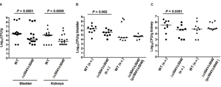
as well as the wild type (WT) in LB, the mutant was significantly outcompeted by the WT, with a nearly 10-fold reduction in the median CFU/g from both bladders and kidneys at 48 hours post-inoculation (Fig. 2A,P,0.05 for both organs). To verify that the impact on colonization is not due to a secondary mutation,in vivo
complementation experiments were performed. A stable low-copy plasmid pGEN-MCS was used as it was shown to be well maintained in CFT073 up to 48 h even in the absence of
antibiotic pressure [39]. Coding regions ofc5041and c5040plus
their predicted promoter region were cloned into pGEN-MCS, and the resultant construct was named pc5041/c5040. As shown in
Fig. 2B and C, theDc5041/c5040 mutant containing the empty
vector (pGEN-MCS) demonstrated an expected colonization defect in bladder and kidneys colonization as compared to the
WT (empty vector) (P,0.05) while mutants harboring the
complementation plasmid (pc5041/c5040) were able to colonize
the bladder and kidneys at wild-type levels at 48 hours post-inoculation. These results clearly demonstrate that c5041/c5040
has a role in UPEC colonization and fitnessin vivo.
Proteomic determination of gene products targeted by C5041/C5040
To determine how C5041/C5040 affects UPEC fitnessin vivo,
2D-DIGE was used to identify differentially expressed gene products in the proteomes of the WT and single deletion mutants of c5041and c5040 grown aerobically in human urine. Human urine was used as the growth medium to simulate the urinary tract
environment [40–42]. c5041 and c5040 single mutants were
Figure 1. Predicted 3-D structures and conserved domains of the HK C5041 and the RR C5040.The exact positions of each domain are described in the text. 3-D structures were predicted by I-TASSER online server. (A) HK C5041. The signature segments (H, N, G1, F, and G2) of the kinase domain (CA, blue) and dimerization and histidine phosphorylation domain (DHp, yellow and purple) were identified by sequence alignment and indicated. The conserved histidine residue is indicated by green. The two transmembrane helices (TMH-1 and TMH-2) were predicted by transmembrane protein topology prediction tool TMMOD. (B) RR C5040. The receiver domain (yellow) containing the putative aspartate residue (Asp, purple) for phosphorylation during signaling cascade, the AAA+ATPase domain (blue), and the helix-turn-helix (HTH, red) domain for DNA binding
are shown.
constructed by replacement with Chlr gene, and the resultant
mutants were named LMP100Chl (CFT073 Dc5041::Chlr) and
LMP101Chl (CFT073 Dc5040::Chlr). Using a cutoff of 1.5-fold
change, seventeen proteins in the mutants were differentially expressed, as compared to the WT (Fig. S1). Of these, five were induced and five were repressed inDc5041::Chlrmutant; while six were induced and five were repressed in theDc5040::Chlrmutant. To determine the identities of the differentially expressed proteins, protein spots in the gel were excised and subjected to enzymatic digestion with trypsin followed by tandem mass spectroscopy. Functions of these proteins primarily fell into three categories: metabolism, cell envelope constituents, and translational machin-ery (Table 1). C5035, which showed a 24.6-fold induction, is a putative 2-oxoglutarate dehydrogenase presumably involved in tricarboxylic acid (TCA) cycle. XylA, a xylose isomerase, was increased 8.7-fold. It catalyzes the interconversion between xylose and xylulose. SitA, induced 4-fold, is a putative periplasmic iron-binding protein. Several outer membrane proteins including OmpA, NmpC, OmpF and OmpX were uniformly induced in
Dc5040::Chlrmutant. 50S ribosomal proteins, such as Rpll, RplQ, and RplF, were also differentially expressed in the mutants as compared to the WT. These results indicate that C5041/C5040 regulate multiple genes, suggesting that they may serve as a pleiotropic two-component system.
Genomic island genes targeted by C5041/C5040 contribute to UPEC fitnessin vivo
The most differentially expressed protein identified by proteo-mic analysis was C5035, which was induced nearly 25-fold in the
Dc5041::Chlrmutant. qRT-PCR results confirmed thatc5035was up-regulated in Dc5041::Chlr mutant, but not in Dc5040::Chlr mutant, as compared to the WT when grown aerobically in human urine (Fig. 3A). To rule out the possibility of polar effects,
the Chlr gene was removed from both Dc5041::Chlr and
Dc5040::Chlr mutants to generate mutants LMP100 (CFT073
Dc5041) and LMP101 (CFT073 Dc5040), respectively. Interest-ingly, the expression ofc5035was detected at extremely low levels not only in the mutant strainsDc5041andDc5040, but also in the
WT. No significant differences in the c5035 expression were
detected among the WT, Dc5041 and Dc5040 mutants when
grown in human urine under aerobic conditions (Fig. 3A). Note that in theDc5041::Chlrmutant,c5041was replaced by the Chlr
gene, which transcribes in the same orientation as its downstream
gene,c5040. We further compared the expression levels of RR
c5040in the WT,Dc5041, andDc5041::Chlrmutants. The results
showed that expression ofc5040was extremely low in the WT and
Dc5041 mutant, but it was significantly upregulated in the
Dc5041::Chlrmutant when grown in human urine under aerobic conditions (Fig. 3B). Therefore, we suspect that the upregulation of
c5035 in Dc5041::Chlr mutant was due to the constitutive
expression of c5040 caused by the upstream Chlr gene. If so,
overexpression ofc5040in theDc5041mutant should induce the
expression ofc5035. Thus,c5040was cloned into plasmid
pMAL-MCS under the control of promoter Ptacand the resultant plasmid
transformed into theDc5041mutant. Indeed, induction ofc5040
significantly upregulated the expression of c5035 in the Dc5041
mutant (Fig. 3C). We also tested such regulation under different culturing condition and the results revealed similar regulatory pattern. In summary, constitutive expression ofc5040induced the expression ofc5035regardless of growth conditions.
Further analysis of genomic localization of c5035in CFT073
revealed that genes c5032-5039 including c5035, together with
TCS genesc5041 and c5040, are encoded by a genomic island
inserted betweenE. coli K12 genestyrBand aphA (Fig. 4A). Not
surprisingly, island genesc5032-5039were strongly associated with UPEC, and about 91.4% of UPEC strains tested containing
c5040/c5041loci also possessc5032-5039genes (data not shown). Prediction by bioinformatics tools (http://linux1.softberry.com/
berry.phtml and http://nostradamus.cs.rhul.ac.uk/,leo/
Figure 2. Deletion ofc5041/c5040reduces the colonization of the murine urinary tract by UPEC strain CFT073.(A) The WT andDc5041/
c5040::Chlrmutant strains were mixed to a 1:1 ratio and approximately 26109CFU were transurethrally inoculated into female mice. Two days after
infection, the mice were sacrificed and their bladders and kidneys were aseptically removed. WT and mutant bacteria were recovered by plating homogenized tissue samples on LB or LB containing chloramphenicol and their viable counts were determined. (B) and (C), for the in vivo
complementation assay, a mixture of type strain containing empty vector (e.v.) pGEN-MCS and mutant strain containing empty vector or wild-type strain containing empty vector and mutant strain containing complementation plasmid (pc5041/c5040) were incolulated to female mice as in (A). Homogenized tissue samples (B, bladder; C, kidneys) were plated on LB with ampicillin or LB with both ampicillin and chloramphenicol. Each dot represents log10CFU/g in the bladder or kidney from an individual animal and the detection limit is 1000 CFU/g. Bars indicate the median log10CFU/g. A two-tailed Wilcoxon matched pairs test was performed, and the difference in colonization levels of WT and mutants was considered statistically significant if P,0.05.
doi:10.1371/journal.ppat.1003428.g002
sak_demo/) followed by reverse transcription PCR indicate that
c5032-5037forms one operon andc5038-5039forms another (Fig. S2).
We then examined if this genomic island contributed to UEPC fitnessin vivo. Anin vivocompetition assay was used to compare the colonization levels ofDc5032-5039mutants with that of wild-type strain.Dc5032-5039mutant was constructed by replacement with thecatcassette. Fig. 4B showed that there is significant reduction in colonization levels ofDc5032-5039mutants in bladder and kidneys
at 48 hours post-inoculation, as compared to the WT (P,0.05).
These results clearly demonstrated that island genesc5032-5039
are involved in UPEC colonization of murine urinary tract and it’s tempting to presume that C5041/C5040 contributes to UPEC fitness through regulation ofc5032-5039genes (The results that all other target genes in the genomic island are regulated by TCS C5041/C5040 were shown later in this paper).
Island genesc5032-5039contribute to anaerobica-KG utilization anda-KG induces their expression under anaerobic conditions
The proteins encoded by c5032, c5034, and c5035 are
homologous to the E1, E2 and E3 components of a-KG
dehydrogenase inE. coli, with similarity of 64%, 69%, and 55%,
respectively; whereas, the proteins encoded byc5036 and c5037
are 70% and 83% similar to the beta- and alpha-subunits of
succinyl-CoA synthetase from E. coli. a-KG dehydrogenase
convertsa-KG to succinyl-CoA, which can be further transformed
to succinate by succinyl-CoA synthetase [43].c5038is predicted to encode a putative dicarboxylate transporter with 13 transmem-brane alpha-helices (TMHMM program [44]), showing 49% similarity to citrate/succinate antiporter CitT, andc5039encodes an enzyme belonging to malate/L-lactate dehydrogenase (Ldh_2) family. To test the hypothesis that these enzymes and the
metabolic transporter are involved in a-KG utilization, we
compared the growth of the WT, the Dc5032-5037mutant, the
Dc5038-5039 mutant, and the Dc5032-5039 mutant in M9
minimal medium with a-KG as the sole carbon source. Under
aerobic conditions, no significant growth differences were observed among these strains (Fig. S3). However, when tested
under anaerobic conditions, deletion of c5032-5039 completely
abolished the growth, and the Dc5032-5037 and Dc5038-5039
mutants also displayed statistically significant growth defects, as compared to the WT (Fig. 5A). We further tested their growth in M9 medium containing glucose, glycerol, or four-carbon dicar-boxylate compounds, including succinate, fumarate, and malate, as the sole carbon source. In these cases, no differences were found Table 1.Differentially expressed proteins identified in mutant strains of UPEC CFT073 as compared to the wild-type when cultured in human urine.
Order Name or ORF Function MWa/pIb
No. of peptides matched
Total ion score
Total ion C.I.c%
Fold-Changed
Differentially expressed proteins inDc5041::Chlrmutant
1 C5035 putative 2-oxoglutarate dehydrogenase 49832/6.1 16 172 100 24.6
2 XylA xylose isomerase 49634/5.63 16 63 100 8.7
3 SitA putative periplasmic iron-binding protein 31544/8.7 12 284 100 4.0
4 TufB EF-Tu 43427/5.3 7 556 .95 2.2
5 GlnA glutamine synthetase 52099/5.26 2 76 .95 2.0
6 YbiS hypothetical protein 33059/5.99 10 232 100 21.9
7 SpeE spermidine synthase 32286/5.33 7 228 100 22.2
8 RplF 50S ribosomal protein L6 17763/9.49 9 208 100 22.2
9 RplQ 50S ribosomal protein L17 14355/11.05 6 115 100 23.2
10 RplI 50S ribosomal protein L9 15759/6.17 14 474 100 24.0
Differentially expressed proteins inDc5040::Chlrmutant
1 OmpA outer membrane protein A precursor 41028/6.24 17 449 100 4.2
2 OmpA outer membrane protein A precursor 41028/6.24 14 317 100 3.0
3 NmpC outer membrane porin protein nmpC precursor 40277/4.64 6 391 .95 2.1
4 OmpF outer membrane protein F precursor 39309/4.76 9 496 .95 1.9
5 OmpX outer membrane protein X precursor 18648/6.56 2 152 .95 1.9
6 RplF 50S ribosomal protein L6 18949/9.71 7 356 .95 1.8
7 RplD 50S ribosomal protein L4 22073/9.72 5 380 .95 21.9
8 FocD F1C fimbrial usher 96379/6.72 1 29 .95 21.9
9 Tig Trigger protein 48163/4.83 4 150 .95 22.0
10 TufB EF-Tu 43427/5.3 11 677 .95 22.3
11 RplQ 50S ribosomal protein L17 14355/11.05 5 211 100 24.3
aMolecular weight. bIsoelectric point.
cConfidence interval (Ion Confidence Interval % greater than 95 were considered significant). dAverage from two biological repeats.
among the strains tested (data not shown). These results indicated that genomic island genesc5032-5039specifically contribute toa -KG utilization under anaerobic conditions but play no or a very limited role ina-KG utilization under aerobic conditions.
The ability ofa-KG to induce expression ofc5032-5039was also examined. Chromosomalc5032-lacZandc5038-lacZ transcriptional
reporter fusions in CFT073DlacZYA were constructed in order to
study the expression of operonc5032-5037and operonc5038-5039, andb-galactosidase activity in M9 medium usinga-KG (40 mM) as the sole carbon source was also determined. Under anaerobic conditions,a-KG significantly induced the expression ofc5032and
c5038, as compared to the rich medium LB or M9 medium containing glycerol or glucose as the sole carbon source (Fig. 5B);
while the analogs of a-KG such as 2-HO-glutarate, glutamate,
glutarate, fumarate, or succinate could not. To verify that other target
genes on the genomic island are also induced bya-KG, qRT-PCR
was performed. RNA was isolated from bacteria grown in M9
medium (glycerol and TMAO) with or without inducera-KG. As
shown in Fig. S4, all of the genes tested were greatly induced in the
presence of a-KG. We then examined the effect of a-KG
concentration on the stimulation ofc5038expression. Bacteria were grown in M9 medium plus glycerol and TMAO in the presence of various amounts ofa-KG. We found that the induction ofc5038by the presence ofa-KG was dose-dependent with concentrations as low as 200mM able to induce their expression under anaerobic conditions
(Fig. 5C). These results indicate that island genes c5032-5039are induced bya-KG and involved in anaerobic utilization ofa-KG.
C5040 directly regulates the expression ofc5032-5039and contributes toa-KG utilization under anaerobic conditions
To determine the regulatory role of C5040 ina-KG induction
of target genes, chromosomal c5032-lacZ and c5038-lacZ
tran-scriptional reporter fusions in theDc5040mutant were construct-ed. Strains were grown in M9 medium containing glycerol and
TMAO in the presence or absence of inducer a-KG under
anaerobic conditions. As shown in Fig. 6A, in wild-type genetic
background, c5032 and c5038 expression was considerably
induced in response to a-KG; in contrast, deletion of c5040
abolished the expression ofc5032 and c5038 to the background
level under anaerobic conditions. Re-introduction of a plasmid construct pc5041/c5040 carrying c5041/c5040 genes with their
native promoter into c5040 mutants (LMP206 and LMP209)
markedly increased expression ofc5032and c5038; whereas, the
empty plasmid vector did not affect the expression ofc5032and
c5038. In addition, the regulation of other target genes (c5034,
c5035,c5036,c5037, andc5039) in the genomic island in response
to a-KG by C5040 was confirmed using qRT-PCR (Fig. S4).
Together, these results indicate that a-KG induction of c5032
-5039is C5040-dependent (Fig. 6A).
To determine if C5040 directly regulates c5032 and c5038
expression, an electrophoretic mobility shift assay (EMSA) was
performed. The promoter regions of c5032 and c5038 were
predicted by BProm program (http://linux1.softberry.com) [45].
The potential binding sites forc5032 and c5038 were identified
and found to be highly similar, with TGTGTG-N13-CGCGCA
for c5032 and TGTGCG-N8-CGCACA for c5038. DNA
frag-ments containing the potential binding sites were then PCR
amplified for use as probes (305 nucleotides in size for c5032,
starting from2272 to+32 relative to translational start codon; 196 nucleotides in size forc5038, starting from2163 to+32 relative to translational start codon). Fragments amplified from the coding
region of c5036 were used as negative controls. Gene c5040
encoding the RR was cloned into the expression vector pMal-c2x
and fused with MBP-His6. MBP-C5040-His6 fusion protein
retained its regulatory biological function since introduction of Figure 3. Overexpression ofc5040upregulatedc5035expression independent of the natural stimulus.Bacteria were grown in human urine under aerobic conditions before being subjected to RNA extraction. qRT-PCR expression values are means plus standard deviations (error bars) of three independent experiments. (A)c5035expression in various TCS mutants compared to WT. (B)c5040expression inDc5041andDc5041::Chlr mutants compared to WT. (C)c5035expression inDc5041mutant carrying the pMAL plasmid withc5040under the control of Ptacpromoter as compared toDc5041mutant carrying the empty vector (e.v.). IPTG is present at 1 mM for induction.
doi:10.1371/journal.ppat.1003428.g003
the plasmid construct expressing MBP-C5040-His6fusion protein
into Dc5040 mutant activated the expression of c5038; whereas, the empty plasmid did not affect expression ofc5038(Fig. S5). As
shown in Fig. 6B, the purified MBP-C5040-His6 fusion protein
was able to shift the promoter fragments of bothc5032andc5038, but not the control fragment. Meanwhile, use of the purified
MBP-His6 fusion protein without C5040 was not associated with a
detectable protein-DNA complex (Fig. 6B). These results demon-strate that C5040 directly binds to the promoters of the two operons.
To determine if C5040 is involved in the utilization ofa-KG as the sole carbon source under anaerobic conditions, the growth
properties of the WT, Dc5040 mutants, and the complemented
strains were compared. When grown in M9 medium witha-KG as
the sole carbon source under anaerobic conditions, the Dc5040
mutant strains showed a growth defect as compared to the WT. Thec5040-complemented strain (Dc5040carrying pc5041/c5040) significantly increased growth in this medium; while the empty
plasmid vector (pGEN-MCS) did not improve growth of the mutant (Dc5040) (Fig. 6C).
The role of C5041 in the expression of island genes
c5032-5039
The regulatory role of HK C5041 was also determined. Chromosomalc5032-lacZandc5038-lacZtranscriptional reporter
fusions in theDc5041mutant (LMP106) were obtained to create
LMP205 and LMP208, respectively. Strains were grown in M9 medium (supplemented with glycerol and TMAO) with or without
inducer a-KG under anaerobic conditions. Deletion of c5041
abolished the expression ofc5032andc5038to background levels
in the presence ofa-KG. Re-introduction of the plasmid pc5041
carrying thec5041gene with its native promoter into LMP205 and
LMP208 mutants significantly increased their expression
(P,0.05), indicating that C5041 positively affects expression of the target genes (Fig. 7A). This suggests that phosphorylation is very important for the regulatory function of RR C5040. In Figure 4.c5032-5039genes are located on a genomic island and contribute to UPEC fitnessin vivo.(A) Similar genomic regions ofE. coli
K12 strain MG1655, EHEC strain EDL933, and UPEC strain CFT073 were compared and it was found thatc5032toc5041genes are inserted between
tyrBandaphAgenes and can be considered as a genomic island. Within the island, three predicted operons shown as white, grey, and black arrows, respectively. Picture was drawn to scale. UPEC, uropathogenicE. coli; EHEC, enterohemorrhagicE. coli. (B) Island genesc5032-5039contribute to UPEC fitnessin vivo. Anin vivocompetition assay was used. Equally mixed WT andDc5032-5039::Chlrmutant bacteria containing approximately 2
6109CFU
were transurethrally inoculated into female mice. Two days after infection, the mice were sacrificed and their bladders and kidneys were aseptically removed. Wild-type and mutant bacteria were recovered by plating homogenized tissue samples on LB or LB containing chloramphenicol and their viable counts were determined. Each dot represents log10CFU/g in the bladder or kidney from an individual animal and the detection limit is 1000 CFU/g. Bars indicate the median log10CFU/g. A two-tailed Wilcoxon matched pairs test was performed, and the difference in colonization levels of WT and mutants was considered statistically significant ifP,0.05.
addition, the expression of other genes (c5034, c5035, c5036,
c5037, and c5039) in the genomic island affected by C5041 was also confirmed using qRT-PCR (Fig. S4). The growth phenotypes
of thec5041mutant and its complementation strain in difference
medium were then tested. Interestingly, the growth of the WT and its derivative mutants (c5041mutant and complementation strain) in all media tested was statistically indistinguishable (Fig. 7B).
Oxygen tension modulates expression ofc5032-5037and
c5038-5039directly via the TCS C5041/C5040
Island genesc5032-5039contribute to anaerobic utilization of
a-KG but play a limited role under aerobic conditions causing us to suspect that oxygen might modulate expression of these genes.
Indeed, the c5032-5037 operon encoding a putative a-KG
dehydrogenase and a putative succinyl-CoA synthetase was
upregulated.5-fold in a reduced oxygen environment. Similarly,
the expression of the c5038-5039 operon, encoding a putative
transporter ofa-KG and a protein with unknown function, was
upregulated 2-fold under anaerobic conditions, as compared to aerobic conditions (Fig. 8A).
To determine if oxygen regulates c5032-5039 expression via
C5041/C5040 but not through other independent pathways, the effects ofc5041/c5040double deletion on the expression of
c5032-5039 were examined. Deletion of c5041/c5040 completely
abolished expression of the two operons under both aerobic and
anaerobic conditions, supporting the hypothesis that oxygen regulates these two operons via TCS C5041/C5040 (Fig. 8A).
In addition, we wished to determine if oxygen deficiency regulates expression of the TCS itself. Under anaerobic conditions,
the expression of c5041 was about 3-fold greater than that
observed under aerobic conditions (Fig. 8B), suggesting thatc5041
expression is significantly induced in response to anaerobiosis.
Similarly, under anaerobic conditions, c5040 expression is
dramatically increased as compared to that under aerobic conditions (Fig. 8B). These results suggested that oxygen deficiency
upregulated the expression of c5041 and c5040 and raised the
possibility that oxygen modulates the expression of c5032-5037
andc5038-5039operons via controllingc5041/c5040expression. However, these results did not rule out the possibility that anaerobiosis stimulates the typical TCS phosphotransfer reactions between C5041 and C5040, leading to increased DNA binding and up-regulation of target gene expression.
Discussion
One of the greatest challenges confronted by all microorganisms is adapting to rapid changes of nutrient availability in different habitats. In the course of evolution, bacteria have developed several mechanisms to sense and utilize available nutrient sources associated with particular niches or to favor the most efficiently metabolizable nutrient sources when exposed to a range of Figure 5. Contribution ofc5032-5039to growth ona-KG and induction ofc5032-lacZorc5038-lacZexpression bya-KG.(A)In vitro
growth of island gene mutants in M9 medium containinga-KG as the sole carbon and energy source. Optical density of the CFT073 WT and mutants during growth in M9 medium containinga-KG as the sole carbon source under anaerobic conditions was determined. Growth bars represent the average measurement at each time point from triplicate experiments. (B) Effects of different substrates onc5032-lacZorc5038-lacZexpression. Gluc, glucose; Glyc, glycerol; TMAO, TrimethylamineN-oxide. (C) The induction ofc5038-lacZexpression bya-KG is dose-dependent. Bacteria were grown in M9 medium containing 0.25% glycerol as the sole carbon source plus different amounts ofa-KG as stimulus under anaerobic conditions. doi:10.1371/journal.ppat.1003428.g005
choices. TCSs are major mechanisms enabling bacteria to couple
environmental stimuli to adaptive responses [46]. The TCSs inE.
coliK12 have been extensively studied [25–27]. Many TCSs that
are common to both commensal and pathogenic E. coli, such as
PhoQ/PhoP [31], QseC/QseB [29,30], BarA/UvrY [28], and the
AirS system [32] have been shown to contribute to virulence by mediating bacterial adaption to the host environment. However, no pathogen-associated TCS has yet been characterized inE. coli. Driven by the hypothesis that UPEC-associated TCSs exist to sense and respond to host environment signals distinct from those Figure 6. The role ofc5040.(A) Effects ofc5040on the expression of target genes in response toa-KG. Bacteria were anaerobically grown in M9 medium containing glycerol as sole carbon source and TMAO as electron acceptor in the absence or presence ofa-KG. The expression ofc5032-lacZ
orc5038-lacZin WT,Dc5040(LMP107), and the complemented strains were represented by theb-galactosidase activity. (B) Non-radioactive EMSA studying the binding of MBP-C5040-His6to the promoter regions. PCR products ofc5032promoter region (left panel) andc5038promoter region (right panel) were used as probes. Purified MBP-C5040-His6fusion protein was added in different concentration in each reaction mixture as indicated and MBP-His6was used as negative control at 300 ng per each reaction. For the lane on the right on each panel, a negative control DNA fragment amplified from coding region ofc5036was used. DNA fragments were stained with SYBR green. (C)In vitrogrowth ofDc5040mutants. Optical density of the CFT073 WT,Dc5040mutant, and the complemented strain during growth in M9 medium containinga-KG as the sole carbon source under anaerobic conditions was measured. Growth bars represent the average measurement at each time point from triplicate experiments.
of intestinal E. coli, we identified a novel TCS C5041/C5040 significantly associated with UPEC strains, but not with EHEC, EPEC or ETEC strains. This TCS activated the expression of a
genomic island involved in transport and metabolism of a-KG
under anaerobic conditions. In view of these findings, C5041 was
renamed KguS (a-ketoglutarate utilization sensor) and C5040
renamed KguR (a-ketoglutarate utilization regulator). These
results indicated that UPEC might actively import a-KG and
thata-KG could be an important carbon source for UPECin vivo. Consistent with this observation, deletion of the TCS or its target island genes resulted in a significant reduction in a UPEC’s
colonization of the murine urinary tract. To our knowledge, this is
the first report of a pathogen-associated TCS in E. coli that
contributes to UPEC pathogenesis.
a-KG is an intermediate in the citric acid/tricarboxylic acid
(TCA) cycle, which is a metabolic pivot for both catabolic and anabolic processes that supply key metabolic intermediates and energy in most eubacteria [47]. Many intermediates in the TCA cycle can be sensed by TCSs, which elicit an adaptive response in
E. coli. Four-carbon (C4) dicarboxylate-sensing DcuS/DcuR was
shown to control fumarate transportation (dcuB) and respiration (frdABCD) under anaerobic conditions, and under aerobic condi-Figure 7. The role ofc5041.(A) Effects ofc5041on the expression of the target genes in response toa-KG. The expression ofc5032-lacZorc5038
-lacZin the WT,Dc5041(LMP106), and the complemented strains were represented by theb-galactosidase activity. (B)In vitrogrowth ofDc5041
mutants. Optical density of CFT073 WT,Dc5041mutant, and the complemented strain during growth in M9 medium containinga-KG as the sole carbon source under anaerobic conditions was measured. Growth bars represent the average measurement at each time point from triplicate experiments.
doi:10.1371/journal.ppat.1003428.g007
tions affect the utilization of most of C4-dicarboxylates like
succinate, fumarate, malate, and aspartate [48,49]. In addition, tricarboxylate-sensing CitA/CitB was demonstrated to regulate
anaerobic citrate fermentation genes citCDEFXGT encoding an
active holo-citrate lyase catalyzing the breakdown of citrate to acetate and oxaloacetate and a citrate carrier [50–52]. This study has demonstrated that a novel TCS KguS/KguR responds to and
only to a-KG, a five-carbon (C5) dicarboxylate by activating
metabolic enzymes and transporter involved in anaerobic
metab-olism of a-KG. This is the first-time that such a TCS has been
identified responding to C5-dicarboxylate inE. coli. Identification
of the TCS regulating utilization of C4, C5-dicarboxylates and
tricarboxylate suggested the tight control of the pathways involved in the metabolism of intermediates inE. coliTCA cycle.
As found in this study,a-KG can induce expression of the TCS
target genesc5032-5039, while deletion ofkguSorkguRabolished their expression in response toa-KG. By extrapolation from what we know of other TCSs and the results obtained in this study, it is
very likely thata-KG serves as the signal molecule and HK KguS
senses it via its periplasmic domain, then phosphorylates itself, and subsequently trans-phosphorylates KguR, which can then induce expression of the target genes. Very interestingly, overexpression of KguR can activate the expression of target genes independent of
inducer a-KG. Similar observations have been reported for
Salmonella where induction of target genes depends on the intracellular concentration of the response regulator PhoP and overexpression can substitute for phosphorylation and enhance the functionality of PhoP [53]. We further confirmed that oxygen tension modulated the expression of this pathway by regulating the expression of KguS/KguR itself. InE. coli, regulation by oxygen is usually mediated by the cytoplasmic regulator FNR and/or by TCS ArcB/A [54]. Currently, it is unknown if FNR and/or ArcB/ A have any effect on the expression of KguS/KguR.
Genome sequencing has shown that the TCA cycle is a readily modified pathway in bacteria with many strains harboring variant TCA cycles. The common feature of these variants is the absence
or repression of the a-KG dehydrogenase with the oxidative
branch terminating in the production ofa-KG and the reducing
branch in succinate [55,56]. Like any otherE. coli, UPEC operate
a complete TCA cycle aerobically, but thea-KG dehydrogenase
shared by all E. coli is neither functional nor expressed under
anaerobic conditions [47]. The KguS/KguR-controlled metabolic
pathway in UPEC includes a putative a-KG dehydrogenase
(c5032-5035) and a putative succinyl-CoA synthetase (c5036-5037),
which were induced and contributed to utilization ofa-KG under
anaerobic conditions. To our knowledge, this is the first variant TCA cycle described inE. colithat links the oxidative and reducing branches under anaerobic conditions. To maintain carbon flux under oxygen-limiting or anaerobic conditions, many
microaero-philic and obligate anaerobic organisms that lacka-KG
dehydro-genase possess similar bypass pathways to metabolically link
oxidative and reducing branches. Helicobacter pylori preferably
growing under microaerophilic conditions use a-KG ferredoxin
oxidoreductase to connect oxidative and reducing half-cycles [57].
Likewise, in Mycobacterium an a-KG decarboxylase structurally
related to the E1 component of 2-oxoglutarate dehydrogenase can produce succinic semialdehyde (SSA), which is subsequently oxidized to succinate by succinic semialdehyde dehydrogenase (SSADH) [58]. This study and other groups’ findings have highlighted the importance of the branched variants in adaptation to oxygen limiting or obligate anaerobic conditions and suggest that UPEC have different metabolic niches and needs compared to otherE. coli.
The acquisition of additional metabolic traits often increases adaptability and competitiveness of pathogens in a new niche [18,22]. This study found that TCS KguS/KguR and its
controlled-anaerobic utilization pathway ofa-KG were encoded
by a genomic island in UPEC, which was absent from the genomes of commensalE. coliand intestinal pathogenicE. coli. These results were consistent with the previous report thatc5032toc5040were among the 131 UPEC-specific genes identified using comparative genomic hybridization andin silicoanalysis of ten UPEC and four fecal/commensal isolates [59], and also suggested that this
genomic island may facilitate UPEC’s adaptation to in vivo
colonization. This was confirmed by the observation that deletion of kguS/kguR resulted in a significant reduction in UPEC Figure 8. Oxygen tension modulates the expression of two operons in genomic island via TCS C5041/C5040.(A) Target genes’ expression inc5041/c5040mutants under aerobic and anaerobic conditions. The expression ofc5032-lacZorc5038-lacZin WT andDc5041/c5040
mutant (LMP108) under aerobic and anaerobic conditions were compared. (B) Induction ofc5041/c5040expression by oxygen deficiency.c5041-lacZ
colonization in the bladders and kidneys of mice. However,
deletion of kguS/kguRor its target genomic island genes did not
affect growth in human urine in vitro under both aerobic and
anaerobic conditions (data not shown). We reasoned that human urine might only be able to partially imitate thein vivoconditions of UPEC infection, whereas the contributions of KguS/KguR to UPEC pathogenesis may be restricted to some particular site of infection or pathogenic process, which cannot be represented by urine.
Interestingly, Chlamydia trachomatis, an obligate intracellular bacterial pathogen of urogenital infections [60], possesses an incomplete TCA cycle. Because of their small genome, chlamydiae lack many metabolic pathways retaining only the functions for performing key steps and interconversions of metabolites from the host cells. The fact that chlamydiae lack
the first three enzymes of the TCA pathway and obtainsa-KG
from their host cell via a membrane transporter [56,61] suggests that utilization of intracellulara-KG is important for the survival of this urogenital intracellular pathogen. It could be inferred from
Chlamydia and hypothesized that a-KG might be one of the preferred carbon and energy sources for UPEC during infection. Previous studies supporting this hypothesis showed that the oxygen tension (PO2) in UPEC-infected tissue dropped to 0 mmHg within 3.5–4 h [62], suggesting UPEC encountered anaerobic conditions during infection. It was also reported that
the concentration of a-KG in human cells and blood is about
10mM, a concentration that was able to induce the expression of
KguS/KguR-controlled pathwayin vitro under anaerobic
condi-tions in this study. The KguS/KguR-controlled transporter (C5038) differs from the constitutively expressed KgtP encoded by allE. coli[63,64] and was induced under anaerobic conditions.
During infection, this transporter allows UPEC to importa-KG
from the infected tissue into the cell, anda-KG dehydrogenase
and succinyl-CoA synthetase allow UPEC to take advantage ofa
-KG under anaerobic conditions, thus contributing to UPEC’sin
vivofitness. More interestingly, proximal tubule cells in the kidney
are able to accumulatea-KG. Proximal tubules are the primary
site where organic anions (OA) of physiological, pharmacological, and toxicological importance are cleared from the body. Their clearance involves uptake of OA via OA-dicarboxylate exchange driven bya-KG gradients. Thus, intracellular concentration ofa
-KG in the proximal tubule cells is,100–400mM, which is 10 to
40-fold higher than that found in any other cells [65–67]. UPEC undergo an intracellular lifestyle where they form biofilm-like communities [68], and UPEC are able to infect proximal tubule cells [62,69–71]. However, very little is known about the metabolism of UPEC within these host cells. It is very likely
that KguS/KguR-controlled anaerobic utilization pathway ofa
-KG would increase UPEC fitnessduring infections of renal proximal tubule cells. Future studies in our laboratory should help clarify these questions.
In summary, our findings suggested a model that describes a novel regulatory and metabolic pathway in UPEC. For optimal
growth during infection, HK KguS may sensea-KG, an abundant
metabolite in UPEC infection site-renal proximal tubules, and then phosphorylate the RR KguR, which finally activates the
import and utilization ofa-KG (Fig. 9). These findings provide
compelling evidence that this first UPEC-associated TCS enables
E. colito sense infection niche-specific stimuli and adapt to local nutrient availability, thus increasing itsin vivo fitness. Hopefully, this and future studies on this regulatory system and metabolism pathway could lead to a better understanding of correlations between bacterial metabolism and virulence, but also the molecular pathogenesis of UPEC.
Materials and Methods
Ethics statement
7All animal procedures were conducted in accordance with NIH guidelines, the Animal Welfare Act and US federal law. The experimental protocol (Protocol number 4-11-7111-Z) for han-dling animals was approved by Institutional Animal Care and Use Committee at Iowa State University (IACUC). All surgery was performed under isoflurane anesthesia, and all efforts were made to minimize suffering.
The study using human urine was approved by Institutional Review Board (IRB ID: 04-171). Written informed consent was obtained from human participants and/or their legal guardians for urine collection.
Bacterial strains and culture conditions
Strains and plasmids used in this study are listed in Table S1. Aerobic growth was achieved by shaking in air at 180 rpm and anaerobic growth by incubating in a Bactron chamber (Sheldon Manufacturing, Inc., OR) filled with gas mixture (N2, 90%; CO2,
5%; H2, 5%). For genetic manipulations, allE. colistrains were grown
routinely in lysogenic broth (LB) medium. For growth and gene expression studies, bacteria were generally grown aerobically or anaerobically in M9 minimal salts with certain carbon sources
indicated, supplemented with 2 mM MgSO4, 0.1 mM CaCl2, and
1 mg/ml vitamin B1. When used, trimethylamine N oxide (TMAO) and di- or tri-carboxylates were present at 40 mM. Glucose (0.5% v/ v) or glycerol (0.25% v/v) was added as energy substrates, as indicated. Fresh mid-stream human urine was collected from six male and female consenting donors, pooled, and filter sterilized. Selective antibiotics and IPTG were added when necessary at the following concentrations: ampicillin (Amp), 100mg ml21; kanamycin (Kan), 50mg ml21;chloramphenicol (Chl), 25mg ml21; IPTG, 1 mM.
Recombinant DNA techniques
Polymerase chain reaction (PCR), DNA ligation, electroporation and DNA gel electrophoresis were performed according to Sambrook and Russel [72] unless otherwise indicated. All oligonucleotide primers were purchased from Integrated DNA Technologies (Iowa) and are listed in Table S2. All restriction and DNA-modifying enzymes were purchased from New England Biolabs and used based on the suppliers’ recommendations. Recombinant plasmids, PCR products, and restriction fragments were purified using QIAquick PCR purification kit or MinElute gel extraction kit (Qiagen, CA) as recommended by the supplier. DNA sequencing was performed at the DNA facility, Iowa State University. DNA and amino acid sequence analyses were performed using CloneManager software (Scientific & Educational Software, NC) and the search engine (http://blast.ncbi.nlm.nih.gov/Blast.cgi.) was used to identify con-served domain structures of the two-component system.
Deletion mutants were constructed using the lambda red recombinase system described by Datsenko and Wanner [73]. Chromosomal transcriptionallacZfusion was constructed by homol-ogous recombination of the suicidal plasmid pVIK112 carrying a fragment of complete 59-region, 39- region, or internal fragment of the target gene [74]. Briefly, PCR fragments of target genes were cloned
into pVIK112 usingEcoRI and XbaI sites. The resultant pVIK112
derivatives were introduced into CFT073DlacZYA::Chlrby conjugation and the integration was allowed to occur. Conjugants were selected
and confirmed by PCR. Chlrwas removed by transforming pCP20
plasmid carrying flippase [75]. For complementation, the coding sequences of genes plus their putative promoter regions were amplified from the CFT073 genome and independently cloned into pGEN-MCS [39] usingEcoRI andSalI restriction sites.
To construct the plasmid overproducing MBP-KguR-His6
fusion protein, a 1.4-kb fragment containing the coding region
ofkguRwas obtained by PCR from genomic DNA using MalE/
c5040Histag-F and MalE/c5040Histag-R carrying codons for
66His and subsequently cloned into pMAL-c2x (New England
Biolabs) using BamHI and HindIII sites. The resultant plasmid
contains MBP-KguR-His6under the control of the Ptacpromoter.
The construction of control vector pMAL-MCS was achieved by replacing the MalE coding sequence with the MCS fragment from
pEGFP plamid usingNdeI andEcoRI sites.
PCR genotyping
The nucleotide sequences publicly available of c5041/c5040
genes were aligned by using the ClustalW2 program. The primers were selected from a relatively conserved region and on the basis of G/C content, annealing temperature and size of the amplicon. Multiplex PCR was carried out according to Johnson et al. [76].
b-Galactosidase assays
Overnight LB cultures ofE. colicontaining the gene of interest-lacZ
fusions were washed with PBS once and then were diluted 1:100 in LB or M9 medium with the carbon sources indicated and grown at 37uC to log phase. These cultures were diluted 1:10 in Z buffer and assayed for b-galactosidase activity using ortho-Nitrophenyl-b -galactoside (ONPG) as a substrate as described previously [77].
2D-DIGE
To prepare bacterial protein samples, isolated colonies were
used to inoculate LB and cultures grown overnight at 37uC, the
culture was further diluted 1:50 to fresh LB medium and
re-incubated; after OD 600 reached 1.0, 2 ml of culture
(approxi-mately 109CFU) was pelleted and the supernatant removed.
Bacterial pellets were re-suspended in 20 ml of human urine and
grown aerobically for 4 hours at 37uC. Bacteria grown in human
urine were then collected by centrifugation and washed 3 times with sterile PBS. The resultant bacterial pellets were snap-frozen at 280uC and submitted to Applied Biomics, Inc. in California for Fluorescence difference in gel electrophoresis (2D-DIGE) [78].
Reverse transcription PCR and quantitative real-time RT-PCR
RNA fromE. coliCFT073 and its derivatives was stabilized by
RNAprotect Bacterial Reagent (QIAGEN) and extracted using an RNeasy Mini Kit (QIAGEN) with a one-hour in-tube DNase digestion (QIAGEN) to remove possible DNA contamination according to the manufacturer’s instructions. Three biological replicates of the each sample were prepared. The concentration of RNA was determined using a Spectrophotometer (ND-1000) (NanoDrop).
For the co-transcription test, one microgram of total RNA was reverse transcribed in triplicate using random hexamers and ImProm-II reverse transcriptase (Promega). Reactions without reverse transcriptase were used as a DNA contamination control. cDNA was then used as template for subsequent PCR reactions.
For quantitative real-time RT-PCR, melting curve analyses were performed after each reaction to ensure amplification specificity. Differences (n-fold) in transcripts were calculated using the relative comparison method, and amplification efficacies of each primer set were verified as described by Schmittgen et al
[79]. RNA levels were normalized using the housekeeping genetus
Figure 9. A schematic model for the regulatory mechanism of C5041/C5040 and its controlled target genes. In certain host environments of UPEC, such as the epithelial cells of renal proximal tubule, where oxygen tension is low anda-KG is abundant, KguS/KguR can sense and respond to these stimuli by activating the expression ofc5038encoding dicarboxylate transporter andc5032-5037genes encoding metabolic enzymes, involved in the utilization ofa-KG as carbon and energy source. Possession and functionality of this system may meet special metabolic needs for UPEC during urinary tract infections. Question mark indicates function is unknown. Blue dots,a-KG (a-ketoglutarate); Succ-CoA, succinyl-CoA; Succ, succinate.
encoding DNA replication terminus site-binding protein as an endogenous control [80]. Quantitative real-time RT-PCR (qRT-PCR) was performed with a Bio-Rad iQ5 iCycler detection system using iScript one-step RT-PCR kit with SYBR Green (Bio-Rad) according to the manufacture’s instruction [81].
Electrophoretic mobility shift assays
To study the binding of KguR to the DNA probe, electropho-retic mobility shift assays (EMSAs) were performed using the commercialized EMSA kit (Invitrogen, California) [82].
MBP-KguR-His6 fusion protein was purified to homogeneity
using Ni-NTA Spin Columns and dialyzed against binding buffer. DNA probes were PCR amplified using specific primers and gel purified using a QIAGEN MinElute gel extraction kit. EMSAs were performed by adding increasing amounts of purified
MBP-KguR-His6fusion protein (0 to 300 ng) to the DNA probe (60 ng)
in binding buffer (10 mM Tris (pH 7.5), 1 mM EDTA, 1 mM
dithiothreitol, 50 mM KCl, 50 mM MgCl2, 1mg ml21 bovine
serum albumin (NEB)) for 30 min at room temperature. Reaction mixtures were then subjected to electrophoresis on a 6%
polyacrylamide gel in 0.56TBE buffer (44.5 mM Tris, 44.5 mM
boric acid, 1 mM EDTA, pH 8.0) at 200 V for 45 min. The gel
was stained in 0.56TBE buffer containing 16SYBR Gold nucleic
acid staining solution for 30 min.
Experimental UTIs
Mouse infection studies were performed according to the methods of Johnson et al [83]. Female CBA/J mice (six to ten weeks of age) were anesthetized and inoculated via transurethral catheterization with a 20ml (26109CFU) challenge inocula per mouse. Overnight LB cultures for CFT073 and the mutant strain were pelleted and resuspended in sterile PBS, mixed in equal number and adjusted to make challenge inocula. To determine the initial CFU/mL, dilutions of each inoculum were plated onto LB plates with and without chloramphenicol. After 48 h, the mice were euthanized and the bladder and kidneys were aseptically removed, weighed, and homogenized in tubes containing PBS. Dilutions of the homogenized tissue were then plated onto duplicate LB plates with and without chloramphenicol or plates with different antibiotics to determine the bacterial concentration (CFU/g) of tissue. After overnight incubation, distinct colonies on plates were enumerated. The numbers of colonies on selective plates were subtracted from those on LB plates to obtain the
number of wild-type bacteria. In the case ofin vivo
complemen-tation studies, recovered bacteria were plated on LB plates with ampicillin and LB with ampicillin and chloramphenicol. Similarly, the numbers of colonies on ampicillin and chloramphenicol plates were subtracted from those on LB plates with ampicillin to obtain the number of wild-type bacteria. A group of 10 mice for each dual-strains challenge were used to determine alterations in fitness.
The WT/Dc5041/c5041::Chlrcompetition assay was performed
twice while other assays were performed once. For statistical analysis, a two-tailed Wilcoxon matched pairs test was used (Prism software, CA) and the threshold for statistical significance was aP
value,0.05.
Supporting Information
Figure S1 Comparison of the proteomes from the WT,
Dc5041 and Dc5040 mutants during growth in human
urine. Whole cell proteins of WT, Dc5041::Chlr, and
Dc5040::Chlr mutants cultured in urine were labeled with cy2, cy3, and cy5 respectively and analyzed by 2D-DIGE in a single gel. Red spots represent proteins induced in the mutant as
compared to the WT; green spots indicate proteins repressed in the mutant as compared to the WT. Differentially expressed proteins were circled and numbered. (A) Comparison between the WT andDc5041::Chlrmutants. Ten proteins were identified to be differentially expressed. (B) Comparison between the WT and
Dc5040::Chlr mutants. Eleven proteins were identified to be
differentially expressed. (TIF)
Figure S2 Operon formation represented by reverse transcription (RT)-PCR results using cDNA and a
negative control without reverse transcriptase. Primers
were designed to span ORFsc5032&c5034,c5034&c5035,c5035
& c5036, c5036 & c5037, c5037 & c5038, c5038 & c5039, and
c5039 & c5040. UPEC CFT073 were cultured in M9 medium containing glycerol as the sole carbon source. RNA was purified and reverse transcribed to cDNA. The RNA that was not reverse transcribed served as a negative control.
(TIF)
Figure S3 In vitrogrowth of island gene mutants in M9
medium containinga-KG as the sole carbon and energy
source under aerobic conditions.Optical density of CFT073
WT and island gene mutants during growth in M9 medium
containing a-KG as the sole carbon source under aerobic
conditions was determined. Growth curves represent the average measurement at each time point from triplicate experiments. (TIF)
Figure S4 Effects of C5041 and C5040 on the induction
of target genes on genomic island bya-KG.Wild-type or
mutant bacteria were anaerobically grown in M9 medium
containing glycerol and TMAO in the absence or presence ofa
-KG. qRT-PCR expression values in the presence of a-KG are
presented as relative values, as compared to that in the absence of
a-KG. Error bars represent the standard deviations for 3
independent experiments. (TIF)
Figure S5 The induction of c5038 expression by
over-produced MalE/C5040 fusion protein. The expression of
c5038-lacZin the WT transformed with the plasmid vector control
(pMAL), pMAL-c5040, or pMAL-malE/c5040 were represented
by theb-Galactosidase activity. IPTG was present at 1 mM for
induction of Ptacpromoter.
(TIF)
Table S1 Strains and plasmids.The genotypes of all strains of E. coli utilized or constructed in this study and information about the plasmids used in this study.
(DOCX)
Table S2 Oligonucleotides.Oligonucleotide sequences used as PCR primers.
(DOCX)
Table S3 Prevalence ofc5041/c5040locus amongE. coli
isolates.
(DOCX)
Acknowledgments
The authors gratefully acknowledge Drs. Michael J. Wannemuehler and Glenn J. Songer for providing access to an anaerobic chamber and Dr. Qijing Zhang for use of the BioRad iQ5 iCycler. We thank Ting Meng and Capricia Thompson for the help in performing transurethral catheteriza-tion of mouse. Also, we appreciate the kind gift of pGEN plasmids from Dr. Harry L. T. Mobley and intestinal pathogenicE. coli strains from Dr. Nancy Cornick.