Persistence in
Staphylococcus aureus
Jian Han1,2, Lili He1, Wanliang Shi2, Xiaogang Xu3, Sen Wang4, Shuo Zhang2, Ying Zhang2,4*
1Department of Pathogenic Biology, School of Basic Medical Sciences, Lanzhou University, Lanzhou, China,2Department of Molecular Microbiology and Immunology, Bloomberg School of Public Health, Johns Hopkins University, Baltimore, Maryland, United States of America,3Institute of Antibiotics, Huashan Hospital, Fudan University, Shanghai, China,4Department of Infectious Diseases, Huashan Hospital, Fudan University, Shanghai, China
Abstract
S. aureusis a significant human pathogen and has previously been shown to form cell wall deficient forms or L-formsin vitro
andin vivoduring infection. Despite many previous studies onS. aureusL-forms, the mechanisms of L-form formation in this organism remain unknown. Here we established the L-form model inS. aureusand constructed a transposon mutant library to identify genes involved in L-form formation. Screening of the library for mutants defective in L-form formation identified glpF involved in glycerol uptake being important for L-form formation in S. aureus. Consistent with this observation,glpFwas found to be highly expressed in L-form S. aureusbut hardly expressed in normal walled form. In addition,glpFmutant was found to be defective in antibiotic persistence. The defect in L-form formation and antibiotic persistence of theglpFmutant could be complemented by the wild typeglpFgene. These findings provide new insight into the mechanisms of L-form formation and persistence inS. aureusand may have implications for development of new drugs targeting persisters for improved treatment.
Citation:Han J, He L, Shi W, Xu X, Wang S, et al. (2014) Glycerol Uptake Is Important for L-Form Formation and Persistence inStaphylococcus aureus. PLoS ONE 9(9): e108325. doi:10.1371/journal.pone.0108325
Editor:Gunnar F. Kaufmann, The Scripps Research Institute and Sorrento Therapeutics, Inc., United States of America
ReceivedJune 8, 2014;AcceptedAugust 18, 2014;PublishedSeptember 24, 2014
Copyright:ß2014 Han et al. This is an open-access article distributed under the terms of the Creative Commons Attribution License, which permits unrestricted use, distribution, and reproduction in any medium, provided the original author and source are credited.
Data Availability:The authors confirm that all data underlying the findings are fully available without restriction. All relevant data are included within the paper.
Funding:These authors have no support or funding to report.
Competing Interests:The authors have declared that no competing interests exist.
* Email: [email protected]
Introduction
L-form bacteria refer to cell wall deficient form of bacteria that were first discovered by Emmy Klienenberger in 1935 [1]. There is a large body of literature on the bacterial L-forms due to their fascinating biology and their potential importance in latent and persistent infections [2,3,4,5]. L-form bacteria do not grow in regular culture medium but require special culture conditions including rich medium, serum, cell wall inhibitors such as penicillin, osmotic protectant such as sucrose or sodium chloride and soft agar. In contrast to normal bacteria with cell wall that divide by binary fission mediated by FtsZ protein, L-form bacteria divide in a FtsZ-independent manner upon cell envelope stress under specialized conditions [6]. Thus L-form bacteria serve as a useful model to study cell division. There is recent interest in the molecular basis of L-form bacteria formation and survival [5,6,7,8,9]. Despite numerous studies, little is known about the mechanisms of L-form formation. Previous studies have mainly identified mutations in genes involved in cell wall synthesis or cell division in stable L-form bacteria being important for L-form formation [6,9,10,11]. UsingE. coli unstable L-form bacteria as a model, we recently systematically examined the molecular basis of L-form formation by microarray analysis and mutant screens and identified a network of genes and pathways involved in L-form formation or survival [7]. These include DNA repair and protection (SOS response), energy production, efflux/transporters, iron homeo-stasis, cell envelope stress, protein degradation such as
trans-translation [7]. These pathways share significant similarity to those involved in bacterial persister and biofilm formation [7]. Despite the above progress, the molecular basis of L-form bacteria formation in other bacteria remains largely unknown.
Materials and Methods
Antibiotics
Penicillin, ampicillin, chloramphenicol, erythromycin, tetracy-cline, and norfloxacin were obtained from Sigma-Aldrich Co., and their stock solutions were freshly prepared, filter-sterilized and used at appropriate concentrations as indicated.
Bacterial strains and culture conditions
Bacterial strains and plasmids used in this study are listed in Table 1. All of theS. aureus strains except strain RN4220 were derivatives of the Newman strain.S. aureusstrains were cultivated in tryptic soy broth (TSB) (BBL, 211768) and tryptic soy agar (TSA) (Difco, 236950) at 37uC. Chloramphenicol, tetracycline and erythromycin were used at concentrations 5, 2.5 and 10mg/ml respectively for generating random transposon mutant library. Tetracycline was used at 5mg/ml for complementation of the glpFmutant.
Construction ofS. aureustransposon mutant library S. aureus transposon mutant library was constructed as described [12]. Briefly, S. aureus RN4220 and Newman strain electrocompetent cells were prepared from log phase cultures grown in TSB medium. pBursa and pFA545 plasmid DNA were
transformed into S. aureus competent cells by electroporation (voltage = 2.5 kV, resistance = 100 V, capacity = 25mF) using MicroPulser Electroporation Apparatus (Bio-Rad). pFA545 plas-mid DNA was transformed intoS. aureusstrain RN4220 and then was isolated from RN4220 for introduction into S. aureus Newman strain. pBursa was transformed intoS. aureusNewman strain carrying pFA545. The cells were spread on TSA plates containing 2.5mg/ml tetracycline and 5mg/ml chloramphenicol followed by incubation at 30uC and then transferred onto a 43uC prewarmed TSA containing 10mg/ml erythromycin and incu-bated at 43uC. About 6000 mutant clones were picked and cultured in 96 wells and then stored as transposon mutant library at280uC until use.
Induction ofS. aureusL-form colonies
S. aureuswas grown in brain heart infusion (BHI) broth (Becton Dickinson, BD) overnight to stationary phase. Undiluted cultures were spotted onto L-form induction media (LIM) which consisted of BHI supplemented with 1% agar (BD), 10% horse serum (Sigma), 3.5% sodium chloride, 20% sucrose, 0.125% magnesium sulfate, and 600mg (1000 units)/ml of Penicillin G (Sigma). After the inoculum was absorbed into the agar, the plates were inverted and incubated at 33uC for 7,10 days. The bacterial colonies were
Table 1.Bacterial strains and plasmids used in this study.
Strain or plasmid Relevant genotype and property Source or reference
S. aureus
Newman Clinical isolate, ATCC 25904, saeS constitutively active ATCC
glpFmutant Derived from strain Newman with transposon insertion inglpFgene This study
glpFmutant glpFmutant transformed with pT181 This study
glpF-pT181-glpF glpFmutant complemented with pT181 plus wild typeglpFgene This study
RN4220 Restriction-deficient shuttle plasmid host ATCC
Plasmids
pBursa Transposon encoding plasmid [12]
pFA545 Transposase encoding plasmid [12]
pT181 Plasmid vector for transformation ofS. aureus [26]
doi:10.1371/journal.pone.0108325.t001
Table 2.Oligonucleotide primers used in this study.
Primer name Sequence Source or reference
ermF 59-TTTATGGTACCATTCATTTTCCTGCTTTTTC-39 [12]
ermR 59-AAACTGATTTTTAGTAAACAGTTGACGATATTC-39
16SF 59-CGTGCTACAATGGACAATACAAA-39 [27]
16SR 59-ATCTACGATTACTAGCGATTCCA-39
glpKF 59-TGGACAAGCTTGCTTCGAAC-39 This study
glpKR 59-GATGGAACCTTCAAGCGCAT-39 This study
glpFF 59-CTGGCGCGAAATTAGGTGTT -39 This study
glpFR 59-CGGACCTAAATCACGTGCTG -39 This study
glpfF 59- ATTGACGGATCCAACGCTTTCATATCG-39 This study#
glpfR 59-CGCTAACCTGCAGCCATTGTACAAAATC-39 This study#
#
The underlined nucleotide sequences GGATCC and CTGCAG representBamHI andPstI restriction sites incorporated for cloning the wild typeglpFgene fromS. aureus Newman into plasmid pT181 for complementation.
detected by inverted microscope (Nikon GM3) and the typical L-form colonies appeared as ‘‘fried egg’’.
Microscopy
S. aureusL-form bacteria were examined using a Nikon GM3 inverted microscope for ‘‘fried egg’’ colonies grown on LIM agar along with normal growth on a control medium without penicillin. The typical colonies were fixed by glutaraldehyde before being processed and examined by electron microscopy (EM). Scanning EM and transmission EM were performed with scanning electron microscope (JSM-6380Lv) and transmission electron microscope (JEM-1230), respectively, using procedures as described [7,21].
Library screen to identify mutants with defect in L-form colony formation
The library screening procedure was similar to that as we previously described [7]. Briefly, the mutant library consisting of 6076 transposon mutants of S. aureus Newman was grown in 200ml BHI medium at 37uC overnight in 96-well plates without shaking. Stationary phase culture of the mutant library was transferred onto L-form medium LIM plates (150 mm) by a 96-well replicator. Plates were allowed to dry before being inverted and incubated at 33uC for up to 7 days before mutants were scored for defect in forming L-form colonies on LIM plates.
Inverse PCR and DNA sequencing of PCR products from L-form mutants
Overnight cultures of L-form deficient mutants and the S. aureusparent strain Newman were centrifuged and bacterial cells were collected for DNA extraction. The genome DNA was isolated by using lysostaphin (Sigma), glass beads (0.1 mm), RNase solution (4 mg/ml), followed by phenol/chloroform extraction and ethanol DNA precipitation. The purified chromosomal DNA was digested by restriction enzyme AciI (New England Biolabs) and DNA restriction fragments were then circularized using T4 DNA ligase (New England Biolabs). The ligated DNA (5ml) was used as template for inverse PCR reaction in a 25ml reaction volume with primers ermF and ermR (Table 2). The PCR cycling parameters were 10 min at 96uC, followed by 40 cycles of 30 s at 94uC, 30 s at 63uC, and 3 min at 72uC. The PCR products were subjected to DNA sequencing with primer ermF. The identity of the DNA sequences was searched in the NCBI database using the BLAST algorithm to identify the gene of interest.
Real-time reverse transcription PCR
Real-time RT-PCR was used to assess the level of expression of glpF transcription in S. aureus L-form versus classical form bacteria. Cultures ofS. aureusstrain Newman grown overnight in TSB were inoculated onto LIM and BHI media with sucrose control respectively. These plates were incubated at 33uC for 7 days. The colonies grown on LIM and BHI with sucrose were collected for RNA isolation. The culture samples were washed once with DEPC-H2O and centrifuged at 8,000 g at 4uC for 5 min. RNA was isolated according to manufacturer’s instruction (Sangon Biotech Co., Ltd., Shanghai). Primers corresponding to the genes of interest were designed using Primer Express software (Version 2.0, Applied Biosystems) (Table 2). Total RNA was converted to cDNA using Super-Script III First-Strand Synthesis (Takara Bio) as described by the manufacturer. The cDNA was used as template to perform real-time RT-PCR per instruction of the reagent kit SYBR Premix Ex Taq II (Takara Bio). The expression of 16S rRNA was used as the control for estimating the fold changes of genes of interest. Cycling parameters were 95uC for 30 s and followed by 40 cycles of 5 s at 95uC, 30 s at 60uC. Relative expression levels were determined by the comparative threshold cycle (ggCt) method.
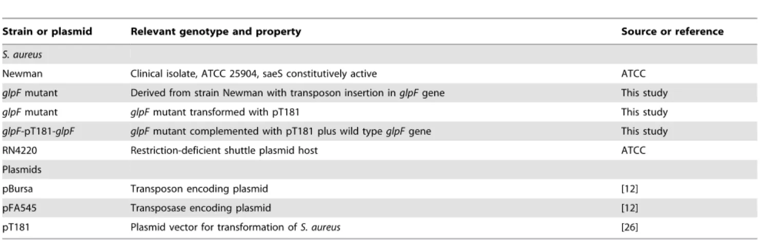
Figure 1. Comparison of L-form and classical form S. aureus
morphologies. (A). S. aureus Newman L-form colony on L-form induction media (LIM) exhibiting typical ‘‘fried egg’’ morphology. (B). Control classicalS. aureuscolony on BHI medium with sucrose control but without penicillin. (C).S. aureusL-form shape and structure (TEM,
6100,000).S. aureusL-form had irregular morphology with larger size
than normalS. aureusand contained a large number of vesicles. The L-form bacteria had deficient or fractured cell wall. (D). NormalS. aureus with spherical shape and thick cell wall structure (TEM,6100,000). (E).S. aureus L-form (TEM,610,000) showing polymorphic L-form bacteria
with varying sizes and shapes. (F).S. aureusnormal classical form (TEM,
610,000) showing regular spherical morphology with homogeneous
size and shapes. (G).S. aureusL-form colony (SEM,6400).S. aureus
L-form colony exhibited typical ‘‘fried egg’’ morphology. (H).S. aureus L-form colony inner structure (SEM,65000). In the inside ofS. aureus
L-form colony, the bacteria exhibited polymorphic shapes of varying sizes with the L-form colony structure showing similarity to a multilayered biofilm structure. The polymorphic bacteria are connected with large amounts of extracellular matrix materials (exopolysaccharide (EPS)) and lysed bacteria (arrow).
Complementation ofS. aureusmutants
The wild typeglpFgene fromS. aureusNewman was amplified by PCR using primers glpfF and glpfR (see Table 2). The PCR primers were taken from 2134 bp and 99 bp upstream and downstream of the glpF gene and contained restriction sites BamHI and PstI respectively. The PCR parameters were: 94uC
15 min, followed by 35 cycles of 94uC 30 s, 55uC 30 s, and 72uC 2 min, and a final extension at 72uC for 10 min. The PCR products were digested withBamHI andPstI and then cloned into plasmid pT181 cut with the same enzymes. The ligation products were electroporated intoS. aureusstrain RN4220 and the positive clones were identified by restriction digestion and PCR. The
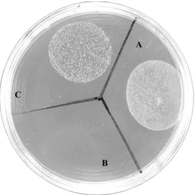
Figure 2. Results ofS. aureusNewman,glpFmutant and the complemented strain in L-form formation.Stationary phase cultures of (A)S. aureusNewman, (B)glpFmutant-pT181 vector control, and (C)glpF-pT181-glpFcomplemented strain were spotted on L-form induction media (LIM) followed by incubation at 33uC for 5 days when the L-form growth was examined.
doi:10.1371/journal.pone.0108325.g002
Figure 3. Relative concentrations ofglpFandglpKexpression in normal growth and L-formS. aureusas assessed by RT-PCR.
recombinant plasmid pT181+glpF and the pT181 vector alone control were then transformed into S. aureus glpF mutant by electroporation as described above under Construction of S. aureustransposon mutant library.
For complementation of theglpFmutant in L-form formation, S. aureusNewman strain was grown in BHI broth, and bothglpF mutant-pT181 vector control and theglpFpT181-glpF comple-mented strain were grown in BHI broth with 5mg/ml tetracycline overnight to stationary phase. The strains were spotted onto LIM and incubated aerobically at 33uC for 5 days followed by detection of typical L-form colonies which appeared as ‘‘fried egg’’ by inverted microscope as described above.
Effect of glycerol on L-form formation
The stationary phase cultures of wild typeS. aureusNewman strain grown in BHI broth and glpF mutant and the comple-mented strain in BHI broth containing 5mg/ml tetracycline were spotted onto LIM without glycerol and with varying concentra-tions (0, 0.1%, 1%, and 10%) of glycerol, respectively. After incubation aerobically at 33uC for 5 days, the ‘‘fried egg’’ colonies
were detected to confirm the effects of glycerol onglpFmutant L-form L-formation.
Persister assays
For antibiotic exposure, S. aureus glpF mutant, the glpF mutant complemented strain, and S. aureus parent strain Newman were grown to stationary phase overnight when antibiotics ampicillin (50mg/ml) and norfloxacin (40mg/ml) were added to undiluted cultures and incubated without shaking for various times up to 7 or 8 days. Aliquots of bacterial cultures exposed to antibiotics were taken at different time points and washed and then plated onto TSA plates for CFU counting.
Results and Discussion
Generation of antibiotic induced unstable L-forms ofS. aureus
L-form induction media (LIM) was tested for its ability to induce S. aureus(Newman) to grow as L-form colonies. Stationary phase cells ofS. aureusNewman strain produced L-form colonies when plated directly onto LIM. The minimum bacterial inoculum
Table 3.Effect of glycerol on L-form formation of theglpFmutant.
Wild type glpFmutant glpFcomplementation
Spotted bacterial number on TSA (cfu/ml) 3.636109 1.736109 5.776109
L-form colony number on LIM (cfu/ml) 3.16105 0 2.156105
L-form colony number on 1% glycerol LIM (cfu/ml) 7.756105 1.036104 2.906105
doi:10.1371/journal.pone.0108325.t003
Figure 4. Effect of glycerol on L-form growth forS. aureusNewman,glpFmutant and the complemented strain.Stationary phase cultures of (A)S. aureusNewman, (B)glpFmutant-pT181 vector control, and (C)glpF-pT181-glpFcomplemented strain were spotted on L-form induction media (LIM) containing 1% glycerol followed by incubation at 33uC for 5 days when the L-form growth was assessed.
required for L-form colony formation was approximately 106–107 bacteria (the maximum dilution for 108–109S. aureusbacteria to form L-form colony was 1:100). This frequency is significantly lower than the L-form formation frequency ofE. coliwhich is 104– 105[7]. The typical S. aureusL-form colonies had ‘‘fried egg’’ morphology (Fig. 1A) under inverted microscope in contrast to smooth colony of the normal form of S. aureus (Fig. 1B). The ‘‘fried-egg’’ L-form colonies had typical embedded growth into the soft agar and could not be scraped off in contrast to the normal classical forms which did not show embedded growth and could be scraped off easily from agar surface. Transmission electron microscopy (TEM) indicated that theS. aureus L-form bacteria had complete or partial loss of cell wall and contained a large number of intracellular vesicles (Fig. 1C) in contrast to the normal
forms with cell wall without obvious vesicles (Fig. 1D).S. aureus L-form (TEM 6 10,000) showed polymorphic sizes and shapes (Fig. 1E) in contrast to normal classical form showing morphology with homogeneous size and round shape with clear and smooth cell boundary (Fig. 1F). Scanning electron microscopy (SEM) indicated thatS. aureus L-form colony exhibited rough surface morphology (Fig. 1G) while in the inside of the L-form colony the bacterial cells exhibited polymorphic morphologies of varying sizes (Fig. 1H) with the L-form colony structure showing similarity to biofilm structure. This is consistent with previous observation that L-form bacteria secrete exopolysaccharide (EPS) to the surface to prevent desiccation similar to biofilms and that defect in genes involved in EPS synthesis can cause lack of L-form growth [7].
Table 4.Survival of stationary phase cultures of theglpFmutant, the complemented strain and the parent strain upon antibiotic exposure over time.
Antibiotics Time No. of bacteria (log CFU ML21, mean6SD)
Newman glpFmutant-pT181 glpF-pT181-glpF
Ampicillin 0 9.8860.20 9.4660.11 9.4860.32
(50mg/ml) 1d 9.7460.24 9.2660.21 9.3060.17
2d 8.6060.09 8.7060.24 9.2860.13
3d 8.3760.23 7.6560.07 8.4160.02
5d 6.9260.11 3.8460.09 7.3760.10
7d 5.2460.34 0 2.6563.74
Norfloxacin 0 9.8860.20 9.4660.11 9.4860.16
(40mg/ml) 1d 8.9060.09 9.260.13 9.060.17
2d 9.0860.22 8.4860.11 8.9060.36
3d 7.9160.29 7.4660.04 8.0660.03
5d 7.2260.02 6.0460.01 7.4160.03
8d 5.0660.16 0 5.2260.10
doi:10.1371/journal.pone.0108325.t004
Figure 5. Survival of stationary phase cultures of theS. aureus glpFmutant transformed with vector pT181, or pT181+wild typeglpF, and the parent strain upon ampicillin (50mg/ml) exposure over time.
Screening for mutants with defect in L-form formation fromS. aureustransposon mutant library
Having established theS. aureusL-form conditions, we wanted to identify genes that are involved in L-form formation. To do this, we first constructed a S. aureus transposon mutant library and then grew the library at 37uC overnight in 96-well plates followed by transfer of the mutants onto LIM plates as described in the Methods. Plates were incubated at 33uC for 7 days when mutants were scored for defect in forming L-form colonies.
To identify the genes whose mutation led to defect in L-form formation, we performed inverse PCR as described in the Methods. Using inverse PCR and DNA sequencing we were able to identify 12 genes from 15 mutants, 3 of which mapped toglpF and NWMN_0623 each, 1 mapped to glpK, 1 mapped to gluconate kinase (gntK), NWMN_0623, NWMN_0872 (GTP pyrophosphokinase), NWMN_1269 (sodium:alanine symporter family protein), 2 mapped to hypothetical proteinsNWMN_0333 andNWMN_0843of unknown function, 3 mapped to intergenic region. Because we found 4 mutants (3 in glpFand 1 in glpK) mapped to glycerol metabolism genes which predominate among these identified genes, we therefore focused and further charac-terized the role of glycerol metabolism genes in this study. To determine if the mutatedglpFis indeed responsible for defective L-form formation, we attempted to complement theglpFmutant with the wild type glpF gene using the plasmid vector pT181. However, the initial attempt was unsuccessful when the comple-mentedglpFmutant was plated directly on LIM plates. Since our previous work withE. coliL-form complementation indicated that inducible expression of the gene involved in L-form formation is critical for successful complementation of L-form defect of the mutants [7], we therefore induced the complemented glpF S. aureusstrain containing tetracycline inducible vector pT181 with tetracycline in liquid culture prior to plating on LIM plates. This led to successful complementation of the glpF mutant with the wild typeglpFgene. However, the effect of the complementation was partial (Fig. 2C) compared with the parent strain Newman
(Fig. 2A), while the glpF mutant transformed with the pT181 vector control did not form any L-form colonies (Fig. 2B).
glpFandglpKwere overexpressed in S. aureusL-form bacteria but not in normal bacteria
Since we identified mutations inglpFandglpKcaused defect in L-form growth, we wanted to know if glpF and glpK are overexpressed inS. aureusL-form bacteria compared with normal classical formS. aureus. To confirm this, we preparedS. aureus L-form bacteria from L-L-form media LIM and normal S. aureus growth as a control on media without penicillin and isolated RNA from both types of the bacterial cells. The isolated RNA samples were then subjected to RT-PCR.glpFandglpKwere found to be expressed at very low levels in normal growth but were significantly induced to 144-fold and 68-fold higher respectively in L-forms than in the normal control growth (P,0.05) (Fig. 3).
Effect of glycerol on restoring L-form formation inglpF
mutant
Since GlpF is involved in glycerol transport, we wanted to address the role of exogenously added glycerol in L-form formation in S. aureus. To do so, we incorporated varying concentrations of glycerol 0, 0.1%, 1% and 10% glycerol into the LIM. In LIM media with 0 and 0.1% glycerol, only wild typeS. aureusNewman strain and the glpF complemented strain grew but glpF mutant failed to grow (Table 3). However, at 1% glycerol, theglpFmutant formed 1.036104L-form colonies but its efficiency was much lower than the wild type (7.756105L-form colonies) and the glpF complemented strain (2.906105 L-form colonies) (Fig. 4, Table 3). The 1% glycerol only marginally increased the number of L-form colonies of the wild type strain by about 2 fold (Table 3). In contrast, at 10% glycerol, none of the strains Newman, the glpF mutant, or the glpF complemented strain formed L-form colonies.
Figure 6. Survival of stationary phase cultures of theglpFmutant, the complemented strain, and the parent strain upon norfloxacin (40mg/ml) exposure over time.
To address if the role of 1% glycerol is to serve as an osmoprotectant in facilitating L-form formation in the glpF mutant, we added known osmoprotectant NaCl at 4.5% to the LIM and assessed if it could allow theglpFmutant to grow as L-forms. However, supplementation of NaCl while allowing L-form formation in wild type and complementedglpFmutant, failed to facilitate L-form formation in theglpFmutant (data not shown).
Defective persistence ofS. aureus glpFmutant in antibiotic exposure assays
In our previous study withE. coli L-form bacteria, we found that genes involved in L-form formation overlapped with those involved in antibiotic persistence [7]. Exposure of the stationary phase culture of glpFmutant to ampicillin (50mg/ml) revealed that the mutant began to show defect in persistence at day 3 but the defect was more obvious after 5 days. After 7 days, no CFU was detectable in the glpF mutant transformed with the vector control pT181; in contrast, the complemented strain (transformed with pT181+glpF) and the parent strain Newman had 100–1000 and 10,000 CFUs remaining (Fig. 5) (Table 4). For exposure to norfloxacin (40mg/ml), the glpF mutant had a similar trend as ampicillin treatment, where it reached zero CFU by day 8.
Complementation of theglpFmutant with the wild typeglpFgene only partially restored the persister levels compared with parent strain for ampicillin exposure (Fig. 5) but caused full restoration of persister levels for norfloxacin exposure (Fig. 6) (Table 4).
Although many previous studies have demonstrated the formation of L-forms by S. aureus [13,14,17], the mechanisms involved in L-form formation has remained unknown in this organism. This study provided the first molecular insight into the mechanism of L-form formation inS. aureusby demonstrating the important role of glycerol uptake in the L-form formation. GlpF is involved in glycerol uptake (Fig. 7), and the observation thatglpF mutation causes defect in L-form growth in S. aureussuggests uptake of glycerol is essential for L-form formation. The mechanism by which GlpF is involved in L-form formation inS. aureusis likely mediated through its role in production of energy via pyruvate entry into TCA cycle and glycolysis or alternatively through cell membrane synthesis via lysophosphatidic acid (LPA) to phospholipids (Fig. 7) to strengthen membrane integrity required for L-form growth in the absence of cell wall. This study represents the first effort at identifying the mechanisms of L-form formation inS. aureus,and further studies are needed to explore other possible mechanisms of L-form formation besides glycerol uptake in future studies.
Figure 7. Glycerol uptake and metabolic pathway.GlpF is involved in uptake of glycerol intoS. aureuswhile GlpK is glycerol kinase and is involved in converting glycerol to glycerol-3-phosphate which then can be used for synthesis of cell membrane phospholipid and also for energy production.
It is interesting to note that supplementation of appropriate concentration of glycerol (1%) partially compensated for the loss of L-form formation toglpFmutant (Fig. 4). GlpF is known to be a member of the aquaporin (AQP) channel family. Some of AQPs channels that conduct water and also conduct glycerol (aqua-glyceroporins) [22]. When the environmental glycerol is increased, the aquaporin (AQP) channel may partially compensate for the loss of GlpF and help theglpFmutant to form a small number of L-form colonies (Fig. 4). We believe the role of glycerol in L-form formation is mediated through its uptake and metabolism rather than an osmoprotectant role in L-form formation for the following reasons. First, we demonstrated in the new experiment that adding 4.5% NaCl as an osmoprotectant did not help theglpFmutant to form L-forms while it allowed wild type and the complemented glpF mutant strains to form L-form colonies. This indicates the defect in glpF mutant cannot be complemented by other osmoprotectant like NaCl. Second, if the role of glycerol were to serve as osmoprotectant, we would expect that higher concentra-tion of glycerol would facilitate L-form formaconcentra-tion. However, we found higher glycerol content, e.g., 10% glycerol did not allow L-forms to form. Third, our finding that mutation inglpKwhich is glycerol kinase involved in glycerol metabolism caused defect in L-form L-formation also does not support glycerol serving as an osmoprotectant in facilitating L-form formation. This is because GlpK is glycerol kinase that converts glycerol to glycerol-3-phosphate which can be used for synthesis of cell membrane phospholipid and also for energy production to facilitate L-form formation. However, high glycerol concentration (10%) inhibited the L-form growth for wild type as well as the mutantS. aureus, presumably because high concentrations of glycerol produce toxic metabolites thus preventing L-form growth.
It is noteworthy that in addition to its role in L-form formation, glpFis also involved in tolerance or persistence to antibiotics inS. aureusas demonstrated by a defect in persistence inglpFmutant upon exposure to ampicillin or norfloxacin (Fig. 5 and Fig. 6). This finding is consistent with the previous observation that genes involved in glycerol metabolism such as glpD encoding
sn-glycerol-3-phosphate dehydrogenase and plsBencoding sn-glyc-erol-3-phosphate acyltransferase (Fig. 7), have been found to be involved in persister formation [23]. Our findings that glycerol uptake is important for both L-form and persistence ofS. aureus and provide further support for the close relationship between the two entities as has been observed forE. coli[7]. This finding is consistent with the recent proposal that L-form bacteria are related to persisters and are part of the heterogeneous persister continuum [24]. The only difference is that frequency of L-form forming bacteria is 2–3 orders of magnitude lower than persister frequency [24] and can be considered ‘‘deep’’ persisters [25]. Although previous study with E. coli L-form bacteria did not identifyglpFbeing critical for L-form formation as inS. aureus, microarray analysis indicated that glycerol metabolism geneglpD encoding sn-glycerol-3-phosphate dehydrogenase is upregulated in E. coliL-form bacteria [7], suggesting glycerol metabolism could be important for L-form bacteria in different bacterial species.
In conclusion, we established an L-form model in S. aureus, characterized their morphologies and optima conditions of their formation, and identified genes involved in glycerol uptake and metabolism being important for L-form formation and persistence inS. aureus.These findings shed new light on the mechanisms of L-form formation and persister biology inS. aureusand may have implications for development of new drugs targeting persisters for improved treatment of persistent bacterial infections.
Acknowledgments
We thank Dominique Missiakas for providing plasmids used in this study. Jian Han was supported by China Scholarship Council.
Author Contributions
Conceived and designed the experiments: YZ. Performed the experiments: JH LLH WLS XGX SW SZ. Analyzed the data: JH YZ. Contributed reagents/materials/analysis tools: JH LLH WLS. Contributed to the writing of the manuscript: YZ JH.
References
1. Klienenberger E (1935) The natural occurrence of pleuropneumonia-like organisms in apparent symbiosis with Streptobacillus moniliforms and other bacteria. J Pathol Bacteriol 40: 93–105.
2. Dienes L, Weinberger HJ (1951) The L forms of bacteria. Bacteriol Rev 15: 245–288.
3. Domingue GJ Sr., Woody HB (1997) Bacterial persistence and expression of disease. Clin Microbiol Rev 10: 320–344.
4. Allan EJ, Hoischen C, Gumpert J (2009) Bacterial L-forms. Adv Appl Microbiol 68: 1–39.
5. Domingue GJ (2010) Demystifying pleomorphic forms in persistence and expression of disease: Are they bacteria, and is peptidoglycan the solution? Discovery medicine 10: 234–246.
6. Leaver M, Dominguez-Cuevas P, Coxhead JM, Daniel RA, Errington J (2009) Life without a wall or division machine in Bacillus subtilis. Nature 457: 849–853. 7. Glover WA, Yang Y, Zhang Y (2009) Insights into the molecular basis of L-form
formation and survival in Escherichia coli. PLoS One 4: e7316.
8. Devine KM (2012) Bacterial L-forms on tap: an improved methodology to generate Bacillus subtilis L-forms heralds a new era of research. Molecular microbiology 83: 10–13.
9. Joseleau-Petit D, Liebart JC, Ayala JA, D’Ari R (2007) Unstable Escherichia coli L forms revisited: growth requires peptidoglycan synthesis. J Bacteriol 189: 6512–6520.
10. Siddiqui RA, Hoischen C, Holst O, Heinze I, Schlott B, et al. (2006) The analysis of cell division and cell wall synthesis genes reveals mutationally inactivated ftsQ and mraY in a protoplast-type L-form of Escherichia coli. FEMS Microbiol Lett 258: 305–311.
11. Dominguez-Cuevas P, Mercier R, Leaver M, Kawai Y, Errington J (2012) The rod to L-form transition of Bacillus subtilis is limited by a requirement for the protoplast to escape from the cell wall sacculus. Molecular microbiology 83: 52– 66.
12. Bae T, Glass EM, Schneewind O, Missiakas D (2008) Generating a collection of insertion mutations in the Staphylococcus aureus genome using bursa aurealis. Methods Mol Biol 416: 103–116.
13. Fuller E, Elmer C, Nattress F, Ellis R, Horne G, et al. (2005) Beta-lactam resistance in Staphylococcus aureus cells that do not require a cell wall for integrity. Antimicrob Agents Chemother 49: 5075–5080.
14. Banville RR (1964) Factors affecting growth of Staphylococcus aureus L forms on semidefined medium. Journal of bacteriology 87: 1192–1197.
15. Michailova L, Kussovsky V, Radoucheva T, Jordanova M, Markova N (2007) Persistence of Staphylococcus aureus L-form during experimental lung infection in rats. FEMS Microbiol Lett 268: 88–97.
16. Tanimoto A, Kitagaki Y, Hiura M, Fujiwara H, Iijima K, et al. (1995) [Methicillin-resistant Staphylococcus aureus forming the fried egg appearance colonies isolated from a patient with septicemia]. Jap J Clin Pathol 43: 1061– 1065.
17. Owens WE (1987) Isolation of Staphylococcus aureus L forms from experimentally induced bovine mastitis. Journal of clinical microbiology 25: 1956–1961.
18. Sears PM, Fettinger M, Marsh-Salin J (1987) Isolation of L-form variants after antibiotic treatment in Staphylococcus aureus bovine mastitis. Journal of the American Veterinary Medical Association 191: 681–684.
19. Lechner S, Lewis K, Bertram R (2012) Staphylococcus aureus persisters tolerant to bactericidal antibiotics. J Mol Microbiol Biotechnol 22: 235–244. 20. Keren I, Kaldalu N, Spoering A, Wang Y, Lewis K (2004) Persister cells and
tolerance to antimicrobials. FEMS Microbiol Lett 230: 13–18.
21. Shingaki R, Kasahara Y, Iwano M, Kuwano M, Takatsuka T, et al. (2003) Induction of L-form-like cell shape change of Bacillus subtilis under microculture conditions. Microbiology (Reading, England) 149: 2501–2511.
23. Spoering AL, Vulic M, Lewis K (2006) GlpD and PlsB participate in persister cell formation in Escherichia coli. J Bacteriol 188: 5136–5144.
24. Zhang Y (2014) Persisters, Persistent Infections and the Yin-Yang Model. Emerging Microbes and Infections (Nature Publishing Group) 3, e3; doi:10.1038/emi.2014.3.
25. Ma C, Sim S, Shi W, Du L, Xing D, et al. (2010) Energy production genes sucB and ubiF are involved in persister survival and tolerance to multiple antibiotics and stresses in Escherichia coli. FEMS Microbiol Lett 303: 33–40.
26. Khan SA, Novick RP (1983) Complete nucleotide sequence of pT181, a tetracycline-resistance plasmid from Staphylococcus aureus. Plasmid 10: 251– 259.