The RecA-Dependent SOS Response Is Active
and Required for Processing of DNA Damage
during
Bacillus subtilis
Sporulation
Fernando H. Ramírez-Guadiana1☯, Rocío del Carmen Barajas-Ornelas1☯, Saúl U. Corona-Bautista1, Peter Setlow2, Mario Pedraza-Reyes1
*
1Department of Biology, Division of Natural and Exact Sciences, University of Guanajuato, Guanajuato, Gto. 36050, México,2Department of Molecular Biology and Biophysics, UConn Health, 06030–3305, Farmington, Connecticut, United States of America
☯These authors contributed equally to this work. *[email protected]
Abstract
The expression of and role played by RecA in protecting sporulating cells ofBacillus subtilis
from DNA damage has been determined. Results showed that the DNA-alkylating agent Mitomycin-C (M-C) activated expression of a PrecA-gfpmut3afusion in both sporulating
cells’mother cell and forespore compartments. The expression levels of arecA-lacZfusion were significantly lower in sporulating than in growing cells. However, M-C induced levels of ß-galactosidase from arecA-lacZfusion ~6- and 3-fold in the mother cell and forespore compartments ofB.subtilissporangia, respectively. Disruption ofrecAslowed sporulation and sensitized sporulating cells to M-C and UV-C radiation, and the M-C and UV-C sensitiv-ity of sporangia lacking the transcriptional repair-coupling factor Mfd was significantly increased by loss of RecA. We postulate that when DNA damage is encountered during sporulation, RecA activates the SOS response thus providing sporangia with the repair machinery to process DNA lesions that may compromise the spatio-temporal expression of genes that are essential for efficient spore formation.
Introduction
Sporulation inBacillus subtilisis a developmental process that is triggered by nutrient depletion and high cell density, and serves as a strategy for population survival. An early morphological event in sporulation is the asymmetric cell division that gives rise to a two-compartment spo-rangium [1]. Distinct programs of gene expression are then directed in these two cells by com-partment-specific RNA polymerase sigma (σ) factors [2]. The spores produced in this process
exhibit minimal if any metabolism, but are highly resistant to heat, radiation, desiccation and chemical agents [3,4]. Consequently, these spores can survive for very long periods [5,6], until they encounter an appropriate environment allowing spore germination and resumption of cell growth [7].
OPEN ACCESS
Citation:Ramírez-Guadiana FH, Barajas-Ornelas RdC, Corona-Bautista SU, Setlow P, Pedraza-Reyes M (2016) The RecA-Dependent SOS Response Is Active and Required for Processing of DNA Damage duringBacillus subtilisSporulation. PLoS ONE 11(3): e0150348. doi:10.1371/journal.pone.0150348
Editor:Claude Prigent, Institut de Génétique et Développement de Rennes, FRANCE
Received:August 10, 2015
Accepted:January 29, 2016
Published:March 1, 2016
Copyright:© 2016 Ramírez-Guadiana et al. This is an open access article distributed under the terms of theCreative Commons Attribution License, which permits unrestricted use, distribution, and reproduction in any medium, provided the original author and source are credited.
Data Availability Statement:All relevant data are within the manuscript.
Spontaneous or induced chromosomal damage in either compartment of the sporangium may compromise the successful accomplishment of the sporulation program inB.subtilis. Con-sequently, sporulating cells deploy a variety of mechanisms to sense and process DNA lesions that could interfere with proper spore formation [8–11]. These mechanisms include: 1) the DNA-damage scanning protein (DisA) which operates early in sporulation delaying this devel-opmental program until genomic lesions have been eliminated; and 2) the nucleotide excision repair (NER) pathway that processes DNA-distorting or -crosslinking lesions produced by ultra-violet irradiation and mitomycin-C (M-C), respectively. Interestingly, the efficiency of the NER system is enhanced by the transcription-coupling repair (TCR) factor, Mfd, duringB.subtilis
sporulation [11]. Moreover, the UV-endonuclease YwjD, which possibly works in concert with the error-prone polymerases YqjH and/or YqjW also operates during sporulation to eliminate UV-promoted DNA damage [9,10]. Of note, genes encoding UvrA and UvrB repair proteins as well as YqjW are part of the SOS regulon, a gene circuitry that is under transcriptional control by RecA and DinR [12,13]. In exponentially growingB.subtiliscells, the presence of strand breaks promote the formation of RecN foci; moreover, viability is compromised in growing RecA-deficient cells [14]. Interestingly, sporangia lacking RecA exhibit aberrant morphologies, which likely affect nucleoid condensation and ultimately result in a low sporulation frequency [15]. Low levels of RecA have been detected in dormant spores; however, the RecA concentra-tion increases during spore outgrowth and its contribuconcentra-tion to DNA repair during this develop-mental stage has been demonstrated [16]. Thus, spores lacking RecA exhibit decreased
resistance to DNA damaging factors such as heat, UV irradiation, hydrogen peroxide, ultrahigh vacuum desiccation and ionizing radiation [16–19]. A recent study revealed that YabT, a Hanks-type protein kinase, phosphorylates RecA during sporulation promoting the formation of RecA foci that can assemble into a nucleofilament in response to DNA damage. Interestingly, persis-tence of this protein structure beyond late sporulation results in abortive spore morphogenesis, suggesting that phosphorylated RecA may contribute to a checkpoint that delays or prevents the completion of spore development in sporangia with severely damaged chromosomes [20].
Although it has been shown that DNA replication is important for assembly of RecA in response to DNA damage and likely for SOS induction [21], there is no current information regarding the role of this protein when the progress of the transcription complex becomes impaired due to DNA damage. This is important since during endospore formation,B.subtilis
does not replicate its chromosomes, although the transcriptional program that is essential for endospore development continues [2,22,23]. We report here that expression ofrecAis induced by DNA-damaging factors during sporulation, and in both sporangium compart-ments, and that the activation of the SOS response by RecA is required to eliminate DNA lesions inflicted by UV-C and M-C inB.subtilissporangia.
Materials and Methods
Bacterial strains and growth conditions
TheB.subtilisstrains and plasmids used in this study are described inTable 1. Bacterial cultures were grown at 37°C in Luria-Bertani medium (LB, [24]), Penassay broth (PAB) (antibiotic medium 3; Difco Laboratories, Sparks, MD) or Difco sporulation medium (DSM, [25]) with shaking at 250 rpm. When appropriate, spectinomycin (100μg/mL), neomycin (12.5μg/mL) or
erythromycin (5μg/mL) was added to media.
Strains construction
Standard techniques were used for strain construction, including isolation of chromosomal and plasmid DNA as well as for bacterial transformation [26–28].
To generate a translational PrecA-gfpmut3agene fusion, a 492-bp EcoRI/BamHI fragment
containing therecApromoter (-345 to +147 relative to therecAstart codon) was PCR ampli-fied using chromosomal DNA fromB.subtilis168. The oligonucleotide primers used for this
reaction were5’-GCGAATTCCAGGACCTGATGCTCAAG-3’(forward) and5’-GCGGAT
CCCAGTGCTGTATCAAGAGC-3'(reverse). Restriction sites (underlined) were included in
the primers for cloning the amplified product between the EcoRI-BamHI sites of plasmid pAD123 to generate plasmid pPERM1236. This latter plasmid was cut with EcoRI and SphI and the resulting PrecA-gfpmut3aDNA fragment was cloned between the same restriction sites
of plasmid pDR111 (a generous gift from David Rudner), giving plasmid pPERM1237 (amyE:: PrecA-gfpmut3a). This plasmid was linearized and independently used to transformB.subtilis
168 to generateB.subtilisstrains PERM1238 and PERM1238a, respectively.
Chromosomal DNA was isolated fromB.subtilisYB3001 (amyE::recA-lacZ) and used to transform competent cells ofB.subtilisstrains LAS600 and LAS523, generatingB.subtilis
PERM1486 and PERM1487, respectively.
Chromosomal DNA fromB.subtilisSL7360 (ΔrecA::neo) was used to transform competent cells ofB.subtilisstrains168, PERM938 and PERM1233, generating strains PERM1030 (ΔrecA), PERM1033 (ΔmfdΔrecA) and PERM1280 (ΔrecA;spoVFA-lacZ), respectively. Proper single- and double-crossover events leading to the integration of vectors and/or to the inactiva-tion of the appropriate genes were confirmed in all cases by PCR (data not shown).
Table 1. B.subtilisstrains and plasmids used in this study.
Strain Genotype and descriptiona Construction or sourceb
168 Wild type,trpC2 Laboratory stock
SL7360 ΔrecA::neo(Neo) Patrick Piggot
PERM938 Δmfd::tet(Tet) [11]
PERM1030 ΔrecA::neo(Neo) SL7360!168
PERM1033 Δmfd::tet,ΔrecA::neo(Tet Neo) SL7360!PERM938
PERM1233 spoVFA::lacZ(Ery) Laboratory stock
PERM1238 amyE::PrecA-gfpmut3a(Spc) pPERM1237!168
PERM1238a amyE::-gfpmut3a(Spc) pPERM1237a!168
PERM1280 ΔrecA::neo,spoVFA::lacZ(Neo Ery) SL7360!PERM1233
YB3001 amyE::recA-lacZ(Cm) Ron Yasbin
LAS600 Δupp [31]
LAS523 +dinR3[lexA(Ind-)] [31]
PERM1486 LAS600amyE::recA-lacZ(Cm) YB3001!LAS600
PERM1487 LAS523amyE::recA-lacZ(Cm) YB3001!LAS523
Plasmid
pAD123 Shuttlegfpmut3afusion vector (Amp Cm) BGSC
pDR111 amyE::Phyper-spankpromoter (Phs) (Amp Spc) David Rudner
pPERM1236 pAD123 containing the PrecA-gfpmut3aconstruct (Amp Cm) This study
pPERM1237 pDR111 containing the PrecA-gfpmut3a construct (Amp Spc) This study
aSelection markers are in parentheses. Antibiotics were used at the following concentrations: Neo, neomycin (12.5μg/mL); Tet, tetracycline (10μg/mL);
Ery, erythromycin (5μg/mL); Spc, spectinomycin (100μg/mL); Amp, ampicillin (100μg/mL) and Cm, chloramphenicol (5μg/mL).
b
“X”!“Y”indicates that“strain Y”was transformed with DNA from“source X”. BGSC,BacillusGenetic Stock Center.
ß-galactosidase assays
ß-galactosidase activities duringB.subtilissporulation were determined as described by [29]. In brief, samples harvested from sporulating cultures were disrupted with lysozyme, centri-fuged and the supernatant fluid was assayed for ß-galactosidase using ortho-nitrophenyl-ß-D-galactopyranoside (ONPG) as the substrate. This activity was assigned as the mother cell frac-tion, and consisted of enzyme from mother cells as well as lysozyme-sensitive forespores. The pellet fraction that contains lysozyme-resistant forespores present from 4 to 8 h after T0(the
time when the slopes of the logarithmic and stationary phases of growth intersected and defined as the time of initiation of sporulation) was subjected to spore coat removal [29], and a second lysozyme treatment was used to allow assay of the ß-galactosidase activity in forespores. All ß-galactosidase activities were expressed in Miller units [24], and the basal specific activities of ß-galactosidase in the RecA strain without alacZfusion during growth and sporulation were determined in parallel and subtracted from the data obtained for the strain carrying the recA-lacZfusion. These corrections were always10%.
Microscopic analysis
B.subtiliscells were grown and induced to sporulate in DSM. 1 hour after T0, sporulating cell
samples were supplemented with FM4-64 (Invitrogen) at a final concentration of 5μg/ml.
Cell samples collected at the appropriate times were processed for microscopy essentially as described previously [11] and samples were observed under a ZEISS Axioscope A1 microscope equipped with an AxioCam ICc1. Image collection was carried out using AxioVision V 4.8.2 software and images were adjusted only for brightness and contrast. Excitation and emission wavelengths employed were 498 nm and 512 nm for GFP and 506 and 750 nm for FM4-64, respectively. More than 400 sporulating cells expressing the GFPmut3a protein (from ~600 cells analyzed) were counted in four different fields at 100☓for each sporulation time point.
Treatment of sporulating cells with DNA-damaging factors
B.subtilisstrains were induce to sporulate in DSM at 37°C. At 4.5h after T0, the cultures were
challenged with M-C or UV-C radiation as described previously [10]. Briefly, different doses of M-C were added to 2 mL cell samples and the cultures were incubated for additional 1 h before plating. Prior to UV-C treatment, cells were collected by centrifugation and washed with cold phosphate-buffered saline (PBS; 0.7% Na2HPO4, 0.3% KH2PO4, 0.4% NaCl [pH 7.5]). Cell
samples (8 mL at an OD600= 0.5) in PBS were stirred continuously and UV-C irradiated at
room temperature. Artificial UV-C light (monochromatic 254 nm UV irradiation) was pro-vided by a commercial low-pressure mercury arc lamp (model UVG-11; UV Products, Upland, CA). Cell survival after these treatments was determined by plating serial dilutions on solid LB medium, and colony-forming units were counted after 16 h of incubation at 37°C.
Results and Discussion
recA
is induced by DNA damage during
B
.
subtilis
sporulation
Since RecA regulates the SOS response inB.subtilis[30], we generated a recombinant strain of
B.subtilisto monitorin situthe transcriptional activation of the SOS response by DNA dam-age. To this end, thegfpmut3a reporter gene was fused to therecApromoter and the resulting PrecA-gfpmut3afragment was recombined into theamyElocus ofB.subtilis.
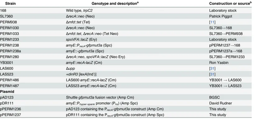
The results of this analysis showed that the DNA damage promoted by M-C induced the syn-thesis of the GFPmut3a protein in the three-sporulation stages analyzed. Interestingly, in all cases, the GFP signal was detected in both the mother cell and the forespore compartments (Fig 1), indicating that DNA damage can induce the RecA-dependent SOS response in both compartments of the sporangium. In contrast, low fluorescence was detected in untreated sporangia of the PrecA-gfpmut3astrain that were collected at the same developmental stages (Fig 1).
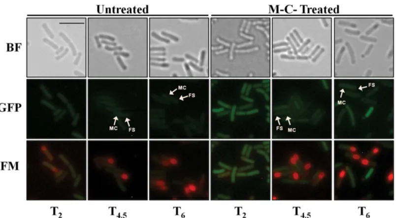
To better assess and quantify therecAexpression during the life cycle ofB.subtilis, we used a strain carrying a transcriptionalrecA-lacZfusion and determined the levels of ß-galactosidase produced during exponential growth and sporulation and in both the mother cell and fore-spore compartments. The results of this analysis revealed that therecA-driven ß-galactosidase activity was present in exponentially growing cells as well as in developing sporangia (Fig 2a). However, the expression levels of therecA-lacZfusion were significantly lower in sporulation than during logarithmic growth (Fig 2a). These results are in agreement with a previous study that detected a lower amount of RecA in dormant spores than in vegetative cells [16]. More-over, as shown inFig 2d, addition of M-C to exponentially growing cells increased ~18 fold the expression levels of therecA-lacZfusion.
A recent report showed that M-C was able to induce the expression of a chromosomal
uvrA-lacZfusion during stage IV of sporulation suggesting that the SOS response may be trig-gered in this stage of sporulation [11]. To further examine this suggestion, therecA-lacZstrain was treated with M-C 4 h after the onset of sporulation. Notably, in comparison with an untreated sporulating culture, addition of this alkylating agent increased the expression of the
recA-lacZ6- and 3-fold in the mother cell and the forespore compartment fractions, respec-tively (Fig 2b and 2c). Taken together, these results support the notion that inB.subtiliscells committed to sporulate, the SOS response can be activated and is possibly required to induce the expression of DNA repair proteins necessary to eliminate genetic insults that may compro-mise spore formation. In support of this contention, expression of therecA-lacZfusion in both sporangia compartments was abolished in a mutant strain ofB.subtilisunable to trigger the SOS response as it carries a non-cleavable form of the SOS-repressor protein DinR [31] (Fig 3).
Of note, in comparison with exponentially growing cells (Fig 2d), the extent of induction of therecA-lacZfusion by M-C in strain YB3001 was ~2 fold lower during sporulation (Fig 2b and 2c) as well as during the stationary phase in vegetative cells propagated in PAB, a rich medium unable to promoteB.subtilissporulation [32] (Fig 2d). Therefore, the SOS-response can be elicited inB.subtilisdespite unfavorable metabolic conditions prevailing in cells com-mitted to sporulation or facing nutritional stress.
Inactivation of
recA
delays sporulation
It has been reported that a RecA-deficientB.subtilisstrain exhibits morphological irregularities and a delayed prespore nucleoid condensation during sporulation [15], suggesting that RecA is required for normal sporulation. Therefore, we investigated further whether the lack of RecA interferes with growth and sporulation inB.subtilis. Our results showed that the growth rate of aΔrecAstrain in DSM at 37°C had a significantly longer doubling time compared to the wild-type strain (33 ± 2 min and 24 ± 2 min, respectively; unpairedt-test,P<0.05). Moreover, as
previously described [15], the sporulation efficiency in the RecA-deficient strain was ~40% lower than that of the wild-type strain (data not shown). To directly examine effects of loss of RecA on sporulation-specific gene expression, we examined the expression of aspoVFA-lacZ
fusion was used as a marker of gene expression late in sporulation of wild-type andΔrecA
and is expressed exclusively in the mother cell compartment reaching its highest expression level at T5of sporulation [33,34]. Analysis ofspoVFA-lacZexpression during sporulation of
the wild-type andΔrecAstrains revealed a ~2 h delay in expression ofspoVFA-lacZfusion in theΔrecAmutant compared to the wild-type parental strain (Fig 4). Therefore, the morpholog-ical abnormalities observed in RecA-deficient sporangia [15] seem to be accompanied and pre-sumably caused by alterations in the program of gene expression that drives the process of sporulation inB.subtilis. Notably, a recent report showed thatB.subtiliscells expressing a non-phosphorylatable RecA protein also exhibited slowed sporulation [20]. Taken together, these results highlight the importance of RecA in this developmental process, as a deficiency in this protein slows sporulation significantly, presumably as a consequence of the resulting tran-scriptional abnormalities.
RecA protects
B
.
subtilis
sporulating cells from DNA damage
In stage T4.5where sporangial cells are commited to sporulate, the mother cell is programmed
to lyse whereas forespores will eventually give rise to a dormant spore [2]. Therefore surviving colonies arising after treatment of T4.5sporulating cells with genotoxic agents are mostly
gener-ated from forespores [10]. Keeping these facts in mind, to investigate whether RecA contributes to the survival of sporulating cells treated with DNA damaging agents, we examined the resis-tance of wild-type andΔrecA B.subtilissporangia to M-C and UV-C radiation.
Fig 1. Monitoring of activation of the SOS response induced by DNA damage using a PrecA-gfpmut3a during sporulation.Sporulating cells ofB.
subtilisPERM1238 (PrecA-gfpmut3a) collected at different times after T0, in the sporulation program (T2, T4.5and T6) were treated with M-C (125 ng/mL),
incubated for additional 1 h and finally observed under fluorescence microscopy. BF, bright field; GFP, GFP channel; FM, FM4-64 staining; MC and FS, mother cell and forespore compartments. The scale bar is 2μm and all images are at the same magnification.
Fig 2. Levels of ß-galactosidase fromrecA-lacZin growth and sporulation and with and without DNA damage.B.subtilisstrain YB3001 containing a
recA-lacZfusion was grown and induced to sporulate in DSM (a-c) The optical densities of cultures were measured without () or after (●) DNA damaging treatment. Samples were also collected at different times during growth and sporulation and were processed and assayed for ß-galactosidase specific activity. In(a)ß-galactosidase fromrecA-lacZwas assayed throughout growth and sporulation (◆). In(b,c)4 h after the onset of sporulation (T0), the culture
was divided into two subcultures; one subculture was challenged with M-C (500 ng/mL) and the other one was untreated. Cells samples from untreated (open symbols) or treated (filled symbols) were collected at the indicated times and ß-galactosidase specific activity in the mother cell (b, triangles) and forespore (c, squares) fractions was determined, all as described in Materials and Methods. In (d),B.subtilisYB3001 was propagated in PAB medium, when the culture reached an OD600nm= 0.5 (Exponential) or 4 h after T0(Stationary), vegetative cells were treated (black bars) or not (gray bars) with M-C (500 ng/mL)
for 1.5 h and then the cultures were processed for determination of ß-galactosidase as described above. Results are the average of values from three independent experiments±standard deviations (SD) of ß-galactosidase specific activity.
As noted above, the RecA-deficient strain is slowed ~2 h in sporulation. Therefore, in these experiments sporulating cells of therecAand parental strains were collected at equivalent mor-phological stages in sporulation (stages IV-V), as assessed by microscopic analysis. As shown inFig 5a and 5b, in comparison with the wild-type strain therecAmutant exhibited an increased susceptibility to both M-C and UV-C radiation during sporulation. These two agents are known to produce lesions that block the progress of transcription elongation complexes [35,36]. Notably, a previous report showed that the TCR factor Mfd plays a key role in protect-ing sporulatprotect-ing cells against M-C and UV-C [11], and the sporangia´s susceptibility to these
Fig 3. Levels of ß-galactosidase fromrecA-lacZwith and without DNA damage during sporulation in SOS-proficient (a and b) and–deficient (c and d)B.subtilisstrains.B.subtilisstrains LAS600 (parental) and LAS523 (SOS-deficient) containing arecA-lacZfusion were grown and induced to sporulate in DSM. The optical densities of cultures were measured without () or after (●) DNA damaging treatment. 4 h after the onset of sporulation (T0), the culture
was divided into two subcultures; one subculture was challenged with M-C (500 ng/mL) and the other one was untreated. Cells samples from untreated (open symbols) or treated (filled symbols) were collected at the indicated times and ß-galactosidase specific activity in the mother cell (a and c, triangles) and forespore (b and d, squares) fractions was determined, all as described in Materials and Methods.
genotoxic agents was greater in theΔrecAmutant than in the Mfd-deficient strain (Table 2). Together, these results strongly suggest that RecA as well as Mfd are necessary during spore morphogenesis to process pyrimidine dimers as well as DNA crosslinks generated by UV-C and M-C, respectively.
Since cells committed to sporulate no longer replicate the chromosomes in the mother cell and the forespore compartments [2,23], a recombination-repair role for RecA in this develop-mental stage can largely be ruled out. Rather, as shown in this work, the function of RecA is most likely to activate the SOS response to provideB.subtilissporangia with the repair machin-ery to process DNA lesions occurring during sporulation. The experiments inFig 5c and 5d
provided further support for this contention by showing that in comparison with its parental strain (B.subtilisLAS600), sporulating cells of the SOS-deficientlexA(Ind-) mutant exhibited a major susceptibility to UV-C and M-C. Consistent with a previous report in dormant spores
Fig 4. Expression ofspoVFA-lacZduring sporulation of wild-type andΔrecA B.subtilisstrains.B.subtilisstrains PERM1233 (wild-type) (open symbols) and PERM1280 (ΔrecA) (closed symbols) containing thespoVFA-lacZfusion were grown and induced to sporulate in DSM and the OD600nm
(circles) was measured. Cells were collected during sporulation at the indicated times, treated with lysozyme and the extracts were assayed for ß– galactosidase in the mother cell fractions only (triangles). Results are the averages of values from three independent experiments±SD of ß-galactosidase specific activity.
treated with ionizing radiation [19], our results revealed that sporangia harboring thelexA
(Ind-) allele were less susceptible to M-C and UV-C than RecA-deficient sporangia (Fig 5). These analyses, also found that the SOS-deficient sporangia exhibited a higher resistance to UV-C than to M-C. However, in addition to the NER pathway sporulating cells rely on Mfd as well as an alternative excision repair pathway to counteract the noxious effects of UV-C [10,11].
Since spore formation inB.subtilisrequires the activation of hundreds of genes, it seems possible that most if not all DNA repair activity during this developmental process will be
Fig 5. Resistance ofΔrecAand SOS-deficient strains to M-C (a, c) and UV-C radiation (b, d) during sporulation.Sporulating cells of strains 168 (wild-type), PERM1030 (ΔrecA), LAS600 (parental) and LAS523 (lexA[ind-]) were treated (open symbols) or not (filled symbols) with increasing doses of M-C (a
and c, circles) or UV-C light (b and d, triangles) at 4.5 h (wild-type, LAS600 and LAS523) or 6.5 h (ΔrecA) after the onset of sporulation, and cell survival was determined as described in Materials and Methods. Data are expressed as the average±SD of at least three independent experiments.
dedicated to eliminate damage from actively transcribing genes. Thus DNA repair proteins induced in the SOS response may work not only alone, but also in concert with Mfd as indi-cated by the following evidence. First,uvrA, a component of the NER machinery and a member of the SOS regulon, was induced by DNA damage in both sporangial compartments [11]. In addition, the absence of this essential NER component sensitized sporulating cells to UV-C and M-C and the inactivation of both Mfd and NER made sporangia even more sensitive to these agents [11]. Second, a UV-endonuclease encoded byywjDoperates through an alterna-tive excision repair mechanism to eliminate UV-induced DNA damage in sporulating cells and dormant spores ofB.subtilis[10]. Of note, a previous report have revealed that in addition to the spatio-temporal program that drivesywjDexpression during sporulation [10], this gene is induced during spore germination by UV light in a RecA-independent manner [37]. Third, a recent study suggested that under conditions that overwhelm the repair capacity of the NER system, YwjD may repair UV-C-promoted lesions in an error-prone manner employing the polymerase activity of the low-fidelity enzymes YqjH and/or YqjW [11]. Indeed, YqjW also belongs to the SOS regulon [38], and is operative duringB.subtilissporulation and protects the forespore genome against mutagenic effects of UV-C irradiation [9].
Interestingly, as shown inTable 2, in comparison with sporangia lacking either RecA or Mfd, the absence of both functions increased the sporangia´s susceptibility to M-C and UV-C irradiation even more. These results strongly suggest that RecA and Mfd work independently to eliminate MC and UV-C promoted lesions; although it is also possible that by triggering the SOS response, RecA collaborates in the Mfd-mediated TCR pathway to eliminate genetic insults that may interfere with spore development.
Finally, as demonstrated in this work, RecA by triggering the SOS response provides the sporulating cells with the repair machinery necessary to eliminate genetic DNA damage. How-ever, DNA lesions that are left unrepaired in the forespore compartment of sporangia or accu-mulated during dormancy are also eliminated during spore germination/outgrowth with participation of RecA in order to guarantee an‘appropriate return’to life of the dormant spores [16,19,39].
Acknowledgments
The authors wish to acknowledge Victor M. Ayala-Garcia for the excellent assistance with microscopic analysis and are deeply grateful to Patrick Piggot (RIP), Lyle A. Simmons and Ralf Moeller for kindly donating strains SL7360, LAS600 and LAS523, respectively. We also thank to anonymous referees for pertinent suggestions.
Table 2. Killing ofB.subtilisforespores of various strains by DNA-damaging treatments.
Strain LD90a
M-C (ng/mL) UV-C (J/m2)
WT (168) 1,347±74.4b 119.2±14.6b
PERM938 (Δmfd) 187.7±13.2c 20.8±3.9c
PERM1030 (ΔrecA) 53.1±5.7d 16.1±3.7c
PERM1PP033 (Δmfd recA) 12.7±2.9e 3.9±1.1d
aB.subtilissporangia from stage T
4.5were exposed to different concentrations of M-C or different doses of UV-C irradiation in order to determine the
lethal dose to kill 90% of initial colony forming units (LD90). Results are expressed as averages±SD of at least three independent experiments.
Superscriptsb,c,dandeindicate statistically significant differences between strains under the same treatment as determined by one-way ANOVA followed by a Tukey’spost hoctest;P<0.05.
Author Contributions
Conceived and designed the experiments: MPR FHRG. Performed the experiments: FHRG RCBO SUCB. Analyzed the data: MPR FHRG PS. Contributed reagents/materials/analysis tools: MPR. Wrote the paper: MPR FHRG PS.
References
1. Stragier P, Losick R. Molecular genetics of sporulation inBacillus subtilis. Annu Rev Genet. 1996; 30: 297–341. doi:10.1146/annurev.genet.30.1.297PMID:8982457
2. Errington J. Regulation of endospore formation inBacillus subtilis. Nat Rev Microbiol. 2003; 1: 117– 126. doi:10.1038/nrmicro750PMID:15035041
3. Setlow P. Mechanisms which contribute to the long-term survival of spores ofBacillusspecies. Soc Appl Bacteriol Symp Ser. 1994; 23: 49S–60S. PMID:8047910.
4. Nicholson WL, Munakata N, Horneck G, Melosh HJ, Setlow P. Resistance ofBacillusendospores to extreme terrestrial and extraterrestrial environments. Microbiol Mol Biol Rev. 2000; 64: 548–572. doi:
10.1128/MMBR.64.3.548-572.2000PMID:10974126
5. Kennedy MJ, Reader SL, Swierczynski LM. Preservation records of micro-organisms: evidence of the tenacity of life. Microbiol. 1994; 140: 2513–2529. PMID:8000524.
6. Pedraza-Reyes M, Ramírez-Ramírez N, Vidales-Rodríguez LE, Robleto EA. Mechanisms of bacterial spores survival. In: Abel-Santos E, editor. Bacterial Spores: Current Research and Applications. Cais-ter Academic Press. Norfolk, UK; 2012. pp. 73–84.
7. Setlow P. Spore germination. Curr Opin Microbiol. 2003; 6 (6): 550–556. PMID:14662349.
8. Bejerano-Sagie M, Oppenheimer-Shaanan Y, Berlatzky I, Rouvinski A, Meyerovich M, Ben-Yehuda S. A checkpoint protein that scans the chromosome for damage at the start of sporulation inBacillus subti-lis. Cell. 2006; 125: 679–690. PMID:16713562
9. Rivas-Castillo AM, Yasbin RE, Robleto E, Nicholson WL, Pedraza-Reyes M. Role of the Y-family DNA polymerases YqjH and YqjW in protecting sporulatingBacillus subtiliscells from DNA damage. Curr Microbiol. 2010; 60: 263–267. doi:10.1007/s00284-009-9535-3PMID:19924481
10. Ramírez-Guadiana FH, Barraza-Salas M, Ramírez-Ramírez N, Ortiz-Cortes M, Setlow P, Pedraza-Reyes M. Alternative excision repair of ultraviolet B- and C-induced DNA damage in dormant and developing spores ofBacillus subtilis. J Bacteriol. 2012; 194: 6069–6104. doi:10.1128/JB.01340-12 11. Ramírez-Guadiana FH, Barajas-Ornelas RC, Ayala-García VM, Yasbin RE, Robleto EA,
Pedraza-Reyes M. Transcriptional coupling of DNA repair in sporulatingBacillus subtiliscells. Mol Microbiol. 2013; 90: 1088–1099. doi:10.1111/mmi.12417PMID:24118570
12. Yasbin RE, Cheo D, Bayles KW. The SOB system ofBacillus subtilis: A global regulon involved in DNA repair and differentiation. Res Microbiol. 1991; 142: 885–892. PMID:1784826
13. Goranov AI, Kuester-Shoeck E, Wang JD, Grossman AD. Characterization of the global transcriptional responses to different types of DNA damage and disruption of replication inBacillus subtilis. J Bacteriol. 2006; 188: 5595–5605. PMID:16855250
14. Sanchez H, Kidane D, Castillo Cozar M, Graumann PL, Alonso JC. Recruitment ofBacillus subtilis
RecN to DNA doublé-strand breaks in the absence of DNA end processing. J Bacteriol. 2006; 188: 353–360. PMID:16385024
15. Sciochetti SA, Blakely GW, Piggot P. Growth phase variation in cell and nucleoid morphology in a Bacil-lus subtilis recAmutant. J Bacteriol. 2001; 183: 2963–2968. doi:10.1128/JB.183.9.2963-2968.2001
PMID:11292820
16. Setlow B, Setlow P. Role of DNA repair inBacillus subtilisspore resistance. J Bacteriol. 1996; 178: 3486–3495. PMID:8655545
17. Hanlin JH, Lombardi SJ, Slepecky RA. Heat and UV light resistance of vegetative cells and spores of
Bacillus subtilisRec-mutants. J Bacteriol. 1985; 163: 774–777. PMID:3926753
18. Ibarra JR, Orozco AD, Rojas JA, López K, Setlow P, Yasbin RE, et al. Role of the Nfo and ExoA apuri-nic/apyrimidinic endonucleases in repair of DNA damage during outgrowth ofBacillus subtilisspores. J Bacteriol. 2008; 190: 2031–2038. doi:10.1128/JB.01625-07PMID:18203828
20. Bidnenko V, Shi L, Kobir A, Ventroux M, Pigeonneau N, Henry C, et al.Bacillus subtilisserine/threonine protein kinase YabT is involved in spore development via phosphorylation of a bacterial recombinase. Mol Microbiol. 2013; 88: 921–935. doi:10.1111/mmi.12233PMID:23634894
21. Simmons LA, Grossman AD, Walker GC. Replication is required for the RecA localization response to DNA damage inBacillus subtilis. Proc Natl Acad Sci USA. 2007; 104: 1360–1365. PMID:17229847 22. Hilbert DW, Piggot PJ. Compartmentalization of gene expression duringBacillus subtilisspore
forma-tion. Microbiol Mol Biol Rev. 2004; 68: 234–62. PMID:15187183
23. Veening JW, Murray H, Errington J. A mechanism for cell cycle regulation of sporulation initiation in
Bacillus subtilis. Genes Dev. 2009; 23: 1959–1970. doi:10.1101/gad.528209PMID:19684115 24. Miller JH. Experiments in Molecular Genetics. Cold Spring Harbor, NY: Cold Spring Harbor Laboratory
Press. 1972.
25. Schaeffer P, Millet J, Aubert JP. Catabolic repression of bacterial sporulation. Proc Natl Acad Sci USA. 1965; 54: 704–711. PMID:4956288
26. Sambrook J, Russell DW. Molecular Cloning: A Laboratory Manual. Cold Spring Harbor Laboratory Press, Cold Spring Harbor, NY. 2001.
27. Cutting SM, Vander Horn PB. Genetic analysis. In Harwood CR C.R. and SM Cutting, editors. Molecu-lar biological methods for Bacillus. John Wiley and Sons, Sussex, England. 1990; p. 27–74.
28. Boylan RJ, Mendelson NH, Brooks D, Young FE. Regulation of the bacterial cell wall: analysis of a mutant ofBacillus subtilisdefective in biosynthesis of teichoic acid. J Bacteriol. 1972; 110: 281–290. PMID:4622900.
29. Nicholson W, Setlow P. Sporulation germination and outgrowth. In Harwood CR and Cutting SM, edi-tors. Molecular Biological Methods forBacillus. John Wiley & Sons Ltd, Chichester, UK. 1990; pp. 391–450.
30. Cheo DL, Bayles KW, Yasbin RE. Cloning and characterization of DNA damage-inducible promoter regions fromBacillus subtilis. J Bacteriol. 1991; 173: 1696–1703. PMID:1847907
31. Simmons LA, Goranov AI, Kobayashi H, Davies BW, Yuan DS, Grossman AD, et al. Comparison of responses to double-strand breaks betweenEscherichia coliandBacillus subtilisreveals different requirements for SOS induction. J. Bacteriol. 2009; 91: 1152–1161.
32. Sung HM and Yasbin RE. Adapative, or stationary-phase, mutagenesis, a component of bacterial dif-ferentiation inBacillus subtilisJ. Bacteriol. 2002; 184: 5641–5653. PMID:12270822
33. Daniel RA, Errington J. Cloning, DNA sequence, functional analysis and transcriptional regulation of the genes encoding dipicolinic acid synthetase required for sporulation inBacillus subtilis. J Mol Biol. 1993; 232: 468–483. PMID:8345520.
34. Setlow B, McGinnis KA, Ragkousi K, Setlow P. Effects of major spore-specific DNA binding proteins on
Bacillus subtilissporulation and spore properties. J Bacteriol. 2000; 182: 6902–6912. doi:10.1128/JB. 182.24.6906-6912.2000
35. Tornaletti S, Hanawalt PC. Effect of DNA lesions on transcription elongation. Biochimie. 1999; 81: 139–46. PMID:10214918.
36. Dronkert ML, Kanaar R. Repair of DNA interstrand cross-links. Mutat Res. 2001; 486: 217–247. doi:
10.1016/S0921-8777(01)00092-1PMID:11516927
37. Moeller R. Characterization of different types of radiation- and pressure-induced DNA damage in Bacil-lus subtilisspores and their global transcriptional response during spore germination. PhD Thesis, Technical University of Braunschweig, Germany. 2008.
38. Duigou S, Ehrlich SD, Noirot P, Noirot-Gros MF. Distinctive genetic features exhibited by the Y-family DNA polymerases inBacillus subtilis. Mol Microbiol. 2004; 54: 439–451. PMID:15469515.