International Journal of Medical Dentistry 225 UTILIZATION OF 940 NM WAVELENGTH DIODE LASERS AND THE MORPHO‑HISTOLOGICAL
MODIFICATIONS IN PERIODONTAL TISSUES
Abstract
Introduction: Non‑conventional techniques represent a more and more frequently employed alternative in medi‑ cine, irstly due to their minimally invasive character. Laser technologies represent forward‑looking methods to which numerous stomatologists resort, mainly because of their multiple applications in periodontology.
The scope of the study was to identify the possible morpho‑histological differences on microscopic preparati‑ ons obtained by the two ‑ conventional and non‑conventi‑ onal – laser‑assisted techniques.
Materials and method: Gingivectomies have been rea‑ lized on a mandible of freshly sacriiced pig, by the classi‑ cal surgical technique, 10 tissue samples of comparable size being taken over. On the same mandible, in the opposite quadrant, gingivectomies were realized by means of a diode‑type laser with a wavelength of 940 nm, followed by taking over of other 10 tissue samples. All specimens were conserved in a ixing solution and histological cups were obtained for subsequent analysis in the laboratory of pathological anatomy.
Results and discussion: Histological evaluation evi‑ denced no signiicant morpho‑histological differences between the two techniques applied. The clinical advanta‑ ges of the photo‑mecanical interactions provided by laser‑assisted periodontal surgery include mainly reduc‑ tion of bleeding, absence of oedema, a higher confort for the patient (who suffers less pain) and a much more rapid healing (by a faster tissular repair).
Conclusions: Laser‑assisted technologies may be the‑ refore viewed as extremely useful alternatives in the new periodontal therapies, which recommends their applica‑ tion in periodontal surgery for at least three reasons: they are minimally invasive, they induce minor morpho‑histo‑ logical modiications and the technique of their application is simple to learn.
Keywords: diode laser, gingivectomy, surface carboniza‑ tion, photo‑thermic effect
UTILIZATION OF 940 NM WAVELENGTH DIODE LASERS
AND THE MORPHO‑HISTOLOGICAL MODIFICATIONS IN
PERIODONTAL TISSUES
I. LUCHIAN1, Ioana MÂRŢU1, Monica TATARCIUC2, Anca SAVA,3 Silvia MÂRŢU4
1. PhD student, “Gr.T. Popa” UMPh Iaşi, Faculty of Dental Medicine, Dept. Periodontology 2. Univ. Prof., “Gr.T. Popa” UMPh Iaşi, Faculty of Dental Medicine, Dept. Dental Technology 3. Univ. Prof., “Gr.T. Popa” UMPh Iaşi, Faculty of Dental Medicine, Dept. Anatomy
4. Univ. Prof., “Gr.T. Popa” UMPh Iaşi, Faculty of Dental Medicine, Dept. Periodontology
Contact person: Luchian Ionut –e‑mail: [email protected]
INTRODUCTION
The irst laser‑assisted interventions per‑ formed in periodontology, initiated as early as 1985, have used a laser with CO2. Nowadays, a large range of lasers for periodontal utilizations are available, such as: Er‑YAG, Er, Cr:YSGG, CO2, Nd:YAG, Diode. Apart from the cutting effect at the level of periodontal tissues, laser radiations signiicantly reduce the microbial populations from the region of their application. [1]
Diodes have different wavelenghts, varying between 810 and 980 nm, being successfully applied in periodontal therapy for soft tissue sur‑ gery, hemostasis decontamination, pain therapy or biostimulation. They have an in‑depth action, being very well absorbed by haemoglobin. [2,3]
SCOPE
The scope of the study was to identify the possible morpho‑histological differences on microscopic preparations obtained by the two surgical ‑ conventional and non‑conventional – laser‑assisted techniques.
MATERIALS AND METHOD
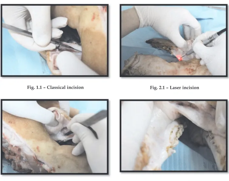
Gingivectomies have been realized on a man‑ dible of freshly sacriiced pig, by the classical surgical technique, 10 tissue samples of compa‑ rable size being taken over. (Figs. 1.1 and 1.2)
226 volume 3 • issue 3 July / September 2013 • pp. 225‑228 I. Luchian, Ioana Mârţu, Monica Tatarciuc, Anca Sava, Silvia Mârţu
Fig. 1.1 – Classical incision
Fig. 1.2 – Tissue prelevation
On the same mandible, in the opposite quad‑ rant, gingivectomies were performed by means of a diode‑type laser (Biolase Epic 10) with a wavelength of 940 nm and a surgical tip with an optical iber size of 400 μm, 10 tissue samples being also collected. The optical iber was previ‑ ously activated, a 2 W power being applied in pulsed mode. During the laser‑assisted interven‑ tion, another operator assured cooling of the tis‑ sues. During the whole surgical intervention, the operators wore protection glasses speciic to a wavelength of 940 cm. (Figs. 2.1 and 2.2)
The samples were stored in a saline solution and transported, over an interval of 30 min after their taking over, to the Laboratory of Patholog‑ ical Anatomy, to be ixed and processed for the realization of the histological cups. (Figs. 3.1 and 3.2)
Fig. 2.1 – Laser incision
Fig. 2.2 – Laser marking of the incision
International Journal of Medical Dentistry 227 UTILIZATION OF 940 NM WAVELENGTH DIODE LASERS AND THE MORPHO‑HISTOLOGICAL
MODIFICATIONS IN PERIODONTAL TISSUES
Fig. 3.2 – Laser and classical prelevation
RESULTS AND DISCUSSION
No morpho‑histological modiications could be observed on the cups obtained from the sam‑ ples taken over through classical incision, with the exception of the microscopic aspect of the incision line. The other tissues, such as the pavi‑ mentous one, showed a normal non‑modiied aspect. (Fig. 4)
Fig. 4 – Histological aspect of a classical incision
At the level of the histological cups realized from the samples taken over by laser incision, minor histological modifications could be observed, caused by the surface carbonization determined by the photo‑thermic effect of the laser. The microscopically‑examined incision line evidenced a homogeneous microscopic aspect. (Fig. 5)
Fig. 5 – Histological aspect of a laser incision Due to thermic shock, supericial necrotic tis‑ sue was identiied in the cups made from the laser samples. (Fig. 6)
Fig. 6 – Necrotic tissue
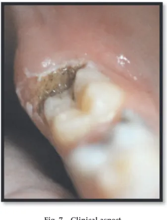
The carbonized tissue resulted from the laser‑assisted intervention produces no signii‑ cant morpho‑histological modiications and has a protecting effect upon the periodontal tissues on which the intervention had been made. (Fig. 7)
The clinical advantages of laser‑assisted peri‑ odontal therapy are: excellent bacterial decon‑ tamination, reduced bleeding, reduced oedemas, reduced post‑surgery discomfort, higher degree of comfort for the patient. [4]
228 volume 3 • issue 3 July / September 2013 • pp. 225‑228 I. Luchian, Ioana Mârţu, Monica Tatarciuc, Anca Sava, Silvia Mârţu
Fig. 7 – Clinical aspect
CONCLUSIONS
Laser‑assisted technologies may be therefore viewed as extremely useful alternatives in the
new periodontal therapies, which recommends their application in periodontal surgery for at least three reasons: they are minimally invasive, they induce minor morpho‑histological modii‑ cations and the technique of their application is simple to learn.
Classical periodontal therapies cannot be wholly substituted by the non‑conventional ones, however the inal result may be optimized by the introduction, in everyday practice, of some speciic protocols which include them or associate them for reaching predictable results.
References
1. Kusek ER, Kusek AJ, Kusek EA., Five‑year retrospec‑
tive study of laser‑assisted periodontal therapy, Gen
Dent. 2012, Nov‑Dec; 60(6):540‑543.
2. Cobb CM, Lasers in periodontics: a review of the litera‑
ture, J Periodontol. 2006 Apr; 77(4):545‑564.
3. Coleton SH, The use of lasers in periodontal therapy,
Alpha Omegan. 2008, Dec; 101(4):181‑187.
4. Coleton S, Lasers in surgical periodontics and oral medi‑
cine, Dent Clin North Am. 2004, Oct; 48(4):937‑962,
vii. Review.
5. Mârţu S, Amălinei C, Tatarciuc M, Rotaru M, Potâr‑
nichie O, Liliac L, Căruntu ID, Healing process and
laser therapy in the supericial periodontium: a histolo‑
gical study. Rom J Morphol Embryol. 2012; 53(1):