OATP1B1 and OATP1B3 Expressing Cancer Cells
Timo H. J. Niedermeyer
1,2*, Abigail Daily
3, Monika Swiatecka-Hagenbruch
1, Jeffrey A. Moscow
31Cyano Biotech GmbH, Berlin, Germany,2Interfaculty Institute for Microbiology and Infection Medicine, University of Tu¨bingen, Tu¨bingen, Germany,3Department of Pediatrics, University of Kentucky, Lexington, Kentucky, United States of America
Abstract
Microcystins are potent phosphatase inhibitors and cellular toxins. They require active transport by OATP1B1 and OATP1B3
transporters for uptake into human cells, and the high expression of these transporters in the liver accounts for their
selective hepatic toxicity. Several human tumors have been shown to have high levels of expression of OATP1B3 but not
OATP1B1, the main transporter in liver cells. We hypothesized that microcystin variants could be isolated that are
transported preferentially by OATP1B3 relative to OATP1B1 to advance as anticancer agents with clinically tolerable hepatic
toxicity. Microcystin variants have been isolated and tested for cytotoxicity in cancer cells stably transfected with OATP1B1
and OATP1B3 transporters. Microcystin variants with cytotoxic OATP1B1/OATP1B3 IC
50ratios that ranged between 0.2 and
32 were found, representing a 150-fold range in transporter selectivity. As microcystin structure has a significant impact on
transporter selectivity, it is potentially possible to develop analogs with even more pronounced OATP1B3 selectivity and
thus enable their development as anticancer drugs.
Citation:Niedermeyer THJ, Daily A, Swiatecka-Hagenbruch M, Moscow JA (2014) Selectivity and Potency of Microcystin Congeners against OATP1B1 and OATP1B3 Expressing Cancer Cells. PLoS ONE 9(3): e91476. doi:10.1371/journal.pone.0091476
Editor:Matias A. Avila, University of Navarra School of Medicine and Center for Applied Medical Research (CIMA), Spain
ReceivedDecember 19, 2013;AcceptedFebruary 13, 2014;PublishedMarch 10, 2014
Copyright:ß2014 Niedermeyer et al. This is an open-access article distributed under the terms of the Creative Commons Attribution License, which permits unrestricted use, distribution, and reproduction in any medium, provided the original author and source are credited.
Funding:This work was supported in part by DanceBlue, a student-run effort that supports clinical care and research in pediatric oncology at the University of Kentucy, and in part by the German Federal Ministry of Economics and Technology (KF2766301SB0). Open Access publishing is supported by Deutsche Forschungsgemeinschaft and Open Access Publishing Fund of Tuebingen University. The funders had no role in study design, data collection and analysis, decision to publish, or preparation of the manuscript.
Competing Interests:Timo Niedermeyer and Monika Swiatecka-Hagenbruch are employees of Cyano Biotech GmbH. There are no marketed products to declare. Cyano Biotech GmbH has given permission to publish the manuscript. This does not alter the authors’ adherence to all the PLoS ONE policies on sharing data and materials.
* E-mail: [email protected]
Introduction
Microcystins (MCs) are cyclic heptapeptides produced by
several cyanobacterial genera such as
Microcystis
,
Oscillatoria
,
Planktothrix
,
Nostoc
, and
Anabaena
. They can be considered to be
among the best studied cyanobacterial secondary metabolites [1–
4]. The common structural feature of microcystins is a polyketide
synthase derived amino acid with the acronym ‘‘Adda’’,
(2S,3S,8S,9S)-3-amino-9-methoxy-2,6,8-trimethyl-10-phenyldeca-4,6-dienoic acid, which is also one of the two mandatory
substructures for the potent protein serine/threonine phosphatase
(PP) 1 and PP2A inhibition observed for the microcystins [5–9].
Not only is the biosynthesis of these compounds by polyketide
synthases and non-ribosomal peptide synthetases remarkably well
understood [10–12], but also more than 90 individual members of
this chemically highly diverse compound family have been
described in the scientific literature to date (Fig. 1) [13–56].
Especially prone to variation are the
L-amino acids situated at
positions 2 and 4 of the MC backbone. Related compounds are
the nodularins, found in
Nodularia
sp., which also contain Adda but
are cyclic pentapeptides instead of heptapeptides (Fig. 1) [57–60].
Being potent protein serine/threonine phosphatase inhibitors,
microcystins and nodularins have a profound effect on cell
signaling and cytoskeleton maintenance, leading to the death of
affected cells [61,62]. However, the relatively large and
amphi-philic MCs are unable to cross cell membranes by passive
diffusion. Instead, they rely on active uptake by cells. Three
members of the organic anion transporting polypeptides (OATP)
family are able to mediate this uptake of MCs, namely OATP1B1,
1B3 and 1A2 [63,64]. OATP1B1 and OATP1B3 are the most
efficient microcystin transporters, and as in healthy humans both
transporters are exclusively found to be expressed in liver tissue
[64,65], microcystins and nodularins are known to cause extensive
liver damage [66–68]. Thus microcystins became infamous as
hepatotoxins causing harm to humans and cattle when these
compounds accumulated in sources of drinking water during algal
water bloom times [69,70]. Inhibitors of these OATP transporters
ameliorate the hepatotoxicity of microcystins and nodularins
[68,71,72]. In contrast to OATP1B1, which is expressed in
hepatocytes throughout the liver lobe, OATP1B3 localization is
restricted around the central vein [64].
OATP1B1 should lead to a decreased hepatic clearance and
increased uptake of MCs in OATP1B3-expressing tumors,
creating a therapeutic window of the respective compound by
decreasing the hepatic clearance rate and toxicity. MCs are
interesting as novel lead structures because they have a mode of
action not yet used but currently discussed for cancer therapy
(phosphatase inhibition) [81–83], and in contrast to the majority of
currently available anticancer drugs, they need active transport
into cells and thus spare all tissues not expressing the mentioned
OATPs.
We have isolated various microcystin congeners from
cyano-bacteria of the genera
Microcystis
,
Planktothrix
, and
Nodularia
to test
the hypothesis whether different transporter selectivities might be
attainable. As tools for selectivity testing, cervical cancer HeLa
cells and colon cancer RKO cells stably transfected with
expression vectors for OATP1B1 and 1B3 have been used. In
the present manuscript, the results of the testing of isolated MCs
on these cell lines are described. The determined IC
50values as
well as the observed selectivity differences clearly show that small
structural differences of the tested MCs indeed have a significant
impact on transporter selectivity and cytotoxic potency.
Results and Discussion
The amino acid (AA) compositions of the tested MCs are
summarized in Table 1
The structures of the microcystin congeners have been
determined based on tandem HRMS [51,52,84], supported by
1H- and COSY-NMR spectroscopy. All MCs contain the
characteristic Adda moiety at position 5 and
D-Ala at position 1
of the molecule. Only one of the isolated congeners features an
O
-methylated
D-iso-Glu instead of
D-iso-Glu at position 6 (
22
).
D-iso-b
-MeAsp and
D-iso-Asp in position 3 are almost equally
distributed, as are Mdha and Dhb in position 7. The
Lamino
acids in position 2 comprise Leu, Tyr, Arg, Hty, Hil (descending
order of count), in position 4 Arg, Tyr, Trp, Phe, Hty, and Har are
found. In addition to 22 MC variants, nodularin (
23
) as a cyclic
Adda containing pentapeptide as well as okadaic acid (
24
) as a
structurally not MC-related PP inhibitor have been tested.
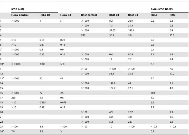
The IC50 values of all isolated MCs against OATP1B1- and
OATP1B3-expressing HeLa or RKO cell lines have been
determined. The results are shown in Table 2.
Interestingly, marked differences for both potency and
selectiv-ity of the individual MC congeners could be observed.
Selectivity
While substitution of
D-iso-
b
-MeAsp
3for
D-iso-Asp
3(e.g.
compounds
1
/
2
and
13
/
14
) seems not to have a significant
influence on neither potency nor selectivity, the influence of the
presence of either Mdha
7or (
E
)-Dhb
7(e.g.
3
/
4
,
5
/
6
,
8
/
9
, and
14
/
15
) is ambiguous: While in the case of
3
/
4
and
5
/
6
substitution of Mdha
7for (
E
)-Dhb
7lead to a profound increase
in selectivity for OATP1B3 over OATP1B1, this effect was not
observed for
8
/
9
and
14
/
15
. As the six least OATP1B3 selective
compounds (
1
,
2
,
3
,
5
,
7
,
23
) all feature Mdha
7or Mdhb
7while
(
E
)-Dhb
7is found in 3 of the 6 most OATP1B3 selective
compounds (
4
,
10
,
12
,
15
,
16
,
18
), it is likely that (
E
)-Dhb
7has
an influence in selectivity but is not the only structural feature
conferring selectivity.
Indeed, especially the amino acid residues at positions 2 and 4 of
the MC core structure seem to be important for both potency and
selectivity. While 80% of the most OATP1B1 selective compounds
feature Leu at position 2, this amino acid is completely absent in
the most OATP1B3 selective compounds. However, Leu is also
found in position 2 of several congeners that are weakly OATP1B3
selective (
6
,
8
,
9
), thus Leu alone does not make a compound
OATP1B1 selective. Arg in position 2 seems to induce OATP1B3
selectivity, while Arg in position 4 does not have any influence on
selectivity. This is obvious with compounds
13
and
16
, where
exchange of the amino acids in positions 2 and 4 has a huge
impact on selectivity. Arg in combination with the aromatic amino
acids Phe, Tyr, and Hty are prominent monomers found in
OATP1B3 selective compounds. Especially combinations of Arg
2and Phe
4(
12
) or Arg
2and Tyr
4(
16
) seem to confer OATP1B3
selectivity. But again, presence of Arg in position 2 seems not to be
mandatory for OATP1B3 selectivity (
4
).
Interestingly, the pentapeptide Nodularin (
23
) is the most
OATP1B1 selective among the Adda containing compounds
examined.
Potency
Potency of MC induced cell death in the test system used for this
study has two facets. On one hand, toxicity of MC congeners
depends on the extent of their PP inhibition [85]. On the other
hand, potency is depending on transporting capacity of the
respective OATP. These two effects have not been distinguished in
the present study. While some previous studies suggest that
in vitro
and
in vivo
toxicity are related to enzyme inhibition rather than
transport [78,85], other reports show comparable PP inhibition of
different microcystin congeners, implying that differences in
transport contribute to
in vivo
toxicity [80,86].
Not surprisingly,
22
, featuring
O
-methylated
D-iso-Glu
6, is one
of the least potent compounds. Modifications of the free carboxylic
acid of Glu are likely to lead to a loss of efficiency due to loss of
interaction with a positively charged arginine in the active center
of the protein phosphatases [7], and Glu(OMe) containing
congeners have been found to inhibit PP1 to a much lesser extent
than analogous Glu containing MCs [87] and have lower
in vivo
toxicity [26]. Interestingly, also
10
,
11
and
21
displayed weaker
toxicity than most other congeners. While
10
inhibits PP2A about
twenty times weaker compared with
1
[78,80,85,86], its toxicity in
the present assay is about 500 times lower. This indicates that in
the case of
10
(and also its analog
11
), it is probable that toxicity is
not only a matter of PP inhibition efficiency, but also of OATP
transporting capability, as MC-RR is a dication under
physiolog-ical conditions and thus less likely to be transported across cell
membranes by anion transporters. In general, cytotoxicity in our
assay does not correlate with protein phosphatase inhibition
potency as described in the literature for compounds
1
,
2
,
6
,
7
,
8
,
9
,
10
,
13
,
18
,
19
,
20
,
23
[56,78,80,85,86]. Our data thus indicate
that differing transport efficiencies have a higher impact on
in vivo
toxicity than differing protein phosphatase inhibition potency.
Interestingly, the most potent MCs, with IC
50values in the
sub-nanomolar range (
5
,
6
,
7
,
8
,
17
,
18
,
19
), all feature combinations
of either Leu
2and Tyr/Phe/Trp
4or Tyr/Hty
2and Tyr/Hty
4.
Arg is absent from the most potent congeners. The least potent
congeners with IC
50values
.
100 nM (
10
,
11
,
21
,
22
) all feature
either Tyr
2and Arg/Har
4or Arg
2and Arg
4. Presence of Arg in
general lowers the potency, again supporting the hypothesis that
the positively charged Arg residues hamper transport efficiency in
addition to reducing PP2A inhibition, and thus lower
in vivo
toxicity.
Summary and Conclusions
In our screening of 23 natural MC variants, we have found
several MCs with an IC
50in the low nanomolar range and with
transporter selectivity that favors OATP1B3 over OATP1B1 by a
factor of up to thirty. While the presence of Arg in general lowers
the potency of MC cytotoxicity, its presence in position 2 of the
MC core structure also seems to be important for OATP1B3
selectivity, implying that this transporter might be more tolerant to
the cationic nature of Arg under physiological conditions than
OATP1B1. Furthermore, the presence of the slightly acidic
aromatic amino acids Tyr or Hty in position 4 in addition to
Arg in position 2 seems beneficial for OATP1B3 selectivity.
Table 1.
Structures of the tested MC congeners originating from
aMicrocystis aeruginosa
,
bPlanktothrix rubescens
, and
cNodularia
sp.
MC Congener AA 1 AA 2 AA 3 AA 4 AA 5 AA 6 AA 7
1a MC-LR Ala Leu MeAsp Arg Adda Glu Mdha
2a [D-Asp3]MC-LR Ala Leu Asp Arg Adda Glu Mdha
3a [D-Asp3]MC-HilR Ala Hil Asp Arg Adda Glu Mdha
4b [D-Asp3,(E)-Dhb7]MC-HilR Ala Hil Asp Arg Adda Glu Dhb
5a MC-LY Ala Leu MeAsp Tyr Adda Glu Mdha
6b [D-Asp3,(
E)-Dhb7]MC-LY Ala Leu Asp Tyr Adda Glu Dhb
7a LF Ala Leu MeAsp Phe Adda Glu Mdha
8a LW Ala Leu MeAsp Trp Adda Glu Mdha
9b [
D-Asp3,(E)-Dhb7]-MC-LW Ala Leu Asp Trp Adda Glu Dhb
10aRR Ala Arg MeAsp Arg Adda Glu Mdha
11b[D-Asp3,(E)-Dhb7]MC-RR Ala Arg Asp Arg Adda Glu Dhb
12aMC-RF Ala Arg MeAsp Phe Adda Glu Mdha
13aYR Ala Tyr MeAsp Arg Adda Glu Mdha
14a[D-Asp3]MC-YR Ala Tyr Asp Arg Adda Glu Mdha
15b[
D-Asp3,(E)-Dhb7]MC-YR Ala Tyr Asp Arg Adda Glu Dhb
16aMC-RY Ala Arg MeAsp Tyr Adda Glu Mdha
17aMC-YY Ala Tyr MeAsp Tyr Adda Glu Mdha
18b[
D-Asp3,(E)-Dhb7]MC-HtyY Ala Hty Asp Tyr Adda Glu Dhb
19b[D-Asp3,(E)-Dhb7]MC-HtyHty Ala Hty Asp Hty Adda Glu Dhb
20b[D-Asp3,(E)-Dhb7]MC-HtyW Ala Hty Asp Trp Adda Glu Dhb
21a[
D-Asp3,MSer7]MC-YHar Ala Tyr Asp Har Adda Glu Mser
22a[D-Glu(OMe)6]MC-YR Ala Tyr MeAsp Arg Adda Glu(OMe) Mdha
23cNodularin MeAsp Arg Adda Glu Mdhb
Hilhomoisoleucine,Htyhomotyrosine,MeAspb-methylaspartic acid,Harhomoarginine,MdhaN-methyl dehydroalanine,Dhbdehydrobutyric acid,MserN-methyl serine,MdhbN-methyl dehydrobutyric acid.
Further studies are needed to discriminate whether the observed
differences in potency are due to differing PP inhibition or
different transport capability of the OATP. However, earlier
findings suggest that
in vivo
toxicity is mainly depending on OATP
transport kinetics than on differences in PP inhibition [80,86].
Although the deduced structure activity relationships are
ambiguous in parts, the structural features observed for OATP1B3
selectivity can be condensed to the at present most likely selective
general
structure
cyclo(-
D-Ala
1-
L-Arg
2-
D-(Me)Asp
3-
L-aromatic
amino acid
4-Adda
5-
D-Glu
6-(
E
)-Dhb
7). This general structure can
be used as a starting point for generating novel compounds e.g. by
precursor feeding or biocombinatorial strategies.
A challenge of using MCs as drug leads still remains: Even with
selective MC variants, liver toxicity still might be a significant
challenge, making it necessary to generate MC variants that could
be metabolically detoxified by healthy liver cells and/or efficiently
effluxed into the bile, and thus take advantage of hepatic
detoxification and clearance mechanisms that would not be found
in tumors.
Materials and Methods
Cyanobacterial Material
Several
Microcystis aeruginosa
and
Planktothrix rubescens
strains as
well as a
Nodularia
strain have been used to produce the studied
microcystin congeners:
2
and
3
,
Microcystis aeruginosa
CBT 265;
4
and
15
,
Planktothrix rubescens
CBT 310;
5
,
16
, and
17
,
Microcystis
aeruginosa
CBT 850,
6
,
9
,
18
,
19
, and
20
,
Planktothrix rubescens
CBT
862,
11
,
Planktothrix rubescens
CBT 329,
12
,
Microcystis aeruginosa
CBT 861,
14
,
21
, and
22
,
Microcystis aeruginosa
CBT 480,
23
,
Nodularia
sp. CBT 786. The strains were classified on the basis of
PCR analysis and sequencing of various marker genes as well as
their morphology, and have been deposited in the Cyano Biotech
(CBT) culture collection under the accession numbers indicated
above (Cyano Biotech, Berlin, Germany). The strains were
cultivated in BG11 medium [88] at 20
u
C under continuous light
(60-80
mmol m
-2s
-1) in 20 L scale photobioreactors and harvested
semi-continuously over a period of several weeks.
Isolation of microcystins
Cyanobacteria strains were screened by
HPLC-DAD/IT-TOF-MS (Kinetex C
18, 2.6
mm, 100
63 mm column, phenomenex,
Torrance, USA) using a linear scouting gradient of aqueous
CH
3CN (5 to 80% within 25 min at 0.6 ml/ min; 0.025% v/v
TFA) to confirm MC presence by their characteristic UV
spectrum as well as the characteristic tandem MS fragment ion
at
m/z
375 [52,89]. Selected positive strains have been cultivated
in BG11 medium [88] at 20
u
C under continuous light
(60-80
mmol m
-2s
-1) in 20 L scale photo bioreactors. After biomass
harvest and freeze-drying, the biomasses were resuspended in 50%
Table 2.
IC
50values of MC congeners (1-23) and okadaic acid (24) against stably transfected HeLa or RKO cell lines expressing
either OATP1B1 or OATP1B3.
IC50 (nM) Ratio IC50 B1/B3
HeLa Control HeLa B1 HeLa B3 RKO control RKO B1 RKO B3 HeLa RKO
1 .1000 1 5.1 .1000 8.2 26.9 0.2 0.3
2 .1000 7.3 31.3 0.2
3 .1000 57.05 142.4 0.4
4 993 56.9 3.8 15.0
5 .10 0.16 0.21 0.8
6 .10 0.47 0.18 2.6
7a
.1000 0.4 0.9 0.4
8 .1000 0.22 0.16 .1000 0.4 0.29 1.4 1.4
9 .1000 11 7.7 1.4
10a
.10000 3800 580 6.6
11 .100 .100 .100 Na
12 .1000 58.3 3.38 17.2
13a
.1000 90 45 2.0
14 .1000 148.8 48 3.1
15 .1000 107.7 27.1 4.0
16 .1000 77 2.5 30.8
17 259 1.5 0.8 1.9
18 .10 0.515 0.078 6.6
19 .10 0.39 0.18 2.2
20 .100 4.9 2.57 1.9
21 .1000 625 385 1.6
22 .1000 595 227 2.6
23 .100 8.4 .100 .100 10 .100 ,0.1 ,0.1
24a 7.8 2.2 3 0.7
aTransiently transfected cell lines; results were reported previously [78].
MeOH (v/v), treated with an ultrasonication rod (Bandelin,
Berlin, Germany) and extracted on a shaker for 30 min. After
centrifugation the biomasses were subsequently extracted using
80% MeOH (v/v). The 50% MeOH and 80% MeOH extracts of
the respective biomasses were combined and dried
in vacuo
. The
crude extracts were fractionated using water-methanol step
gradients on C
18cartridges on a VersaFlash system (supelco,
Bellefonte, USA), and the fractions containing MCs (monitored by
HPLC) were dried
in vacuo
. After reconstitution, these fractions
were subjected to semi-preparative HPLC. For a detailed
description of the isolation procedures see File S1. Microcystins
RR, LW, and LF and okadaic acid were obtained from Axxora,
LLC (San Diego, CA), and microcystins LR and YR were
obtained from Sigma-Aldrich (St. Louis, MO).
Structure elucidation
The structures of the isolated MCs have been determined by
high-resolution tandem mass spectrometry [51,52,84], and
con-firmed by one- and two-dimensional NMR spectroscopy. Samples
for NMR spectroscopy were dissolved in 600
mL d
6-DMSO.
NMR spectra were recorded at 600 MHz (
1H frequency) on a
Bruker AV-III spectrometer using cryogenically cooled 5 mm
TCI-triple resonance probes equipped with one-axis self-shielded
gradients. DQF-COSY spectra [90] were recorded using
20486
512 complex data points using 8 scans. Spectra were
referenced indirectly to tetramethylsilane via the residual signals of
d
6-DMSO (2.5 ppm for
1H). Tandem MS data have been
acquired using an HPLC coupled to an IT-TOF mass
spectrom-eter (Shimadzu Europe GmbH, Duisburg, Germany) with
electrospray ionization in positive mode and were evaluated using
the vendor’s software LCMSSolution version 3.60.361 with
Formula Predictor version 1.13. For the calculation of sum
formulae, the monoisotopic mass averaged from at least three
scans has been used. The compounds were separated on a Kinetex
C
18column (2.6
mm, 1006
3 mm, phenomenex, Torrance, USA)
using a gradient ranging from 5 to 80% CH
3CN in water over 25
min (0.1% formic acid added as modifier). Precursor ions
corresponding to [M+H]
+were isolated in the ion trap,
fragmented by collision induced dissociation (CID) using argon
as collision gas (collision energy set to 150%, collision gas to 100%,
and q(Frequency) to 45.0 kHz), and separated in the TOF
analyzer. MS/MS scans were averaged and converted to the
mzXML format using the vendor’s software and evaluated using
the software mMass (calculated fragment ions: M, a, b, c, and z;
modifications: -H
2O, -NH
3,
+CO, defined, combinations;
match-ing with a tolerance of 0.01 Da) [91]. Annotated tandem HRMS
and
1H-NMR spectra of all isolated compounds can be found in
File S2.
Cell lines and cytotoxicity assays
Primary hepatocytes are not suitable for studying microcystin
toxicity and transport, as OATP transporters are downregulated
within hours upon placing cells in culture [92]. Thus HeLa and
RKO cell lines were obtained from ATCC and transfected with
expression vectors for OATP1B1 and OATP1B3. Individual
clones were isolated by a limiting dilution, and isolated clones
showing high levels of expression by PCR were further
charac-terized as previously described [72,93]. Individual HeLa and
RKO clones were selected for further study by demonstration of
increased uptake [
3H]-BQ123, a substrate for both transporters.
Further, the OATP1B3 clones demonstrated increased uptake of
[
3H]-CCK-8, a specific substrate for OATP1B3 but not
OATP1B1, while the OATP1B1 clones did not show uptake of
[
3H]-CCK-8. In addition, RKO and HeLa cells stably transfected
with an empty expression vector were used as controls. For
cytotoxicity assays, the cells were plated in triplicate in 96-well
microtiter plates in a medium containing 5% fetal bovine serum at
densities of 1000 cells/well. After 24 hours, medium containing
MC structural variants was added to the cells. After another 3
days, cell survival was determined with a sulforhodamine-based
assay as we have previously described [93–95]. As the dose
response curves for the apoptosis causing microcystins are steep
and go down to 0 within 72 hours, the sulforhodamine assay is
indicative of cell death [72,93]. The IC
50was calculated from the
dose response curve as the concentration of drug that produced a
50% decrease in the mean absorbance compared to the untreated
wells and reported as the average of at least three independent
determinations performed in triplicate using Prism software.
Supporting Information
File S1
Details on the Isolation of the Microcystin Congeners.
(PDF)
File S2
Annotated tandem HRMS and
1H-NMR spectra of all
isolated compounds. Raw NMR and MS data of these compounds
are available free of charge via the Internet at http://dx.doi.org/
10.6084/m9.figshare.880755.
(PDF)
Acknowledgments
TN and MSH thank Cyano Biotech GmbH for support and permission to publish this manuscript. We thank R. Lethaus-Weiß for her help in cultivating the cyanobacteria strains and extracting the biomass, and P. Schmieder and B. Schlegel (FMP Berlin-Buch) for recording the NMR spectra.
Author Contributions
Conceived and designed the experiments: THJN JAM. Performed the experiments: THJN AD MSH. Analyzed the data: THJN AD JAM. Wrote the paper: THJN JAM.
References
1. Watanabe MF, Harada K, Carmichael WW, Fujiki H (1995) Toxic Microcystis. CRC Press.
2. Dittmann E, Wiegand C (2006) Cyanobacterial toxins – occurrence, biosynthesis and impact on human affairs. Mol Nutr Food Res 50: 7–17.
3. Van Apeldoorn ME, van Egmond HP, Speijers GJ a, Bakker GJI (2007) Toxins of cyanobacteria. Mol Nutr Food Res 51: 7–60.
4. Pearson L, Mihali T, Moffitt M, Kellmann R, Neilan B (2010) On the Chemistry, Toxicology and Genetics of the Cyanobacterial Toxins, Microcystin, Nodularin, Saxitoxin and Cylindrospermopsin. Mar Drugs 8: 1650–1680. 5. Goldberg J, Huang H, Kwon Y, Greengard P, Nairn AC, et al. (1995)
Three-dimensional structure of the catalytic subunit of protein serine/threonine phosphatase-1. Nature 376: 745–753.
6. Taylor C, Quinn RJ, Suganuma M, Fujiki H (1996) Inhibition of protein phosphatase 2A by cyclic peptides modelled on the microcystin ring. Bioorg Med Chem Lett 6: 2113–2116.
7. Bagu JR, Sykes BD, Craig MM, Holmes CFB (1997) A Molecular Basis for Different Interactions of Marine Toxins with Protein Phosphatase-1. J Biol Chem 272: 5087–5097.
8. Gulledge BM, Aggen JB, Eng H, Sweimeh K, Chamberlin AR (2003) Microcystin Analogues Comprised Only of Adda and a Single Additional Amino Acid Retain Moderate Activity as PP1/PP2A Inhibitors. Bioorg Med Chem Lett 13: 2907–2911.
phosphatases 1 and 2A from both mammals and higher plants. FEBS 264: 187– 192.
10. Tillett D, Dittmann E, Erhard M, von Do¨hren H, Bo¨rner T, et al. (2000) Structural organization of microcystin biosynthesis in Microcystis aeruginosa PCC7806: an integrated peptide-polyketide synthetase system. Chem Biol 7: 753–764.
11. Christiansen G, Fastner J, Erhard M, Bo¨rner T, Dittmann E (2003) Microcystin Biosynthesis in Planktothrix: Genes, Evolution, and Manipulation. J Bacteriol 185: 564–572.
12. Hicks LM, Moffitt MC, Beer LL, Moore BS, Kelleher NL (2006) Structural characterization of in Vitro and in Vivo Intermediates on the Loading Module of Microcystin Synthetase. ACS Chem Biol 1: 93–102.
13. Botes DP, Tuinman AA, Wessels PL, Viljoen CC, Kruger H, et al. (1984) The Structure of Cyanoginosin-LA, a Cyclic Heptapeptide Toxin from the Cyanobacterium Microcystis aeruginose. J Chem Soc Perkin Trans I: 2311– 2318.
14. Botes DP, Wessels PL, Kruger H, Runnegar MTC, Santikarn S, et al. (1985) Structural Studies on Cyanoginosins-LR, -YR, -YA, and -YM, Peptide Toxins from Microcystis aeruginosa. J Chem Soc Perkin Trans I 1: 2747–2748. 15. Gathercole PS, Thiel PG (1987) Liquid chromatographic determination of the
cyanoginosins, toxins produced by the cyanobacterium Microcystis aeruginosa. J Chromatogr 408: 435–440.
16. Kusumi T, Ooi T, Watanabe MM, Takahasi H, Kakisawa H (1987) Cyanoviridin RR, a toxin from the cyanobacterium (blue-green alga) Microcystis viridis. Tetrahedron Lett 28: 4695–4698.
17. Krishnamurthy T, Szafraniec L, Hunt DF, Shabanowitz J, Yates III JR, et al. (1989) Structural characterization of toxic cyclic peptides from blue-green algae by tandem mass spectrometry. Proc Natl Acad Sci U S A 86: 770–774. 18. Meriluoto JAO, Sandstro¨m A, Eriksson JE, Remaud G, Craig AG, et al. (1989)
Structure and toxicity of a peptide hepatotoxin from the cyanobacterium Oscillatoria agardhii. Toxicon 27: 1021–1034.
19. Stoner RD, Adams WH, Slatkini DN, Siegelman HW (1989) The effects of single L-amino acid substitution on the lethal potencies of the microcystins. Toxicon 27: 825–828.
20. Harada K, Ogawa K, Matsuura K, Murata H, Suzuki M, et al. (1990) Structural Determination of Geometrical Isomers of Microcystins LR and RR from Cyanobacteria by Two-Dimensional NMR Spectroscopic Techniques. Chem Res Toxicol 3: 473–481.
21. Sivonen K, Carmichael WW, Namikoshi M, Rinehart KL, Dahlem AM, et al. (1990) Isolation and Characterization of Hepatotoxic Microcystin Homologs from the Filamentous Freshwater Cyanobacterium Nostoc sp. Strain 152. Appl Environ Microbiol 56: 2650–2657.
22. Harada K, Ogawa K, Kimura Y, Murata H, Suzuki M, et al. (1991) Microcystins from Anabaena flos-aquae NRC 525-17. Chem Res Toxicol 4: 535–540.
23. Harada K, Ogawa K, Matsuura K, Nagai H, Murata H, et al. (1991) Isolation of two toxic heptapeptide microcystins from an axenic strain of Microcystis aeruginosa, K-139. Toxicon 29: 479–489.
24. Kiviranta J, Namikoshi M, Sivonen K, Evans WR, Carmichael WW, et al. (1992) Structure determination and toxicity of a new microcystin from Microcystis aeruginosa strain 205. Toxicon 30: 1093–1098.
25. Namikoshi M, Sivonen K, Evans WR, Carmichael WW, Rouhiainen L, et al. (1992) Structures of three new homotyrosine-containing microcystins and a new homophenylalanine variant from Anabaena sp. strain 66. Chem Res Toxicol 5: 661–666.
26. Namikoshi M, Rinehart KL, Sakai R, Stotts RR, Dahlem AM, et al. (1992) Identification of 12 Hepatotoxins from a Homer Lake Bloom of the Cyanobacteria Microcystis aeruginosa, Microcystis viridis, and Microcystis wesenbergii: Nine New Microcystins. J Org Chem 57: 866–872.
27. Namikoshi M, Sivonen K, Evans WR, Carmichael WW, Sun F, et al. (1992) Two new L-serine variants of microcystins-LR and -RR from Anabaena sp. strain 202 A1 and 202 A2. Toxicon 30: 1457–1464.
28. Namikoshi M, Sivonen K, Evans WR, Sun F, Carmichael WW, et al. (1992) Isolation and structures of microcystins from a cyanobacterial water bloom (Finland). Toxicon 30: 1473–1479.
29. Sivonen K, Namikoshi TM, Evans WR, Fa¨rdig M, Carmichael WW, et al. (1992) Three New Microcystins, Cyclic Heptapeptide Hepatotoxins, from Nostoc sp. Strain 152. Chem Res Toxicol 5: 464–469.
30. Sivonen K, Namikoshi M, Evans WR, Gromov BV, Carmichael WW, et al. (1992) Isolation and structures of five microcystins from a Russian Microcystis aeruginosa strain CALU 972. Toxicon 30: 1481–1485.
31. Sivonen K, Skulberg OM, Namikoshi M, Evans WR, Carmichael WW, et al. (1992) Two methyl ester derivatives of microcystins, cyclic heptapeptide hepatotoxins, isolated from Anabaena flos-aquae strain CYA 83/1. Toxicon 30: 1465–1471.
32. Craig M, McCready TL, Luu HA, Smillie MA, Dubord P, et al. (1993) Identification and characterization of hydrophobic microcystins in canadian freshwater cyanobacteria. Toxicon 31: 1541–1549.
33. Luukkainen R, Sivonen K, Namikoshi M, Fa¨rdig M, Rinehart KL, et al. (1993) Isolation and identification of eight microcystins from thirteen Oscillatoria agardhii strains and structure of a new microcystin. Appl Environ Microbiol 59: 2204–2209.
34. Azevedo SMFO, Evans WR, Carmichael WW, Namikoshi M (1994) First report of microcystins from a Brazilian isolate of the cyanobacterium Microcystis aeruginosa. J Appl Phycol 6: 261–265.
35. Luukkainen R, Namikoshi M, Sivonen K, Rinehart KL, Niemela¨ SI (1994) Isolation and identification of 12 microcystins from four strains and two bloom samples of Microcystis sp.: structure of a new hepatotoxin. Toxicon 32: 133–139. 36. Bateman KP, Thibault P, Douglas DJ, White RL (1995) Mass spectral analyses of microcystins from toxic cyanobacteria using on-line chromatographic and electrophoretic separations. J Chromatogr A 712: 253–268.
37. Namikoshi M, Sun F, Choi BW, Rinehart KL, Carmichael WW, et al. (1995) Seven More Microcystins from Homer Lake Cells: Application of the General Method for Structure Assignment of Peptides Containing.alpha.,.beta.-Dehy-droamino Acid Unit(s). J Org Chem 60: 3671–3679.
38. Sano T, Kaya K (1995) A 2-Amino-2-Butenoic Acid(Dhb)-Containing Microcystin Isolated from Oscillatoria agardhii. Tetrahedron Lett 36: 8603– 8606.
39. Beattie KA, Kaya K, Sano T, Codd GA (1998) Three dehydrobutyrine-containing microcystins from Nostoc. Phytochemistry 47: 1289–1292. 40. Lee TH, Chen YM, Chou HN (1998) First report of microcystins in Taiwan.
Toxicon 36: 247–255.
41. Namikoshi M, Yuan M, Sivonen K, Carmichael WW, Rinehart KL, et al. (1998) Seven New Microcystins Possessing Two L-Glutamic Acid Units, Isolated from Anabaena sp. Strain 186. Chem Res Toxicol 11: 143–149.
42. Sano T, Kaya K (1998) Two New (E)-2-Amino-2-Butenoic Acid (Dhb)-Containing Microcystins Isolated from Oscillatoria agardhii. Tetrahedron 54: 463–470.
43. Sano T, Beattie KA, Codd GA, Kaya K (1998) Two (Z)-dehydrobutyrine-containing microcystins from a hepatotoxic bloom of Oscillatoria agardhii from Soulseat Loch, Scotland. J Nat Prod 61: 851–853.
44. Brittain S, Mohamed ZA, Wang J, Lehmann VKB, Carmichael WW, et al. (2000) Isolation and characterization of microcystins from a River Nile strain of Oscillatoria tenuis Agardh ex Gomont. Toxicon 38: 1759–1771.
45. Lee T-H, Chou H-N (2000) Isolation and identification of seven microcystins from a cultured M.TN-2 strain of Microcystis aeruginosa. Bot Bull Acad Sin 41: 197–202.
46. Matthiensen A, Beattie KA, Yunes JS, Kaya K, Codd GA (2000) [D-Leu1]Microcystin-LR, from the cyanobacterium Microcystis RST 9501 and from a Microcystis bloom in the Patos Lagoon estuary, Brazil. Phytochemistry 55: 383–387.
47. Robillot C, Vinh J, Puiseux-Dao S, Hennion M-C (2000) Hepatotoxin Production Kinetics of the Cyanobacterium Microcystis aeruginosa PCC 7820, as Determined by HPLC-Mass Spectrometry and Protein Phosphatase Bioassay. Environ Sci Technol 34: 3372–3378.
48. Grach-Pogrebinsky O, Sedmak B, Carmeli S (2004) Seco[D-Asp3]microcystin-RR and [D-Asp3,D-Glu(OMe)6]microcystin-Seco[D-Asp3]microcystin-RR, Two New Microcystins from a Toxic Water Bloom of the Cyanobacterium Planktothrix rubescens. J Nat Prod 67: 337–342.
49. Oksanen I, Jokela J, Fewer DP, Wahlsten M, Rikkinen J, et al. (2004) Discovery of Rare and Highly Toxic Microcystins from Lichen-Associated Cyanobacte-rium Nostoc sp. Strain IO-102-I. Appl Environ Microbiol 70: 5756–5763. 50. Sano T, Takagi H, Kaya K (2004) A Dhb-microcystin from the filamentous
cyanobacterium Planktothrix rubescens. Phytochemistry 65: 2159–2162. 51. Diehnelt CW, Dugan NR, Peterman SM, Budde WL (2006) Identification of
microcystin toxins from a strain of Microcystis aeruginosa by liquid chromatography introduction into a hybrid linear ion trap-Fourier transform ion cyclotron resonance mass spectrometer. Anal Chem 78: 501–512. 52. Mayumi T, Kato H, Imanishi S, Kawasaki Y, Hasegawa M, et al. (2006)
Structural Characterization of Microcystins by LC/MS/MS under Ion Trap Conditions. J Antibiot 59: 710–719.
53. Christiansen G, Yoshida WY, Blom JF, Portmann C, Gademann K, et al. (2008) Isolation and Structure Determination of Two Microcystins and Sequence Comparison of the McyABC Adenylation Domains in Planktothrix Species. J Nat Prod 71: 1881–1886.
54. Del Campo FF, Ouahid Y (2010) Identification of microcystins from three collection strains of Microcystis aeruginosa. Environ Pollut 158: 2906–2914. 55. Okello W, Portmann C, Erhard M, Gademann K, Kurmayer R (2010)
Occurrence of Microcystin-Producing Cyanobacteria in Ugandan Freshwater Habitats. Environ Toxicol 25: 367–380.
56. Niedermeyer THJ, Kurmayer R, Schmieder P (2014) Isolation of microcystins from the cyanobacterium Planktothrix rubescens strain No80. Nat Products Bioprospect, accepted.
57. Mazur-Marzec H, Meriluoto J, Plinski M, Szafranek J (2006) Characterization of nodularin variants in Nodularia spumigena from the Baltic Sea using liquid chromatography/ mass spectrometry/mass spectrometry. Rapid Commun Mass Spectrom 20: 2023–2032.
58. Rinehart KL, Harada K, Namikoshi M, Chen C, Harvis CA, et al. (1988) Nodularin, Microcystin, and the Configuration of Adda. J Am Chem Soc 110: 8557–8558.
59. Beattie KA, Kaya K, Codd GA (2000) The cyanobacterium Nodularia PCC 7804, of freshwater origin, produces [L-Har2] nodularin. Phytochemistry 54: 57–61.
61. Toivola DM, Eriksson JE (1999) Toxins Affecting Cell Signalling and Alteration of Cytoskeletal Structure. Toxicol Vitr 13: 521–530.
62. Campos A, Vasconcelos V (2010) Molecular Mechanisms of Microcystin Toxicity in Animal Cells. Int J Mol Sci 11: 268–287.
63. Ko¨nig J, Seithel A, Gradhand U, Fromm MF (2006) Pharmacogenomics of human OATP transporters. Naunyn Schmiedebergs Arch Pharmacol 372: 432– 443..
64. Hagenbuch B, Gui C (2008) Xenobiotic transporters of the human organic anion transporting polypeptides (OATP) family. Xenobiotica 38: 778–801. 65. Kalliokoski A, Niemi M (2009) Impact of OATP transporters on
pharmacoki-netics. Br J Pharmacol 158: 693–705.
66. Zurawell RW, Chen H, Burke JM, Prepas EE (2005) Hepatotoxic cyanobac-teria: a review of the biological importance of microcystins in freshwater environments. J Toxicol Environ Health Part B 8: 1–37.
67. Pegram RA, Humpage AR, Neilan BA, Runnegar MT, Nichols T, et al. (2008) Cyanotoxins Workgroup Report. Adv Exp Med Biol 619: 317–381. 68. Dawson RM (1998) The Toxicology of Microcystins. Toxicon 36: 953–962. 69. Nishiwaki-Matsushima R, Ohta T, Nishiwaki S, Suganuma M, Kohyama K, et
al. (1992) Liver tumor promotion by the cyanobacterial cyclic peptide toxin microcystin-LR. J Cancer Res Clin Oncol 118: 420–424.
70. Butler N, Carlisle JC, Linville R, Washburn B (2009) Microcystins - A brief overview of their toxicity and effects, with special reference to fish, wildlife, and livestock. California Environmental Protection Agency.
71. Herfindal L, Myhren L, Kleppe R, Krakstad C, Selheim F, et al. (2011) Nostocyclopeptide-M1: A Potent, Nontoxic Inhibitor of the Hepatocyte Drug Transporters OATP1B3 and OATP1B1. Mol Pharm 8: 360–367.
72. Daily A, Monks NR, Leggas M, Moscow JA (2009) Abrogation of microcystin cytotoxicity by MAP kinase inhibitors and N-acetyl cysteine is confounded by OATP1B1 uptake activity inhibition. Toxicon 55: 827–837.
73. Sainis I, Fokas D, Vareli K, Tzakos AG, Kounnis V, et al. (2010) Cyanobacterial cyclopeptides as lead compounds to novel targeted cancer drugs. Mar Drugs 8: 629–657.
74. Buxhofer-Ausch V, Secky L, Wlcek K, Svoboda M, Kounnis V, et al. (2013) Tumor-Specific Expression of Organic Anion-Transporting Polypeptides: Transporters as Novel Targets for Cancer Therapy. J Drug Deliv 2013: 1–12. 75. Hays A, Apte U, Hagenbuch B (2013) Organic anion transporting polypeptides expressed in pancreatic cancer may serve as potential diagnostic markers and therapeutic targets for early stage adenocarcinomas. Pharm Res 30: 2260–2269. 76. Liu T, Li Q (2014) Organic anion-transporting polypeptides: a novel approach
for cancer therapy. J Drug Target 22: 14–22.
77. Lee W, Belkhiri A, Lockhart AC, Merchant N, Glaeser H, et al. (2008) Overexpression of OATP1B3 Confers Apoptotic Resistance in Colon Cancer. Cancer Res 68: 10315–10323.
78. Monks NR, Liu S, Xu Y, Yu H, Bendelow AS, et al. (2007) Potent cytotoxicity of the phosphatase inhibitor microcystin LR and microcystin analogues in OATP1B1- and OATP1B3-expressing HeLa cells. Mol Cancer Ther 6: 587– 598.
79. Muto M, Onogawa T, Suzuki T, Ishida T, Rikiyama T, et al. (2007) Human liver-specific organic anion transporter-2 is a potent prognostic factor for human breast carcinoma. Cancer Sci 98: 1570–1576.
80. Fischer A, Hoeger SJ, Stemmer K, Feurstein DJ, Knobeloch D, et al. (2010) The role of organic anion transporting polypeptides (OATPs/SLCOs) in the toxicity of different microcystin congeners in vitro: a comparison of primary human hepatocytes and OATP-transfected HEK293 cells. Toxicol Appl Pharmacol 245: 9–20.
81. McConnell JL, Wadzinski BE (2009) Targeting Protein Serine/Threonine Phosphatases for Drug Development. Mol Pharmacol 75: 1249–1261. 82. McCluskey A, Sim ATR, Sakoff JA (2002) Serine-Threonine Protein
Phosphatase Inhibitors: Development of Potential Therapeutic Strategies. J Med Chem 45: 1151–1175.
83. Colby DA, Chamberlin AR (2006) Pharmacophore Identification: The Case of the Ser/Thr Protein Phosphatase Inhibitors. Mini-Reviews Med Chem 6: 109– 120.
84. Diehnelt CW, Peterman SM, Budde WL (2005) Liquid chromatography– tandem mass spectrometry and accurate m/z measurements of cyclic peptide cyanobacteria toxins. Trends Anal Chem 24: 622–634.
85. Chen Y-M, Lee T-H, Lee S-J, Huang H-B, Huang R, et al. (2006) Comparison of protein phosphatase inhibition activities and mouse toxicities of microcystins. Toxicon 47: 742–746.
86. Blom JF, Ju¨ttner F (2005) High crustacean toxicity of microcystin congeners does not correlate with high protein phosphatase inhibitory activity. Toxicon 46: 465–470.
87. An J, Carmichael WW (1994) Use of a Colorimetric Protein Phosphatase Inhibition Assay and Enzyme Linked Immunosorbent Assay for the Study of Microcystins and Nodularins. Toxicon 32: 1495–1507.
88. Andersen R (2005) Algal Culturing Techniques. 1st ed. Elsevier Academic Press. 89. Harada K (2004) Production of Secondary Metabolites by Freshwater
Cyanobacteria. Chem Pharm Bull (Tokyo) 52: 889–899.
90. Piantini U, Sorensen OW, Ernst RR (1982) Multiple quantum filters for elucidating NMR coupling networks. J Am Chem Soc 104: 6800–6801. 91. Niedermeyer THJ, Strohalm M (2012) mMass as a software tool for the
annotation of cyclic peptide tandem mass spectra. PLoS One 7: e44913. 92. Boaru DA, Dragos¸ N, Schirmer K (2006) Microcystin-LR induced cellular
effects in mammalian and fish primary hepatocyte cultures and cell lines: A comparative study. Toxicology 218: 134–148.
93. Tsakalozou E, Adane ED, Kuo K-L, Daily A, Moscow JA, et al. (2013) The Effect of Breast Cancer Resistance Protein, Multidrug Resistant Protein 1, and Organic Anion-Transporting Polypeptide 1B3 on the Antitumor Efficacy of the Lipophilic Camptothecin 7-t-Butyldimethylsilyl-10-Hydroxycamptothecin (AR-67) In Vitro. Drug Metab Dispos 41: 1404–1413.
94. Moscow JA, Connolly T, Myers TG, Cheng CC, Paull K, et al. (1997) Reduced Folate Carrier Gene (RFC1) Expression and Anti-Folate Resistance in Transfected and Non-Selected Cell Lines. Int J Cancer 72: 184–190. 95. Moscow JA, Swanson CA, Cowan KH (1993) Decreased melphalan