Outbreak Strain Is Related to the 2011 German O104:H4
Outbreak Strain
Trine M. L’Abe´e-Lund, Hannah J. Jørgensen¤, Kristin O’Sullivan, Jon Bohlin, Goro Liga˚rd, Per Einar Granum, Toril Lindba¨ck*
Department of Food Safety and Infection Biology, Norwegian School of Veterinary Science, Oslo, Norway
Abstract
In 2006, a severe foodborne EHEC outbreak occured in Norway. Seventeen cases were recorded and the HUS frequency was 60%. The causative strain,Esherichia coliO103:H25, is considered to be particularly virulent. Sequencing of the outbreak strain revealed resemblance to the 2011 German outbreak strainE. coli O104:H4, both in genome and Shiga toxin 2-encoding (Stx2) phage sequence. The nucleotide identity between the Stx2 phages from the Norwegian and German outbreak strains was 90%. During the 2006 outbreak,stx2-positive O103:H25E. coliwas isolated from two patients. All the other outbreak associated isolates, including all food isolates, werestx-negative, and carried a different phage replacing the Stx2 phage. This phage was of similar size to the Stx2 phage, but had a distinctive early phage region and nostxgene. The sequence of the early region of this phage was not retrieved from the bacterial host genome, and the origin of the phage is unknown. The contaminated food most likely contained a mixture ofE. coliO103:H25 cells with either one of the phages.
Citation:L’Abe´e-Lund TM, Jørgensen HJ, O’Sullivan K, Bohlin J, Liga˚rd G, et al. (2012) The Highly Virulent 2006 Norwegian EHEC O103:H25 Outbreak Strain Is Related to the 2011 German O104:H4 Outbreak Strain. PLoS ONE 7(3): e31413. doi:10.1371/journal.pone.0031413
Editor:Niyaz Ahmed, University of Hyderabad, India
ReceivedOctober 24, 2011;AcceptedJanuary 10, 2012;PublishedMarch 5, 2012
Copyright:ß2012 L’Abe´e-Lund et al. This is an open-access article distributed under the terms of the Creative Commons Attribution License, which permits unrestricted use, distribution, and reproduction in any medium, provided the original author and source are credited.
Funding:This work was supported by The Research Council of Norway (project 178274/I10). The funders had no role in study design, data collection and analysis, decision to publish, or preparation of the manuscript.
Competing Interests:The authors have declared that no competing interests exist.
* E-mail: [email protected]
¤ Current address: Norwegian Veterinary Institute, Oslo, Norway
Introduction
Enterohaemorrhagic Escherichia coli (EHEC) can cause serious disease in humans. Infection manifests itself as diarrhoea or haemorrhagic colitis. The life threatening haemolytic uraemic syndrome (HUS) is a potential sequelae. Previously, EHEC isolates belonging to serogroups O157, O26, O111, O145 and O103 were most frequently isolated in food borne outbreaks [1]. Recently, less common serotypes and pathotypes, have received more attention. This is illustrated by the enteroaggregativeE. coli(EAEC) O104:H4 causing a large European outbreak in 2011 involving more than 4000 diseased patients, a 22% HUS incidence, and 50 fatalities [2,3].
EHEC virulence is mainly attributed to the production of Shiga toxins (Stx), which are regarded as essential in EHEC disease. Shiga toxins are divided into two major families, Stx1 and Stx2. Toxins belonging to the Stx2 group are the most heterogenic and also include the most potent variants [4]. InE. coli, Stx are usually encoded by temperate, lamdoid bacteriophages whose genomes are mosaic in structure, and may integrate at several sites in theE. colichromosome [5–7]. Upon certain stimuli of the bacterial cell, the phage enters a lytic cycle inducing production of Stx and phage particles. This culminates in bacterial cell lysis and release of toxin and infectious phages [8,9]. Released Stx phages may infect new bacterial cells, playing an important role in the evolution of EHEC [10].
During the spring of 2006, Norway experienced a national disease outbreak caused by EHEC O103:H25. The outbreak was
characterized by an extraordinary high frequency of HUS. Of seventeen recorded cases sixteen had diarrhoea, ten developed HUS and one case was fatal [11]. Stool cultures for E. coli
O103:H25 were positive in 11 of the patients, but only two of the retrieved isolates werestx2-positive while the remaining nine were
stx-negative [11]. The absence of stx genes in EHEC serotypes isolated from patients is not uncommon, and such strains are believed to be offspring of EHEC present at an earlier phase of the infection [12]. The more surprising finding in the Norwegian O103:H25 outbreak was that none of the food isolates hadstx -genes [13]. Genotyping and MLVA revealed that thestx-negative isolates from food were indistinguishable or closely related to the two stx2-positive patient isolates [13,14], and these results
combined with epidemiologic investigation led to the conclusion that fermented sausage with thestx-negative isolates was the culprit of the outbreak [11,13].
In this study, the genome of the outbreak strain was sequenced to elucidate its high virulence and examine potential relationship to other virulent EHEC strains. In addition, we aimed to characterize the Stx2 phage demonstrated in the stx2-positive
isolates.
Results
Genome sequencing and phylogeny
5.2 Mb. Figure 1A shows a BRIG diagram comparing the EHEC O103:H25 NOS genome against theE. coligenomes of O104:H4 GOS1, O103:H2 str 12009, O104:H4 str 55989, O111:H- str 11128, O157:H7 and O26:H11 str 11368, revealing that EHEC O103:H25 NOS shows considerable resemblance to the German outbreak strain, EAEC O104:H4 GOS1.The Mauve alignment [15] shown in Figure 1B supports this finding. EHEC O103:H25 NOS was assigned to the sequence type 2523 (ST2523) complex (adk 77, fumC 7, gyrB 7, icd 151, mdh 65, purA 56, recA 7). A phylogenetic tree of allele sequences supports a close relationship between EHEC O103:H25 NOS and EAEC O104:H4 GOS (ST678 [16]) (Figure 1C).
Virulence features
All five LEE operons (LEE1 to LEE5) comprising the LEE core of 35 kb [17] were present in the EHEC O103:H25 NOS. The intimin gene,eaeh(theta), was present in LEE5. The sequence of the five LEE operons showed 97% and 86% identity to EHEC 0111:H- str 11128 and EHEC O103:H2 str 12009, respectively.
A complete OI122, including the genes sen (Shigella flexneri enterotoxin 2), pagC (required for bacterial survival within macrophages) andefa(EHEC factor for adherence – an adhesin) [18], was present in all of the Norwegian O103:H25 isolates listed in Table 1. The OI122 in these isolates exhibits the large version of efa/lifA gene, an orf of 9672 bp, which encodes a protein inhibiting lymphocyte activation and lymphokine production [19]. The genes encoding enterohemolysin (ehxCABD) were all present in the O103:H25 NOS genome, and ehxA was detected in all Norwegian strains listed in Table 1. Neither this subtilase cytotoxin genes subA-subB, nor the genes pic, aggA, aggR, aatA
associated with enteroaggregative E. coli, was present in EHEC O103:H25 NOS or any of the other outbreak isolates included in this study.
Characteristics and virulence features of EHEC O103:H25 NOS compared to one of the first described EHEC isolates O157:H7 EDL933 and the related strains EAEC O104:H4 GOS [3,16] and EHEC O103:H2 str 12009 are listed in Table 2.
PFGE analysis
To distinguish between the stx2-positive and the stx-negative
isolates the two stx2-positive isolates from patients and two stx
-negative isolates, one from a patient and one from food (isolate NVH-848 and NVH-760, respectively), were analyzed by PFGE using the restriction enzymeAvrII. The PFGE patterns fromXbaI digestion were indistinguishable as previously described by Sekse et al. [13] (Figure 2A), whereas the AvrII digestion exposed a difference in the PFGE pattern between thestx2-positive isolates
and thestx-negative isolates (Figure 2B).
Stx2 phage
To retrieve the genome sequence of the Stx2 phage, contigs from the EHEC O103:H25 NOS genome sequence containing Stx2 phage DNA were assembled, followed by gap closure PCRs and sequencing. The Stx2 phage genome is 60595 bp in length with a G+C content of 50%. A schematic view of the Stx2 phage is shown in Figure 3. The entire phage sequence was blasted against the sequence of the Stx2 phages of EAEC O104:H4 str C227-11, EHEC O103:H2 str 12009 and EHEC O157:H7 EDL933, and an ACT comparison is shown in Figure 4. The nucleotide identity to the Stx2 phages of EAEC O104:H4 str C227-11 [2] and EHEC O103:H2 str 12009 [7] is 90 and 88%, respectively, and covers the entire sequence, while the similarity to the reference EHEC O157:H7 EDL933 phage is limited to the 35 kb late region (Figure 4).
Stx2-related phage
In plaque hybridization assays, all plaques produced by EHEC O103:H25 NOS werestx2positive, while all plaques produced by
E. coliO103:H25 NVH-848 werestx2negative. DNA from these
latter phages (hereafter referred to as stx-negative phage) was isolated via plaque assay. RFLP patterns showed that phages from fivestx-negative isolates were identical, but the restriction pattern differed from that of the phages of the two stx2-positive patient
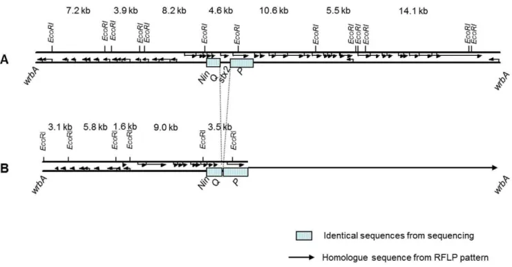
isolates (Figure 5). The integration site for thestx-negative phage waswrbA, which is the same site as the Stx2 phage [26].
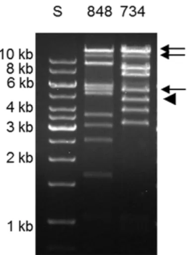
Hybridization located stx2 to a 4.5 kb EcoRI fragment not
present in the stx-negative phage RFLP (Figure 5). Still, RFLP analysis revealed three bands of about 14, 11 and 5.5 kb that were common between thestx2-positive and -negative phages (Figure 5).
The estimated sizes of the three bands correspond to the three largeEcoRI fragments comprising the late region of the sequenced Stx2 phage NOS (Figure 3), indicating that the late region of the two phages are homologue sequences.
To sequence the early region of the stx-negative phage, two fragments of 3.2 and 1.6 kb (Figure 5) were cloned and sequenced. Primer walking was used to amplify PCR products from the stx -negative phage DNA to complete the sequence. The 23 kb sequence of the early region of thestx-negative phage is not related to the Stx2 phage from O103:H25 NOS and a schematic view of the two phages is shown in Figure 3. The sequence of thestx-negative phage revealed an
AvrII restriction site not present in the Stx2 phage genome. The early phage regions of the two phages were blasted against each other and a Dot matrix view is shown in Figure 6.
Contigs from the EHEC O103:H25 NOS genome did not contain the sequence of thestx-negative phage, and also PCR run on total DNA from EHEC O103:H25 NOS with primers specific for the early region of thestx-negative phage were negative or gave products of incorrect size.
Cloning of phage DNA
An attempt to isolate and clone phage DNA directly from EHEC O103:H25 NOS resulted in the discovery of another phage in this strain. This phage is 45 kb, has a G+C content of 47% and the sequence is 53% identical to bacteriophageWV10 which is a temperate phage that specifically infects E. coli serogroup O157:H7 [20]. The insertion site of the phage is within theguaA
gene, and the phage does not infectE. coliDH5a.
Colicin and plasmids
Colicin from EHEC O103:H25 NOS in culture supernatants was phenotypically observed as clear zones in anE. coliDH5acell lawn.In silicoanalysis of the sequenced genome demonstrated the presence of a colicin E2 gene and its associated immunity and lysis genes on a 6744 bp contig. A gap-closure PCR confirmed the contig to be a complete plasmid. Hybridization with acolE2 pcr template identified the presence of the colicin E2 carrying plasmid in outbreak-associated O103:H25 isolates from both patients and food, including the isolates from 2003 and 2005, while non-outbreak O103:H25 isolates associated with Norwegian sheep did not harbour the plasmid. The entire colicin plasmid was 97% identical to pO111_4 fromE. coliO111:H- str 11128 [7].
EHEC O103:H25 NOS and E. coli O103:H25 NVH-848 share identical plasmid profiles and harbour one large plasmid of approximately the same size as pO157 ofE. coliO157:H7 EDL 933 (Figure 7).
Discussion
sporadic disease cases [21,22]. A fewE. coliO103:H25 strains have been characterized and found to carrystx1, but they have not been
associated with severe disease [18]. The Norwegian 2006 outbreak caused by EHEC O103:H25 had a 60% HUS frequency, and the strain is considered to be particularly virulent [11].
We have shown that the genome of EHEC O103:H25 NOS resemble the EAEC O104:H4 GOS and EHEC O103:H2 str 12009. This is supported in a pan genomic study of 61 sequenced
E. coli genomes, showing that theE. coli O103 Oslo (O103:H25 NOS) clusters closely together with the EHEC O103:H2 str 12009 [23], and a more recent study shows the similarities between the EHEC O103:H2 str 12009 and the EAEC O104:H4 GOS genomes [2]. EAEC O104:H4 GOS caused diarrhoea in approximately 4000 individuals, 22% of which developed HUS, and the strain is notably more virulent than most EHEC [2]. The
E. coli O104:H4 outbreak began in Germany in May 2011, but was later identified in other European countries [2,24]. Due to phenotypic and genotypic characteristics, the German O104:H4 outbreak strain is not classified as an EHEC, but rather as a Shiga toxin producing enteroaggregativeE. coli(EAEC) [2,3,25]. Despite the lack of genes characteristic of EAEC in EHEC O103:H25 NOS, and thus differing in both pathotype and serotype, the genomes of the Norwegian and the German outbreak strains are highly similar, as illustrated in Figures 1A and 1B. The close relationship between the two strains is supported by MLST analysis (Figure 1C). Also the Stx2 phages in these two strains show a striking homology with a DNA sequence identity of 90% (Figure 4) [2]. The identity includes a 1 bp silent nucleotide mutation in thestx2Agene [3,26] which is rare in otherstx2genes.
This indicates a common origin for the two phages. The finding of closely related Stx2 phages and genomes in two outbreak strains of
different serotypes and pathotypes, but with a high HUS incidence in common, is remarkable and will be investigated further.
Other strains with related Stx2 phages include EHEC O103:H2 str 12009 from a sporadic case of diarrhoea in Japan in 2001, and
E. coliO111:H- str 11128. Similar Stx2 phages to the Norwegian outbreak strain are thus present inE. coli strains of serogroups O103, O104 and O111, and the phage seems to be rather promiscuous in nature. The similarity between the Stx2 phage of EHEC O103:H25 NOS and the reference Stx2 phage 933W from O157:H7 EDL933 is high in the 38 kb late region of the phages where thestxgenes are located (95%), however, the early regions of these phages differ in composition (Figure 4). In EHEC O103:H25 NOS the Stx2 phage is inserted intowrbA, a previously described integration site of Stx2 phages, e.g. in EHEC O157:H7 EDL933 and Sakai strains [27,28]. The wrbA had not been observed as an integration site in serogroup O103 prior to the Norwegian outbreak [26]. The closely related Stx2 phage in O104:H4 GOS is also inserted inwrbA, while the Stx2 phage in EHEC O103:H2 str 12009 is located within theargWgene.
The only observed feature distinguishing between the Norwe-gian outbreak strain and stx-negative isolates from the 2006 outbreak is the presence of either the stx2-positive or the stx
-negative phage, respectively. These two phages are related and share parts of their sequences and insertion site. While the 30 kb late regions are similar in the two phages, the early regions are completely different. Interestingly, the shift between the similar and dissimilar parts is abrupt and in proximity of thestx genes (Figure 3). As Stx2 phages are mosaic by nature and rearrange-ments are not uncommon [6], thestx-negative phage could have developed from the Stx2 phage by acquiring its distinctive sequence from the chromosome of the E. coli O103:H25 host.
Table 1.Bacterial isolates included in the study.
Strain Synonym Year Stx Source Origin
NVH-734a NIPH-11060424 2006 stx
2 Patient no 2 Norway
NVH-847 NIPH-11060708 2006 stx2 Patient no 9 Norway
NVH-848 NIPH-11060707 2006 - Patient no 9 Norway
NVH-849 NIPH-11060747 2006 - Patient no 10 Norway
NVH-760 625/06 2006 - Fermented sausage, home of patient 7 Norway
NVH-737 2006 - Fermented sausage Norway
NVH-763 2006 - Food Norway
NVH-661 NIPH-10306923 2003 stx2 Patient Norway
NVH-731 NIPH-11051601 2005 stx2 Patient Norway
cdc-08-201 Not known Maine, USA
cdc-08-202 Not known Virginia, USA
All isolates areE. coliO103:H25.
aReferred to as Norwegian outbreak strain (NOS). doi:10.1371/journal.pone.0031413.t001
Figure 1. Genome comparisons of EHEC O103:H25 NOS and relatedE. colistrains.A. BRIG blast atlas of EHEC O103:H25 NOS compared to EAEC O104:H4 GOS, EHEC O103:H2 str 12009, EAEC O104:H4 str 55989, EHEC O111:H- str 11128, EHEC O157:H7 EDL933 and EHEC O26:H11 str 11368. The white regions represent absent genetic regions. B. Whole genome alignment of EHEC O103:H25 NOS, EAEC O104:H4 GOS1, EHEC O103:H2 str 12009, EAEC O104:H4 str 55989, EHEC O111:H- str 11128, EHEC O157:H7 EDL933 and EHEC O26:H11 str 11368 (top to bottom) genomes using Mauve [15]. Each chromosome has been laid out horizontally and homologous blocks in each genome are shown as identically colored regions linked across genomes. C. Phylogenetic analysis of concatenated MLST gene alleles (adk,fumC,gyrB,icd,mdh,purA,recA) of EHEC O103:H25 NOS (ST2523), EAEC O104:H4 GOS (ST678), EAEC O104:H4 str 55989 (ST678), EHEC O103:H2 str 12009 (ST17), EHEC O26:H11 str 11368 (ST21), EHEC O111:H- str 11128 (ST16), EHEC O157:H7 EDL933 (ST11) and EHEC O128:H2 str 3171/00 (ST25) obtained from GenBank.
However, the distinctivestx-negative phage sequence has not been identified in the EHEC O103:H25 NOS genome neither by in silicoanalysis nor by PCR. This indicates that this phage, or at least part of it, has another origin. The distinctive sequence of thestx -negative phage shows some similarities to phage related sequences in a BLAST search, but it seems to have a rather unique construction.
The 2006 EHEC O103:H25 outbreak is remarkable because all food isolates werestx-negative, and only two of 11 isolates from patients werestx2-postive [11,13]. The lack ofstxgenes in EHEC
serotypes isolated from patients is not uncommon and has been reported in both O157 and non-O157 isolates [12,29]. In studies where the mechanism ofstxgene loss has been investigated, it has been shown that the stx-negative strains lack the entire Stx-encoding bacteriophage. This has been demonstrated by the presence or reappearance of an intact integration site for the Stx phage and an altered PFGE pattern [29–32]. Such stx-negative isolates from patients are believed to be progenies of an EHEC that lost thestxgenes during the course of illness, and might be referred to as EHEC-LST (lost Shiga toxin) [12]. The EHEC-LST model is supported by the finding that EHEC are difficult to isolate from patients late in illness [33], and the theory is further confirmed by Mellmann and Karch who demonstrated the presence of stx-negative strains of O26:H11/NM and sorbitol fermenting (SF) O157:NM subsequent to stx2-positive isogenic
isolates in the same patients [12,34,35].
In contrast to the EHEC-LST phenomenon, we find that the integration site of the Stx2 phage is occupied in the stx-negative isolates by a partly related phage but withoutstxgenes. The stx -negative isolates thus differ from thestx2-positive isolates not only
by the lack of the Stx2 phage but also by the presence of thisstx -negative phage. The similar size and the lack of anXbaI restriction site in both the Stx2 phage and thestx-negative phage explain the finding of identicalXbaI digested PFGE profiles of thestx2-positive
andstx-negative isolates shown by Sekse et al. [13]. Digestion with
AvrII, however, revealed a difference in PFGE pattern, and sequencing of the two phage genomes identified an AvrII restriction site in the Stx2 phage which is not present in thestx -negative phage and that most likely explains the observed difference.
Only two stx2-positive patient isolates were retrieved in 2006,
and as nostx2-positive isolates were retrieved from food, it could be
speculated that thestx-negativeE. coliacquired the Stx2 phage in the patients’ gut. However, the Stx2 phages from the two patient isolates have identical RFLP pattern and a rare silent nucleotide mutation instxA, which both are found in two EHEC O103:H25 isolates from sporadic cases in Norway in 2003 and 2005 [26]. This strongly indicates that the Stx2 phages in these four isolates are epidemiologically linked and that thestx2-positive isolates from
2006 originates from the same source.
The peculiar circumstance is that both stx2-positive and stx
-negative O103:H25 E. coli cells must have been present in the contaminated fermented sausage in 2006. Which of the two variants is the ancestor is difficult to predict, but there is reason to believe that thestx2-positive clone preceded thestx-negative clone
because the related Stx2 phage was identified in the sameE. coli
serotype three years prior to the 2006 outbreak. On the other hand,stx-negative isolates could have been overlooked in earlier cases, and the origin and history of this clone is difficult to evaluate. The hypothesis that bacterial cells withstx-negative or Table 2.Characteristics of EHEC O103:H25 NOS, the related strains O104:H4 GOS and O103:H2 str 12009, and the EHEC reference strain O157:H7 EDL933.
Characteristics O157:H7 EDL933
O103:H25
NOS O104:H4 GOS
O103:H2 str 12009
Outbreak
Year of isolation 1982 2006 2011 2001
Country USA Norway Germany Japan
Pathotype EHEC EHEC EAEC EHEC
No of diseased 47 16 4075 1
No of HUS (%) 0 10 (62.5%) 908 (22%) 0
No of deaths 1 50 0
Stx2 phage
Insertion site wrbA wrbA wrbA argW
Stx2A, nucleotide position 867 T C C T
LEE
LEE operons five five none five
Intimin type gamma theta not present epsilon
OI122
sen (Shet2) yes yes no yes
pagC yes yes no yes
efa1/lifA 2.1 kb 9.7 kb not present 9.7 kb
Accessory virulence
set1A (shet1) no no yes no
Colicin E2
ehxA yes yes not present yes
stx2-positive phage were both present in the food is strengthened
by the finding of Sekse et al. [26]. They were not able to isolate infective phage particles from food samples in the 2006 outbreak, butstx2was detected in food samples by PCR. Although the food
most likely contained a mixture of stx2-positive and stx-negative
bacterial cells, the two clones may not have been present in equal numbers. As only thestx-negative clone is isolated from food the proportion ofstx2-positiveE. coliwas probably considerably lower.
In the diseased patients, this imbalance might have been reversed during the course of illness. All investigated non-stx2isolates from
patients have been shown to be the samestx-negative clone as is isolated from food, and not offspring from thestx2-positive clone in
form of EHEC-LST.
Several factors may contribute to the virulence of EHEC including factors within the individual (age, microbiota, number of Gb3 receptors), the bacterial host (intimin type), the toxin itself (subtype, synergy, Stx toxin levels), and other virulence factors (e.g. subtilase cytotoxin) [4,18,36–38]. The pathogenicity island locus of enterocyte effacement (LEE) is associated with virulence, and encodes the intimin gene (eae), the translocated intimin receptor (Tir) and a type III secretion system (TTSS). These are all involved in the intimate attachment of EHEC to enterocytes [39]. All five LEE operons are present in EHEC O103:H25 NOS, however, LEE is not part of the EAEC O104:H4 GOS genome. Another genomic island present in the 2006 outbreak strain is the putative
pathogenicity island OI122 with the genessen,pagCandefa1which have been strongly correlated with virulence and disease severity [18,40]. The complete efa1/lifA gene is 9672 kb and is a bifunctional protein for adherence and inhibition of lymphocyte activation [19]. The distribution of this large toxin is limited to less than 30 strains of heterologous serogroups (BLAST search), only one of the sequenced O157:H7 strains and two O157:NM strains exhibit it [41,42]. EAEC O104:H4 GOS does not exhibit any version of efa/lifA, while O103:H2 str 12009 exhibits the large version ofefa/lifA. Enterohemolysin (Ehx) is regarded a virulence factor of EHEC and the genes are located on large plasmids like pO157 [43–45]. One large plasmid is detected in O103:H25 NOS, in contrast to O104:H4 GOS which carries two large plasmids [25].
The colicin production ofE. coliO103:H25 may have provided an advantage for the bacterial cells in the harsh competition of the gut, making the colonization more efficient. In addition, the colicin E2 is a DNAse colicin which has been found to increase thein vitro
production of Stx in EHEC cells exposed to it [46]. It is however unlikely that colicin E2 can affect the production of Stx when the genes coexist in the same cell, but it has been shown that intestinal
E. colicells can act as chaperones and contribute to the production of Stx [47] and the colicin could possibly play a role here. Similar colicin plasmids are found in EHEC O26:H11 and EHEC O111:H- [7], and in a study by Karama et al. [48], 38.4% ofE. coli
O103:H2 strains were found to have a colicin producing phenotype.
The phi-like phage isolated from the wild-type EHEC O103:H25 NOS has an unknown function in the E. coli
O103:H25 host. However, the number of the phi-like phage particles released from EHEC O103:H25 NOS after induction with Mitomycin C is estimated to be approximately ten times the number of Stx2 phage particles released (data not shown), and hence this abundant phi-like phage is a practical challenge as it complicates the isolation of the Stx2 phage andstx-negative phage directly from wild types.
Conclusion
Both the Stx2 phage and the bacterial genome from EHEC O103:H25 NOS are related to the Shiga toxin producing EAEC O104:H4 that caused a large EuropeanE. colioutbreak in 2011. Two patient isolates from the Norwegian O103:H25 outbreak carry an Stx2 phage, while other outbreak associated isolates carry a related phage in the same insertion site. The two variants,E. coli
O103:H25 with the Stx2 phage or the stx-negative phage, have probably both been present in the contaminated food which caused the Norwegian outbreak.
Materials and Methods
Bacterial isolates
The E. coli O103:H25 isolates included in the study are presented in Table 1. NVH-734 is the outbreak reference strain and is also referred to as the Norwegian outbreak strain (NOS).E. coliDH5awas used as recipient strain in the plaque assay.E. coli strains were cultured in Luria-Bertani (LB) broth or on LB agar plates (LB containing 1% agar).
Whole genome sequencing, alignments and Multilocus sequence typing (MLST)
Isolate NVH-734 (EHEC O103:H25 NOS) was sequenced using 454 technology (454 Life Sciences, Branford, Connecticut, USA). Initial genome analysis revealed similarity to the E.coli
O103:H2 str 12009 (accession: AP010958.1), and this strain was
Figure 2. PFGE of outbreak associated isolates.PFGE ofE. coli O103:H25 NOS (lane 2), 847 (lane 3), 848 (lane 4), and NVH-760 (lane 5). Lambda ladder is used as marker (lane 1). Digestion with XbaI (A) showed indistinguishable PFGE patterns, while digestion with AvrII (B) exposed a difference between thestx2-positive and thestx
-negative isolates.
used as template for the alignment of contigs from the EHEC O103:H25 Norwegian Outbreak Strain (NOS) using Mauve [15]. Subsequently, genome alignment of EHEC O103:H25 NOS and the EAEC O104:H4 str German Outbreak Strain (GOS)1 (distributed on 208 contigs, accessions: AFWO01000001.1-AFWO01000208.1) was also carried out with Mauve, using the progressive alignment option. Whole genome BLAST-comparison was performed using the BRIG software package [49] on the following genomes: EHEC O103:H25 NOS, EAEC O104:H4 GOS1, EHEC O103:H2 str 12009, EAEC O104:H4 str 55989, EHEC O111:H- str 11128, EHEC O157:H7 EDL933 and EHEC O26:H11 str 11368. MLST was performed according to Wirth et al. [50], using the seven housekeeping genesadk,fumC, gyrB, icd,
mdh,purA, andrecA. A maximum likelihood test using PhyML [51] was carried out to assess the best nucleotide substitution matrix in R (http://www.R-project.org/) with the package ‘ape’ [52]. Based on this, Tamura-Nei with invariant sites was shown to be the best model, and subsequently MEGA 5 [53] was used to generate a maximum likelihood tree, which was bootstrapped 500 times. Sequence type (ST) of EHEC O103:H25 NOS was obtained from MLST Databases at the ERI, University College Cork (http:// mlst.ucc.ie). The O103:H25 NOS, O104:H4 str C227-11 (originating from the German outbreak), O103:H2 str 12009 and O157:H7 EDL933 phages were BLASTed against each other and compared using the ACT tool [54].
PCR and sequencing
Primers for gap-closure PCR for completing the sequence of the Stx2 phage, and for detection of genes in other isolates than EHEC O103:H25 NOS were designed on the basis of the genome sequence. All PCRs were carried out in an Eppendorf Mastercy-cler gradient (Eppendorf AG, Hamburg, Germany). DyNAzyme II DNA polymerase (supplied with 106buffer) and dNTP Mix from Finnzymes (Vantaa, Finland) were used as instructed by the
Figure 3. Comparison of Stx2 phage andstx-negative phage.Comparison of Stx2 phage andstx-negative phage from EHEC O103:H25 NOS (A) andE. coliO103:H25 NVH-848 (B), respectively. The Stx2 phage is sequenced, while the illustration of thestx-negative phage is based on sequence (23 kb) and RFLP pattern (illustrated by arrow).
doi:10.1371/journal.pone.0031413.g003
Figure 4. Stx2 phage from EHEC O103:H25 NOS compared to Stx2 phages from EAEC O104:H4, EHEC O103:H2 and EHEC O157:H7.ACT visualization betweenstx2-positive phages from EHEC
O103:H25 NOS, EAEC O104:H4 str C227-11, EHEC O103:H2 str 12009 and reference Stx2 phage 933W from EHEC O157:H7 EDL933. The phage genomes are compared using BLAST and the red regions represent hits. The white regions indicate absent genetic regions, which is especially noticeable in the comparison between the O103:H2 phage and the O157:H7 phage.
manufacturer. The standard program was as follows: 95uC for 1 min, 30 cycles of 95uC for 1 min, 52uC for 1 min and 72uC for 1 min, and finally 72uC for 5 min. Sequencing of PCR products was performed by Source BioScience geneservice (United Kingdom), and DNA sequences were analyzed using Vector NTI Advance 11 (Invitrogen, Carlsbad, USA) and BLAST. Primers used for PCR detection of other virulence genes are listed in Table 3.
Plaque assay
E. coli O103:H25 LB broth cultures were incubated at 37uC with shaking at 200 rpm to an OD600 of 0.3–0.5 (,2 h). To
induce phage production Mitomycin C (Sigma-Aldrich, St. Louis, MO, USA) was added to a final concentration of 0.5mg/ml and incubation was continued overnight in the dark. The cultures were centrifuged (20006g, 10 min) to remove bacterial cells and debris,
and the supernatants (phage lysates) were sterile filtered (0.22mm;
Minisart, Sartorius Stedim Biotech). As the wildtype strains also produced colicin (see below) that lysed the DH5acell lawn, lysates were treated with 100mg/ml trypsin (Sigma) at 37uC for
60 minutes prior to use to destroy colicin. The E. coli DH5a recipient was grown in LB broth to OD 0.4–0.6 at 37uC and shaking at 200 rpm. For determination of infectious phage particles, 100ml of tenfold dilutions of phage lysates were added
900mlE. coliDH5a. CaCl2was added to a final concentration of
10 mM. The phage-recipient mixture was incubated for 30 min at 37uC before 2.5 ml molten soft agar (0.7% LB broth) was added and the mixture poured onto LB agar plates with CaCl2(10 mM).
The plates were incubated at 37uC overnight, plaques were counted by visual examination and phage titres were calculated.
Isolation and RFLP analysis of phage DNA
To ensure that DNA was isolated from only the Stx2- or Stx-related phage, DNA extraction was performed via the plaque assay. This was necessary because an abundant phi like phage is also present in the wild type strains, but this phage is not able to infectE. coliDH5a. Phage isolation was done from seven strains from the 2006 outbreak, in which five were stx-negative strains from either food or patients, and two were stx2-positive isolates
from patients. The isolates are listed in Table 1. Briefly, 0.1 ml from an overnight starter culture ofE. coliDH5awas transferred to 10 ml LB- broth and incubated at 37uC with shaking until bacterial growth reached mid log phase. Approximately 50 plaque were picked (from the plaque assay described above), dissolved in 50ml MQ, and added to 0.5 ml of the E. coli DH5a culture together with 12.5ml 1 M CaCl2 to facilitate bacteriophage
infection. The culture was incubated at 37uC for two hours before 9.5 ml of LB-broth was added and then incubated at 37uC overnight. The overnight culture was centrifuged at 20006gfor 10 minutes and the supernatant containing phage particles was sterile filtered (0.22mm, Minisart, Sartorius Stedim Biotech, Aubagne, France), precipitated with 0.186PEG 8000/NaCl at
Figure 6. Early region of Stx2 phage compared to early region ofstx-negative phage.Dot matrix view of 24 kb early region of the Stx2 phage from EHEC O103:H25 NOS and the 23 kb early region of the stx-negative phage fromE. coliO103:H25 isolate NVH-848. The position of the stx2 gene in EHEC O103:H25 NOS is marked (o). Regions of
similarity are based upon the BLAST results. Alignments are shown in the plot as lines. Plus strand and protein matches are slanted from the bottom left to the upper right corner, minus strand matches are slanted from the upper left to the lower right. The number of lines shown in the plot is the same as the number of alignments found by BLAST. doi:10.1371/journal.pone.0031413.g006
Figure 7. Plasmid profiles ofE. coliO103:H25.Comparison of large plasmids in EHEC O103:H25 NOS (lane 4) andE. coliO103:H25 NVH-848 (lane 5) with EHEC O157:H7 EDL 933 (lane 3). GeneRuler 1 kb ladder (lane 2) and Lambda ladder (lane 1) were used as molecular size marker. doi:10.1371/journal.pone.0031413.g007
Figure 5. RFLP of phage from stx2-positive and stx-negative
isolates.EcoRI restriction fragment length polymorphism analysis of Stx2 phage andstx-negative phage in EHEC O103:H25 NOS andE. coli O103:H25 NVH-848, respectively. The arrow indicates the EcoRI fragments that are indistinguishable between the strains, arrowhead indicates theEcoRI fragment ofE. coliO103:H25 NOS where thestx2
gene is located.
4uC for 2 hours, and centrifuged at 10 0006gfor 1 hours. The pellet
was dissolved in 0.5 ml TE buffer with proteinase K (Sigma-Aldrich, 50 mg/ml) and SDS (final concentration of 0.5%) and incubated for one hour at 56uC. The DNA was extracted using phenol/chloroform/ isoamylalcohol (25:24:1), and the DNA was precipitated using equal amounts of isopropanol. Phage DNA was used as template in restriction fragment length polymorphism (RFLP) analysis and in PCR reactions. Phage DNA was digested with restriction enzymesEcoRI (New England BioLabs, Hertfordshire, England) and the restriction fragments separated by 1% agarose gels. A probe for detection ofstx2A
in RFLP hybridization was made using primers listed in Table 3. To complete the phage genomes of the stx2-positive and stx-negative
phages, phage DNA from O103:H25 NOS and NVH-848, respectively, were used as templates in PCR reactions.
Colicin production
Colicin production was detected by observation of a lytic effect of sterile filtered supernatant from overnight culture of EHEC O103:H25 NOS onE. coliDH5 cell lawns. The identification of a colicin E2 encoding gene was performed byin silicoanalysis of the genome sequence, and gap closure PCR was performed to confirm a plasmid configuration of the contig. PrimerscolE2F andcolE2R (Table 3) and standard PCR conditions were used to generate a probe for plasmid hybridization.
Pulsed-Field Gel Electrophoresis (PFGE) and plasmid isolation
TheE. coliisolates associated with the outbreak were analyzed by PFGE as described previously [13] using the restriction enzymes XbaI and AvrII (New England BioLabs). DNA for detection of colicin E2 encoding plasmids were isolated by Qiagen plasmid purification kit (Qiagen, Hilden, Germany), and separated by electrophoresis in 2% agarose gels. Large plasmids were isolated by the method of Kado and Liu [55] with modifications. Bacteria were grown in 5 ml LB broth (Oxoid) over night at 37uC, 250 rpm of which 1.5 ml were centrifuged. The pellet was suspended in 200 ml cold (4uC) TAE-buffer (40 mM Tris-acetate, 2 mM EDTA, pH 7.6) and 400ml 50 mM Tris/3% SDS,
pH 12.55 were added prior to incubation at 55uC for 60 min.
Plasmid DNA was extracted twice with phenol-chloroform (1:1, vol/vol) and 25ml were applied directly to 0.7% agarose gel.
Plasmids were separated by electrophoresis at 120 V for 3 h at 4uC.
Southern blotting and hybridization
Plasmid DNA and digested phage DNA were transferred to nylon membranes (Hybond-N, Amersham International plc, Amersham, United Kingdom) by Southern blotting [56]. For detection of the colicin E2 encoding gene and phage genes probes were labelled with digoxigenin (DIG) and hybridized with a DIG DNA labelling and detection kit (Roche Diagnostics, Basel, Switzerland) according to the instructions by the manufacturer.
Cloning of phage DNA
Phage DNA isolated directly from EHEC O103:H25 NOS culture supernatants was digested with EcoRI and cloned in pUC18, and the clones were subsequently sequenced.
GenBank accession numbers
The genome sequence of EHEC O103:H25 NOS has been deposited at DDBJ/EMBL/GenBank under the accession no AGSG00000000. The version described in this paper is the first version, AGSG01000000. The 61 kb Stx2 phage genome has accession no JQ011318 and the 24 kb early region of the stx -negative phage has accession no JQ011316. The 45 kb phi-like phage has accession no JQ011317.
Acknowledgments
We thank Evangeline Sowers at the Centers for Disease Control and Prevention (USA) for providing control isolates.
Author Contributions
Conceived and designed the experiments: TMLL HJJ JB PEG TL. Performed the experiments: TMLL HJJ JB KOS GL TL. Analyzed the data: TMLL HJJ KOS JB GL TL. Contributed reagents/materials/ analysis tools: TMLL HJJ KOS GL TL. Wrote the paper: TMLL HJJ JB TL.
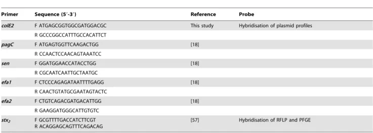
Table 3.Primers used in the study.
Primer Sequence (59-39) Reference Probe
colE2 F ATGAGCGGTGGCGATGGACGC This study Hybridisation of plasmid profiles
R GCCCGGCCATTTGCCACATTCT
pagC F ATGAGTGGTTCAAGACTGG [18]
R CCAACTCCAACAGTAAATCC
sen F GGATGGAACCATACCTGG [18]
R CGCAATCAATTGCTAATGC
efa1 F CTCCCAGAGATAATTTTGAGG [18]
R CAACTGTATGCGAATAGTACTC
efa2 F CTGTCAGACGATGACATTGG [18]
R GAAGGATGGGCATTGTGTC
stx2 F GCGTTTTGACCATCTTCGT R ACAGGAGCAGTTTCAGACAG
[57] Hybridisation of RFLP and PFGE
References
1. Gyles CL (2007) Shiga toxin-producingEscherichia coli:an overview. J Anim Sci 85: E45–E62. jas.2006-508 [pii];10.2527/jas.2006-508 [doi].
2. Rasko DA, Webster DR, Sahl JW, Bashir A, Boisen N, et al. (2011) Origins of the E. coli strain causing an outbreak of hemolytic-uremic syndrome in Germany. N Engl J Med 365: 709–717. 10.1056/NEJMoa1106920 [doi]. 3. Bielaszewska M, Mellmann A, Zhang W, Kock R, Fruth A, et al. (2011)
Characterisation of theEscherichia coli strain associated with an outbreak of haemolytic uraemic syndrome in Germany, 2011: a microbiological study. Lancet Infect Dis 11: 671–676. S1473-3099(11)70165-7 [pii];10.1016/S1473-3099(11)70165-7 [doi].
4. Fuller CA, Pellino CA, Flagler MJ, Strasser JE, Weiss AA (2011) Shiga toxin subtypes display dramatic differences in potency. Infect Immun 79: 1329–1337. IAI.01182-10 [pii];10.1128/IAI.01182-10 [doi].
5. Fogg PC, Gossage SM, Smith DL, Saunders JR, McCarthy AJ, et al. (2007) Identification of multiple integration sites for Stx-phage Phi24B in theEscherichia coligenome, description of a novel integrase and evidence for a functional anti-repressor. Microbiology 153: 4098–4110.
6. Johansen BK, Wasteson Y, Granum PE, Brynestad S (2001) Mosaic structure of Shiga-toxin-2-encoding phages isolated fromEscherichia coliO157:H7 indicates frequent gene exchange between lambdoid phage genomes. Microbiology 147: 1929–1936.
7. Ogura Y, Ooka T, Iguchi A, Toh H, Asadulghani M, et al. (2009) Comparative genomics reveal the mechanism of the parallel evolution of O157 and non-O157 enterohemorrhagicEscherichia coli. Proc Natl Acad Sci U S A 106: 17939–17944. 8. Gamage SD, Patton AK, Hanson JF, Weiss AA (2004) Diversity and host range of Shiga toxin-encoding phage. Infect Immun 72: 7131–7139. 72/12/7131 [pii];10.1128/IAI.72.12.7131-7139.2004 [doi].
9. Johnson AD, Poteete AR, Lauer G, Sauer RT, Ackers GK, et al. (1981) lambda repressor and cro-components of an efficient molecular switch. Nature 294: 217–223.
10. Allison HE (2007) Stx-phages: drivers and mediators of the evolution of STEC and STEC-like pathogens. Future Microbiol 2: 165–174. 10.2217/ 17460913.2.2.165 [doi].
11. Schimmer B, Nygard K, Eriksen HM, Lassen J, Lindstedt BA, et al. (2008) Outbreak of haemolytic uraemic syndrome in Norway caused bystx2-positive Escherichia coliO103:H25 traced to cured mutton sausages. BMC Infect Dis 8: 41.
12. Mellmann A, Bielaszewska M, Zimmerhackl LB, Prager R, Harmsen D, et al. (2005) EnterohemorrhagicEscherichia coliin human infection:in vivoevolution of a bacterial pathogen. Clin Infect Dis 41: 785–792. CID36325 [pii];10.1086/ 432722 [doi].
13. Sekse C, O’Sullivan K, Granum PE, Rørvik LM, Wasteson Y, et al. (2009) An outbreak of Escherichia coli O103:H25 - Bacteriological investigations and genotyping of isolates from food. Int J Food Microbiol 133: 259–264. 14. Lindstedt BA, Brandal LT, Aas L, Vardund T, Kapperud G (2007) Study of
polymorphic variable-number of tandem repeats loci in the ECOR collection and in a set of pathogenic Escherichia coliand Shigellaisolates for use in a genotyping assay. J Microbiol Methods 69: 197–205.
15. Darling AC, Mau B, Blattner FR, Perna NT (2004) Mauve: multiple alignment of conserved genomic sequence with rearrangements. Genome Res 14: 1394–1403. 10.1101/gr.2289704 [doi];14/7/1394 [pii].
16. Brzuszkiewicz E, Thurmer A, Schuldes J, Leimbach A, Liesegang H, et al. (2011) Genome sequence analyses of two isolates from the recentEscherichia coli
outbreak in Germany reveal the emergence of a new pathotype: Entero-Aggregative-HaemorrhagicEscherichia coli(EAHEC). Arch Microbiol. 10.1007/ s00203-011-0725-6 [doi].
17. McDaniel TK, Jarvis KG, Donnenberg MS, Kaper JB (1995) A genetic locus of enterocyte effacement conserved among diverse enterobacterial pathogens. Proc Natl Acad Sci U S A 92: 1664–1668.
18. Karmali MA, Mascarenhas M, Shen S, Ziebell K, Johnson S, et al. (2003) Association of genomic O island 122 of Escherichia coli EDL 933 with verocytotoxin-producing Escherichia coli seropathotypes that are linked to epidemic and/or serious disease. J Clin Microbiol 41: 4930–4940.
19. Klapproth JM, Scaletsky IC, McNamara BP, Lai LC, Malstrom C, et al. (2000) A large toxin from pathogenicEscherichia colistrains that inhibits lymphocyte activation. Infect Immun 68: 2148–2155.
20. Perry LL, SanMiguel P, Minocha U, Terekhov AI, Shroyer ML, et al. (2009) Sequence analysis of Escherichia coli O157:H7 bacteriophage PhiV10 and identification of a phage-encoded immunity protein that modifies the O157 antigen. FEMS Microbiol Lett 292: 182–186.
21. Mackenzie AM, Lebel P, Orrbine E, Rowe PC, Hyde L, et al. (1998) Sensitivities and specificities of premier E. coli O157 and premier EHEC enzyme immunoassays for diagnosis of infection with verotxin (Shiga-like toxin)-producingEscherichia coli. J Clin Microbiol 36: 1608–1611.
22. Rivas M, Miliwebsky E, Chinen I, Rolda´n CD, Balbi L, et al. (2006) Characterization and epidemiologic subtyping of Shiga toxin-producing
Escherichia colistrains isolated from hemolytic uremic syndrome and diarrhea cases in Argentina. Foodborne Pathog Dis 3: 88–96. 10.1089/fpd.2006.3.88 [doi].
23. Lukjancenko O, Wassenaar TM, Ussery DW (2010) Comparison of 61 sequenced Escherichia coli genomes. Microb Ecol 60: 708–720. 10.1007/ s00248-010-9717-3 [doi].
24. Wu CJ, Hsueh PR, Ko WC (2011) A new health threat in Europe: Shiga toxin-producing Escherichia coli O104:H4 infections. J Microbiol Immunol In-fectS1684–1182(11)00152-6 [pii];10.1016/j.jmii.2011.07.001 [doi].
25. Mellmann A, Harmsen D, Cummings CA, Zentz EB, Leopold SR, et al. (2011) Prospective genomic characterization of the German enterohemorrhagic
Escherichia coli O104:H4 outbreak by rapid next generation sequencing technology. PLoS One 6: e22751. 10.1371/journal.pone.0022751 [doi]; PONE-D-11-11826 [pii].
26. Sekse C, Muniesa M, Wasteson Y (2008) Conserved Stx2 phages fromEscherichia coliO103:H25 isolated from patients suffering from hemolytic uremic syndrome. Foodborne Pathog Dis 5: 801–810.
27. Makino K, Yokoyama K, Kubota Y, Yutsudo CH, Kimura S, et al. (1999) Complete nucleotide sequence of the prophage VT2-Sakai carrying the verotoxin 2 genes of the enterohemorrhagicEscherichia coliO157:H7 derived from the Sakai outbreak. Genes Genet Syst 74: 227–239.
28. Perna NT, Plunkett G, III, Burland V, Mau B, Glasner JD, et al. (2001) Genome sequence of enterohaemorrhagicEscherichia coliO157:H7. Nature 409: 529–533. 10.1038/35054089 [doi].
29. Bielaszewska M, Prager R, Ko¨ck R, Mellmann A, Zhang W, et al. (2007) Shiga toxin gene loss and transfer in vitro and in vivo during enterohemorrhagic
Escherichia coliO26 infection in humans. Appl Environ Microbiol 73: 3144–3150. 30. Bielaszewska M, Prager R, Zhang W, Friedrich AW, Mellmann A, et al. (2006) Chromosomal dynamism in progeny of outbreak-related sorbitol-fermenting enterohemorrhagic Escherichia coli O157:NM. Appl Environ Microbiol 72: 1900–1909. 72/3/1900 [pii];10.1128/AEM.72.3.1900-1909.2006 [doi]. 31. Feng P, Dey M, Abe A, Takeda T (2001) Isogenic strain ofEscherichia coli
O157:H7 that has lost both Shiga toxin 1 and 2 genes. Clin Diagn Lab Immunol 8: 711–717. 10.1128/CDLI.8.4.711-717.2001 [doi].
32. Murase T, Yamai S, Watanabe H (1999) Changes in pulsed-field gel electrophoresis patterns in clinical isolates of enterohemorrhagicEscherichia coli
O157:H7 associated with loss of Shiga toxin genes. Curr Microbiol 38: 48–50. 33. Tarr PI, Neill MA, Clausen CR, Watkins SL, Christie DL, et al. (1990)
Escherichia coliO157:H7 and the hemolytic uremic syndrome: importance of early cultures in establishing the etiology. J Infect Dis 162: 553–556. 34. Karch H, Mellmann A, Bielaszewska M (2009) Epidemiology and pathogenesis
of enterohaemorrhagicEscherichia coli. Berl Munch Tierarztl Wochenschr 122: 417–424.
35. Mellmann A, Lu S, Karch H, Xu JG, Harmsen D, et al. (2008) Recycling of Shiga toxin 2 genes in sorbitol-fermenting enterohemorrhagicEscherichia coli
O157:NM. Appl Environ Microbiol 74: 67–72.
36. de Sablet T, Bertin Y, Vareille M, Girardeau JP, Garrivier A, et al. (2008) Differential expression ofstx2variants in Shiga toxin-producingEscherichia coli belonging to seropathotypes A and C. Microbiology 154: 176–186. 154/1/176 [pii];10.1099/mic.0.2007/009704-0 [doi].
37. de Sablet T, Chassard C, Bernalier-Donadille A, Vareille M, Gobert AP, et al. (2009) Human microbiota-secreted factors inhibit shiga toxin synthesis by enterohemorrhagic Escherichia coli O157:H7. Infect Immun 77: 783–790. IAI.01048-08 [pii];10.1128/IAI.01048-08 [doi].
38. Paton AW, Paton JC (2010)Escherichia colisubtilase cytotoxin. Toxins (Basel) 2: 215–228.
39. Schmidt MA (2010) LEEways: tales of EPEC, ATEC and EHEC. Cell Microbiol 12: 1544–1552. CMI1518 [pii];10.1111/j.1462-5822.2010.01518.x [doi].
40. Wickham ME, Lupp C, Mascarenhas M, Va´zquez A, Coombes BK, et al. (2006) Bacterial genetic determinants of non-O157 STEC outbreaks and hemolytic-uremic syndrome after infection. J Infect Dis 194: 819–827. JID36198 [pii];10.1086/506620 [doi].
41. Bielaszewska M, Ko¨ck R, Friedrich AW, von Eiff C, Zimmerhackl LB, et al. (2007) Shiga toxin-mediated hemolytic uremic syndrome: time to change the diagnostic paradigm? PLoS One 2: e1024. 10.1371/journal.pone.0001024 [doi]. 42. Friedrich AW, Zhang W, Bielaszewska M, Mellmann A, Ko¨ck R, et al. (2007) Prevalence, virulence profiles, and clinical significance of Shiga toxin-negative variants of enterohemorrhagicEscherichia coliO157 infection in humans. Clin Infect Dis 45: 39–45. CID50097 [pii];10.1086/518573 [doi].
43. Caprioli A, Morabito S, Brugere H, Oswald E (2005) Enterohaemorrhagic
Escherichia coli: emerging issues on virulence and modes of transmission. Vet Res 36: 289–311. 10.1051/vetres:2005002 [doi];v4055 [pii].
44. Schmidt H, Beutin L, Karch H (1995) Molecular analysis of the plasmid-encoded hemolysin ofEscherichia coliO157:H7 strain EDL 933. Infect Immun 63: 1055–1061.
45. Schmidt H, Kernbach C, Karch H (1996) Analysis of the EHEChlyoperon and its location in the physical map of the large plasmid of enterohaemorrhagic
Escherichia coliO157:H7. Microbiology 142(Pt 4): 907–914.
46. Toshima H, Yoshimura A, Arikawa K, Hidaka A, Ogasawara J, et al. (2007) Enhancement of Shiga toxin production in enterohemorrhagicEscherichia coli
serotype O157:H7 by DNase colicins. Appl Environ Microbiol 73: 7582–7588. AEM.01326-07 [pii];10.1128/AEM.01326-07 [doi].
48. Karama M, Johnson RP, Holtslander R, Gyles CL (2008) Phenotypic and genotypic characterization of verotoxin-producing Escherichia coli O103:H2 isolates from cattle and humans. J Clin Microbiol 46: 3569–3575.
49. Alikhan NF, Petty NK, Ben Zakour NL, Beatson SA (2011) BLAST Ring Image Generator (BRIG): simple prokaryote genome comparisons. BMC Genomics 12: 402. 1471-2164-12-402 [pii];10.1186/1471-2164-12-402 [doi].
50. Wirth T, Falush D, Lan R, Colles F, Mensa P, et al. (2006) Sex and virulence in
Escherichia coli: an evolutionary perspective. Mol Microbiol 60: 1136–1151. MMI5172 [pii];10.1111/j.1365-2958.2006.05172.x [doi].
51. Guindon S, Delsuc F, Dufayard JF, Gascuel O (2009) Estimating maximum likelihood phylogenies with PhyML. Methods Mol Biol 537: 113–137. 10.1007/ 978-1-59745-251-9_6 [doi].
52. Paradis E, Claude J, Strimmer K (2004) APE: Analyses of phylogenetics and evolution in R language. Bioinformatics 20: 289–290.
53. Tamura K, Peterson D, Peterson N, Stecher G, Nei M, et al. (2011) MEGA5: molecular evolutionary genetics analysis using maximum likelihood, evolution-ary distance, and maximum parsimony methods. Mol Biol Evol 28: 2731–2739. msr121 [pii];10.1093/molbev/msr121 [doi].
54. Carver T, Berriman M, Tivey A, Patel C, Bohme U, et al. (2008) Artemis and ACT: viewing, annotating and comparing sequences stored in a relational database. Bioinformatics 24: 2672–2676. btn529 [pii];10.1093/bioinformatics/ btn529 [doi].
55. Kado CI, Liu ST (1981) Rapid procedure for detection and isolation of large and small plasmids. J Bacteriol 145: 1365–1373.
56. Sambrook J, Fritsch EF, Maniatis T (1989) Molecular cloning. A laboratory manual. New York: Cold Spring Harbor Laboratory Press.
57. Muniesa M, Jofre J (1998) Abundance in sewage of bacteriophages that infect