Gene Expression Profiling of Human Vaginal
Cells
In Vitro
Discriminates Compounds with
Pro-Inflammatory and Mucosa-Altering
Properties: Novel Biomarkers for Preclinical
Testing of HIV Microbicide Candidates
Irina A. Zalenskaya1*, Theresa Joseph1¤a, Jasmin Bavarva2¤b, Nazita Yousefieh1, Suzanne
S. Jackson1, Titilayo Fashemi3, Hidemi S. Yamamoto3, Robert Settlage2, Raina N. Fichorova3, Gustavo F. Doncel1*
1CONRAD, Department of Obstetrics and Gynecology, Eastern Virginia Medical School, Norfolk, Virginia, United States of America,2Virginia Bioinformatics Institute, Virginia Polytechnic Institute and State University, Blacksburg, Virginia, United States of America,3Laboratory of Genital Tract Biology, Department of Obstetrics, Gynecology and Reproductive Biology, Brigham and Women’s Hospital, Harvard Medical School, Boston, Massachusetts, United States of America
¤a Current address: Department of Pharmacology and Toxicology, Massey Cancer Center, Virginia Commonwealth University, Richmond, Virginia, United States of America
¤b Current address: Biospecimen Research Group, Leidos Biomedical Research, Inc. for the Frederick National Laboratory for Cancer Research, Rockville, Maryland, United States of America
*[email protected](IAZ); [email protected](GFD)
Abstract
Background
Inflammation and immune activation of the cervicovaginal mucosa are considered factors that increase susceptibility to HIV infection. Therefore, it is essential to screen candidate anti-HIV microbicides for potential mucosal immunomodulatory/inflammatory effects prior to further clinical development. The goal of this study was to develop anin vitromethod for pre-clinical evaluation of the inflammatory potential of new candidate microbicides using a mi-croarray gene expression profiling strategy.
Methods
To this end, we compared transcriptomes of human vaginal cells (Vk2/E6E7) treated with well-characterized pro-inflammatory (PIC) and non-inflammatory (NIC) compounds. PICs included compounds with different mechanisms of action. Gene expression was analyzed using Affymetrix U133 Plus 2 arrays. Data processing was performed using GeneSpring 11.5 (Agilent Technologies, Santa Clara, CA).
Results
Microarraray comparative analysis allowed us to generate a panel of 20 genes that were consistently deregulated by PICs compared to NICs, thus distinguishing between these two
OPEN ACCESS
Citation:Zalenskaya IA, Joseph T, Bavarva J, Yousefieh N, Jackson SS, Fashemi T, et al. (2015) Gene Expression Profiling of Human Vaginal CellsIn VitroDiscriminates Compounds with Pro-Inflammatory and Mucosa-Altering Properties: Novel Biomarkers for Preclinical Testing of HIV Microbicide Candidates. PLoS ONE 10(6): e0128557. doi:10.1371/journal.pone.0128557
Academic Editor:J. Gerardo Garcia-Lerma, Centers for Disease Control and Prevention, UNITED STATES
Received:March 6, 2015
Accepted:April 28, 2015
Published:June 8, 2015
Copyright:© 2015 Zalenskaya et al. This is an open access article distributed under the terms of the Creative Commons Attribution License, which permits unrestricted use, distribution, and reproduction in any medium, provided the original author and source are credited.
groups. Functional analysis mapped 14 of these genes to immune and inflammatory re-sponses. This was confirmed by the fact that PICs induced NFkB pathway activation in Vk2 cells. By testing microbicide candidates previously characterized in clinical trials we demon-strated that the selected PIC-associated genes properly identified compounds with muco-sa-altering effects. The discriminatory power of these genes was further demonstrated after culturing vaginal cells with vaginal bacteria.Prevotella bivia, prevalent bacteria in the dis-turbed microbiota of bacterial vaginosis, induced strong upregulation of seven selected PIC-associated genes, while a commensalLactobacillus gasseriassociated to vaginal health did not cause any changes.
Conclusions
In vitroevaluation of the immunoinflammatory potential of microbicides using the PIC-asso-ciated genes defined in this study could help in the initial screening of candidates prior to en-tering clinical trials. Additional characterization of these genes can provide further insight into the cervicovaginal immunoinflammatory and mucosal-altering processes that facilitate or limit HIV transmission with implications for the design of prevention strategies.
Introduction
According to the latest global estimates from UNAIDS, there were about 35 million people liv-ing with HIV in 2013 (UNAIDS. GAP report; 2014,www.unaids.org). During the same time, there were around 2.1 million new HIV infections, and 1.5 million people died of HIV-related causes. Women represent slightly more than half of the infected population worldwide and ap-proximately 60% in sub-Saharan Africa, where gender inequalities increase women’s vulnera-bility to HIV. In some regions young women and adolescent girls account for a
disproportionate number of new infections among young people and those living with HIV (UNAIDS. GAP report, 2014,www.unaids.org).
The main route of male-to-female HIV transmission is through the epithelium of the female genital tract exposed to virus-containing semen [1]. Vaginal topical antiviral microbicides rep-resent a novel female-controlled method of prevention of sexual transmission of HIV. During the past 20 years, numerous microbicide products have been developed, with a handful of them reaching advanced clinical trials [2–5]. Thus far, only the nucleotide reverse transcriptase inhibitor (NRTI), tenofovir, tested in the CAPRISA 004 study demonstrated a moderate reduc-tion in HIV acquisireduc-tion [6]. All other candidate microbicides were ineffective in preventing pri-mary HIV infection. Moreover, one of the first candidate microbicides, a non-ionic detergent nonoxynol-9 (N-9), increased the risk of HIV infection among frequent users. It was later dem-onstrated that this compound altered epithelial integrity and induced a mucosal immunoin-flammatory response leading to recruitment of immune cells, which are potential targets for HIV infection [7]. Two other microbicide candidates, C31G (Savvy) and cellulose sulfate (CS) which failed in phase III clinical trials also proved to alter the mucosal microenvironment [8,9].
Currently, there are multiple candidate microbicides in preclinical development and a hand-ful of products undergoing clinical studies [3,10]. The new candidates have diverse modes of action, including inhibition of HIV attachment, fusion and entry, replication, integration, and other less defined mechanisms. Preclinical studies of products with multiple mechanisms of
Funding:This work was supported by CONRAD funds from a cooperative agreement with USAID GPO-A-00-08-00005-00 with additional funds from an interagency agreement with NIH/NIAID YI-AI-1756-01 (GFD), and grant 41266 from the Bill and Melinda Gates Foundation(GFD). The views of the authors expressed in this manuscript do not necessarily reflect those of the funding agencies. The funders had no role in study design, data collection and analysis, decision to publish, or preparation of the manuscript. Leidos Biomedical Research, Inc. provided support in the form of salaries for author JB but did not have any additional role in the study design, data collection and analysis, decision to publish, or preparation of the manuscript.
action are also in progress [3,10]. Lessons learned from the failures of the first generation mi-crobicides underscore the need for early testing of candidate mimi-crobicides for their possible ad-verse effects on the cervicovaginal and rectal mucosae.
The healthy female genital epithelium presents a strong barrier to viral invasion. HIV trans-mission is estimated to be 1–2 cases per 1000 coital acts [11,12]. However, affected by complex host and viral factors, transmission rates can be much higher [11]. Increased HIV-1 cervicova-ginal transmission is strongly associated with inflammation and general immune activation of the cervicovaginal mucosa—conditions that stimulate influx of HIV target immune cells to the mucosal surface, increase the receptivity of these cells to HIV-1, and are often accompanied by epithelial lesions, which facilitate HIV-1 access to its target cells [13–24]. To avoid these ad-verse effects, the proinflammatory potential of microbicide candidates should be evaluated early during their primary screening [25,26]. Currently, cell-based, explant-based and animal-based models are used to define and preclinically assess potential biomarkers of inflammation, which include cytokines, chemokines and some other molecules [25,27–32]. Biomarker selec-tion in these assays relies on prior knowledge of their involvement in inflammatory responses.
In this study, we set out to identify novel biomarkers for preclinicalin vitroassessment of the pro-inflammatory/immunomodulatory potential of microbicides using microarray hybrid-ization technology (gene expression profiling). Our goal was to generate unique gene expres-sion profiles for compounds that were known to cause mucosal inflammatory/
immunomodulatory response in order to use these profiles for the characterization of microbi-cide candidates. To this end, transcriptomes of immortalized human vaginal epithelial (Vk2/ E6E7) cells exposed to well-known pro-inflammatory/immunomodulatory compounds (PIC) and non-inflammatory compounds (NIC) were compared. Using microarray technology we identified 20 genes that were consistently deregulated by treatment with PICs but not with NICs, thus presenting a signature specific to compounds with potential pro-inflammatory/ immunomodulatory effects on the cervicovaginal mucosa.
Materials and Methods
Materials
TLR ligands, Pam3CSK4 (Pam), imiquimod (IMQ) were purchased from Invivogen (San Diego, CA), macrophage activating lipopeptide 2 (MALP2) was purchased from Alexis Bio-chemicals (Enzo Life Sciences, Plymouth Meeting, PA). TNF-αwas purchased from R&D sys-tems (Minneapolis, MN). N-9 was a gift from OrthoMcNeil Corporation. Other compounds/ candidate microbicides were acquired as follows: hydroxyethyl cellulose (HEC)—from Hercu-les (Hopewell, VA), cellulose sulfate (CS) and dextran sulfate (DS)—from Dextran Products (Scarborough, Ontario), PRO2000—from Indevus Pharmaceuticals (Lexington, MA), tenofovir (TFV)—from Gilead (Foster City, CA), C31G from Biosyn Inc. (Huntington Valley, PA), UC-781—from Regis (Morton Grove, IL), emtricibine (FTC)—gift from Dr. Parang (Keykavous Parang, University of Rhode Island).
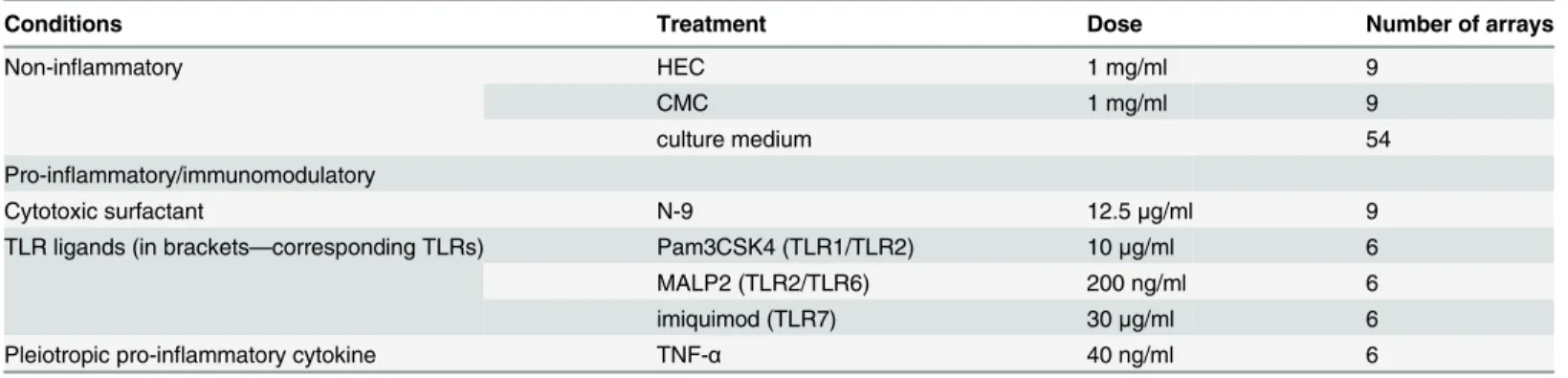
Doses of compounds (Table 1) have been defined in our previous studies such that their cy-totoxicity, if any, would not exceed 15% [33,34].
Cell culture, treatment with compounds
penicillin-streptomycin (1%), and CaCl2 (0.4 mM). Cells were grown to ~70–80% confluence and treated for 6 h with agents presented inTable 1. Antibiotics were omitted in bacterial colo-nization experiments. Cells were collected and RNA extracted for gene expression analysis as described below.
Bacterial colonization
Bacterial colonization of Vk2/E6E7 cells was performed in the Laboratory of Genital Tract Bi-ology, Brigham and Women’s Hospital (BWH) as described in detail elsewhere [41].
Lactobacillusisolate andPrevotella biviawere obtained from vaginal swabs and phenotypi-cally characterized by classic microbiology techniques as described before [42]. Because con-ventional methods of phenotyping cannot identifyLactobacillito the species level, sequencing of the 16S rRNA gene ofLactobacilluswas performed at the Center for Clinical and Transla-tional Metagenomics at BWH. The genetic analysis classified theLactobacillusisolate asL. gas-seriwith 99% identity. Vaginal epithelial cultures (Vk2/E6E7 cells) were colonized withL. gasseri, representing the Gram-positive facultative anaerobes typical for the normal vaginal flora [43–46], orPrevotella (P.) biviarepresenting the anaerobic Gram-negative rods associated with bacterial vaginosis (BV) [47–49].
RNA isolation
Prior to RNA isolation, Vk2 cells were rinsed three times with cold PBS. Total RNA was then extracted with Trizol (Invitrogen Life Technologies, Carlsbad, CA) and purified using RNeasy mini kit columns from Qiagen Sciences (Qiagen, Valencia, CA) according to the
manufacturer’s instructions.
The integrity of total RNA was qualified by Agilent Bioanalyzer 2100 capillary electrophore-sis and input amount quantified by Nanodrop ND-1000 Spectrophotometer.
Microarray expression profiling
Microarray mRNA expression profiling was performed by Asuragen, Inc. (Austin, TX). The mRNA was amplified into cRNA and biotin-labeled using modified MessageAmp-based proto-cols (Ambion Inc., Austin, TX). Labeled cRNA was fragmented, and hybridized to Affymetrix HG-U133 Plus 2.0 arrays (Affymetrix) according to the standard Affymetrix protocol. The U133 Plus 2.0 chip contains more than 56,000 probesets and includes 38,500 well characterized
Table 1. Compounds used for Vk2 cells genome profiling.
Conditions Treatment Dose Number of arrays
Non-inflammatory HEC 1 mg/ml 9
CMC 1 mg/ml 9
culture medium 54
Pro-inflammatory/immunomodulatory
Cytotoxic surfactant N-9 12.5μg/ml 9
TLR ligands (in brackets—corresponding TLRs) Pam3CSK4 (TLR1/TLR2) 10μg/ml 6
MALP2 (TLR2/TLR6) 200 ng/ml 6
imiquimod (TLR7) 30μg/ml 6
Pleiotropic pro-inflammatory cytokine TNF-α 40 ng/ml 6
Doses for candidate microbicides (and placebo) were: for CS, FTC, PRO-2000, TFV, DS, HEC—all 1mg/ml; for UC-781–10μg/ml, for N-9–12.5 -μg/ml,
and for C31G–6.125μg/ml.
human genes and expressed sequence tags. Affymetrix raw data were acquired using GeneChip operating software (GCOS 1.3) to yield CEL files.
Data normalization and statistical analysis
Data were processed and analyzed using GeneSpring 11.5 (Agilent Technologies, Santa Clara, CA). The background subtraction, normalization, and log base 2 transformation of gene signals were carried out using the Robust Multi-array Analysis (RMA) summarization algorithm [50]
For statistical analysis, one-way ANOVA was used for multiple group comparison, followed by multiple testing correction setting the false discovery rate (FDR) at 0.05 using the Benjamini and Hochberg method [51]. Genes/probesets with FDR corrected p-values<0.05 were
consid-ered statistically significant. Next, pair-wise comparisons were performed on probesets having statistical significance to detect how these probesets differ in a treatment condition versus con-trol. Probesets showing fold change differences>2 were considered as differentially expressed.
Only those probes that were consistently detected as differential and statistically significant were considered as altered.
Quantitative real-time PCR (qPCR)
Microarray data for selected genes were validated using quantitative real-time PCR. For the cDNA synthesis 1μg of total RNA was reverse transcribed using the Reverse Transcription Sys-tem kit from Promega Corp. (Madison, WI USA). Reverse transcription was primed with Oligo(dT)15in a total volume of 20μl according to the manufacturer’s protocol.
Quantitative real-time PCR (qPCR) was performed on Roche LightCycler Carousel-based sys-tem using 4.05 software. PCR was performed in 20μl reaction volume containing 1μl cDNA using LightCycler FastStart DNA Master SYBR Green I (Roche, Indianapolis, IN) according to manufac-turer’s instructions. The sequences of the primers used for PCR are presented inS1 Table.
Thermocycler parameters were 95°C for 10 min followed by 45 cycles at 95°C for 10 s, 55°C for 5 s, 72°C for 15 s. Each sample was run in triplicates, and normalized to GAPDH RNA used as the endogenous control. The threshold cycle (Ct) of GAPDH was used to normalize target gene expression (ΔCt). The relative change in gene expression was calculated using the 2-ΔΔCt method [52].
Experiments were performed at least three times, the mean and SD were calculated using Graph Pad software (version 5.01).
Nuclear and cytoplasmic fractions separation, NFkB activation assay
Results
Identification of genes discriminating between PIC and NIC by gene
profiling of Vk2 cells
To develop biomarkers that may discriminate pro-inflammatory/ immunomodulatory com-pounds (PICs) from non-inflammatory comcom-pounds (NICs) inin vitrostudies, we employed comparative transcriptional profiling using a microarray technique (Affymetrix U133 Plus 2.0). Well-characterized PICs and NICs (Table 1) were applied to human immortalized vaginal epithelial cells (Vk2/E6E7) that have been selected as anin vitrotest model. Vk2 cells are simi-lar in characteristics to the cells of the tissue of origin, and have proved to be an adequate model to study vaginal epithelial responses to topical agents [29,35,40]. With the goal of identi-fying genes that are consistently and significantly changed by PIC as compared to NIC treat-ments, Vk2 cells were exposed to the following compounds. PICs included a pleiotropic proinflammatory cytokine TNF-α, TLR ligands (TLR-L) such as Pam3CSK4, MALP2, and imi-quimod, and the detergent N-9 (for a total of 33 arrays), while NICs were represented by HEC and CMC (18 arrays) as well as growth medium control (GM) (54 arrays) (Table 1). A total of 105 Affymetrix U133 Plus 2.0 arrays were processed and analyzed as described in the Materials and Methods section.
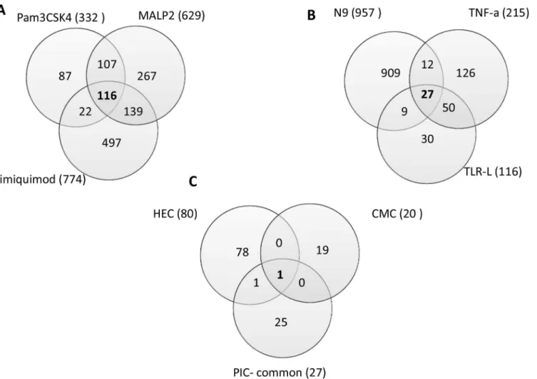
The numbers of differentially expressed probesets (fold change cut off = 2, FDR adjusted p-value<0.05) revealed by microarray analysis in the PIC group were: 215 for TNF-α, 332 for Pam3CSK4, 629 for MALP2, 774 for imiquimod, and 957 for N-9. In the NIC group deregula-tion was much more modest: 80 probesets for HEC and 20 probesets for CMC (Fig 1,Table 2;
S2–S8Tables).
Furthermore, the fold changes induced by NICs did not exceed 3.3, while the fold changes in the PIC group reached values>25 for TNF-α,>30 for Pam3CK4,>48 for MALP2,>22 for
imiquimod, and>23 for N-9 (S2–S8Tables).
With the aim of identifying a group of shared deregulated genes, we first analyzed differen-tially expressed genes within the TLR-L group. There were 116 differendifferen-tially expressed probe-sets in common among Pam3CSK4, MALP2 and imiquimod (Fig 1A;S9 Table).
Next, we compared gene expression after all PIC treatments, TNF-a, TLR-L, and N-9. As a result, 27 probesets with significantly altered expression in all PIC treatments were identified (Fig 1B,S10 Table). These 27 probesets encompass 22 genes (some genes are represented by more than one probeset). Next, the expression profile of PICs was compared against that of NICs. Overlap between the PIC-common probesets and probesets differentially expressed by NICs revealed two genes from the PIC list that were also differentially expressed in NICs (Fig 1C,S10 Table). Upregulated by PICs, MMP1 was also upregulated in HEC treatments (fold change 3.3). COL8A1 was downregulted by PICs and both NICs, HEC and CMC (fold change -3.0 and -2.3 respectively). Therefore these genes were excluded from the final list of the PIC-specific probesets. The final list of the probesets altered by PIC but not NIC treatments con-tains 25 probesets mapped to 20 PIC discriminatory genes (PIC-DG) (Table 3,S10 Table).
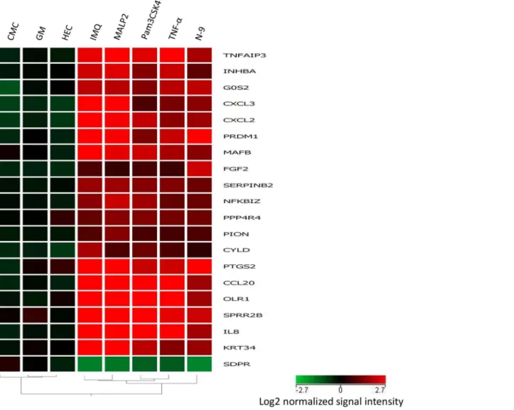
Of these genes, 19 genes are upregulated, and one is downregulated. A heatmap and hierar-chical clustering generated from expression profiles of 20 discriminatory genes common to all PIC treatments of Vk2 cells clearly demonstrates segregation of PICs from NICs (Fig 2).
Functional analysis of the discriminatory genes
Inflammatory response was indicated as the most significant biofunction (p value 3.05E-07– 2.05E-0.3), thus supporting pro-inflammatory activity as the principal common feature for the selected PICs, in spite of belonging to diverse molecular and mechanistic categories. Inflamma-tory/immune modulating response function was attributed by IPA to twelve genes from the PIC-DG list.
Fig 1. Diagrams showing the number of significantly altered probesets indentified by microarray gene profiling of Vk2 cells exposed to PIC and NIC.Total number of the altered probeserts for each treatment/category is shown in brackets (a gene can be represented by more than one probeset).
doi:10.1371/journal.pone.0128557.g001
Table 2. Number of differentially expressed probesets in treatment groups compared to control (growth medium).
Treatment Total Upregulated Downregulated
HEC 80 29 51
CMC 20 14 6
TNF-α 215 200 15
Pam3CSK4 332 309 23
MALP2 629 486 143
IMQ 774 493 281
TLR-L shared 116 106 10
N9 957 549 408
We extended the IPA-generated list of genes belonging to this category by including PIC-upregulatedMAFBandSERPINB2(see below andDiscussion) based on recently published studies indicating their link to the inflammatory/ immunomodulatory processes [54,55]. Four genes in the category of inflammatory/immune response,CCL20,IL8,CXCL2,CXCL3, encode for chemokines that are involved in immune cells trafficking. IL8 is also known as one of the major mediators of inflammatory responses. Other genes in this category includeOLR1, PTGS2,TNFAIP3,CYLDandNFKBIZ. OLR1 is a receptor for oxidatively modified low density lipoprotein (oxLDL) that upon binding by its ligand induces activation of NFκB (nuclear factor kappa B), the master complex in immunoinflammatory response. PTGS2 (prostaglandin synthase-2) also called COX-2 (cyclooxygenase-2) is one of the key enzymes involved in in-flammatory processes [56,57]. TNFAIP3 (A20) and CYLD are deubiquitinating enzymes that play a prominent role in inflammatory signaling by regulating NFkB activation. Another mole-cule that is tightly associated with NFκB regulation is NFKBIZ, NFκB inhibitor-zeta, also known as I kappa B zeta.
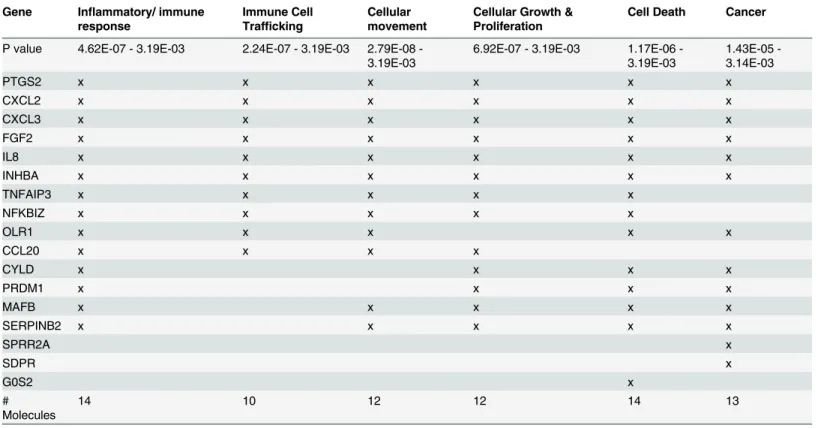
Other important top biofunctions/diseases include immune cell trafficking, cellular move-ment, cellular growth and proliferation, cancer, and cell death (Table 4).
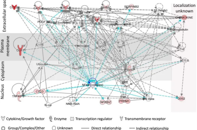
Functional analysis (by IPA) of relationships between PIC-DGs highlighted inflammatory response and cellular movement as the highest scored hypothetical gene networks (Fig 3).
Products of six of these genes are localized to the extracellular space, with functions involved in communication with other cells including chemotaxis of immune cells (Fig 3). Of impor-tance, a factor assigned by IPA as a central mediator for most of the PIC-DGs is nuclear factor
Table 3. Genes differenially expressed in VK2 cells treated with proinflammatory/immunomodulatory compounds.
Fold change—treatment vs GM
Gene Symbol
UniGene ID
TNF-α
Pam3CK4 MALP2 imiquimod N9 Gene name
TNFAIP3 Hs.211600 10.4 5.3 9.4 5.8 3.6 tumor necrosis factor, alpha-induced protein 3
INHBA Hs.583348 4.8 2.9 5.1 4.8 2.1 inhibin, beta A
G0S2 Hs.432132 4.1 2.8 4.1 4.2 4.3 G0/G1switch 2
CXCL3 Hs.89690 3.7 2.7 8.9 11.5 4.1 chemokine (C-X-C motif) ligand 3 CXCL2 Hs.75765 3.9 5.5 7.9 9.9 4.3 chemokine (C-X-C motif) ligand 2 PRDM1 Hs.436023 3.9 2.3 5.5 6.4 5.9 PR domain containing 1, with ZNF domain
MAFB Hs.169487 3.3 4.3 5.0 7.1 2.7 v-maf musculoaponeuroticfibrosarcoma oncogene homolog B (avian) FGF2 Hs.284244 2.2 2.4 2.1 2.5 6.0 fibroblast growth factor 2 (basic)
SERPINB2 Hs.594481 3.1 3.2 3.7 3.6 2.7 serpin peptidase inhibitor, clade B (ovalbumin), member 2 NFKBIZ Hs.319171 2.4 3.6 4.8 3.1 2.5 nuclear factor of kappa light polypeptide gene enhancer in B-cells
inhibitor, zeta
PPP4R4 Hs.259599 2.3 2.5 2.6 2.1 2.2 protein phosphatase 4, regulatory subunit 4
PION Hs.186649 2.0 2.1 3.0 2.2 2.1 pigeon homolog (Drosophila)
CYLD Hs.578973 2.4 3.0 2.4 4.6 2.0 cylindromatosis (turban tumor syndrome)
PTGS2 Hs.196384 3.7 3.5 7.5 7.1 12.5 prostaglandin-endoperoxide synthase 2 (prostaglandin G/H synthase and cyclooxygenase)
CCL20 Hs.75498 9.2 19.0 22.2 22.8 3.4 chemokine (C-C motif) ligand 20
OLR1 Hs.412484 25.5 30.4 38.3 22.2 3.9 oxidized low density lipoprotein (lectin-like) receptor 1 SPRR2B Hs.568239 3.5 9.9 14.4 4.8 2.9 small proline-rich protein 2B
IL8 Hs.624 8.8 16.4 40.2 16.1 4.2 interleukin 8
KRT34 Hs.296942 2.3 3.4 20.0 15.4 2.7 keratin 34
SDPR Hs.26530 -2.9 -3.1 -4.0 -4.4 -4.8 serum deprivation response (phosphatidylserine binding protein)
kB (NFκB), which is a key molecule in the control of inflammatory and immune responses [58–62].We have earlier demonstrated that N-9 activates NFκB signaling pathway in Vk2 cells [34]. In the present study we validate the IPA-identified central position of NFκB in gene de-regulation induced by TNF-αand TLR ligands in human vaginal cells. We demonstrate that following the treatments, fast degradation of IκB-αtakes place in the cytoplasm accompanied by release and translocation of p65/NFκB to the nucleus indicating NFkB activation (Fig 4).
Real-time qPCR analysis of selected genes
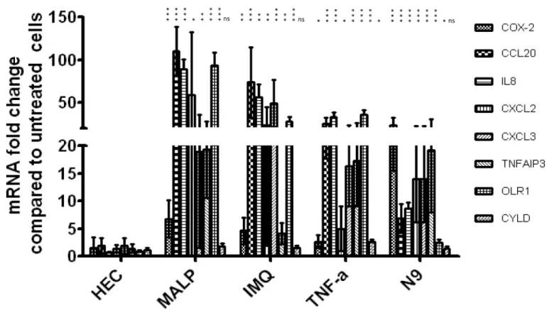
Based on their functions and level of expression, eight genes,PTGS2,CCL20,IL8,CXCL2, CXCL3,TNFAIP3,OLR1andCYLD, were selected for validation by qPCR. All selected genes, except forCYLDshowed significant upregulation (Fig 5), thus confirming the microarray data.
Generally, most of the genes tested demonstrated higher fold changes with qPCR compared to microarray. A similar trend was also described by others [63,64].
Fig 2. Hierarchical clustering of the 20 discriminatory genes was performed using GeneSpring 11.5 as described in Materials and Methods. Columns represent treatments, rows represent genes. Gene expression levels are indicated by color: red is for upregulation and green is for downregulation. Expression data are averages from at least six experiments/microarrays for each treatment. Hierarchical clustering based on the discriminatory genes demonstrates sharp segregation of the PIC vs NIC treatments.
Evaluation of candidate microbicides using discriminatory genes
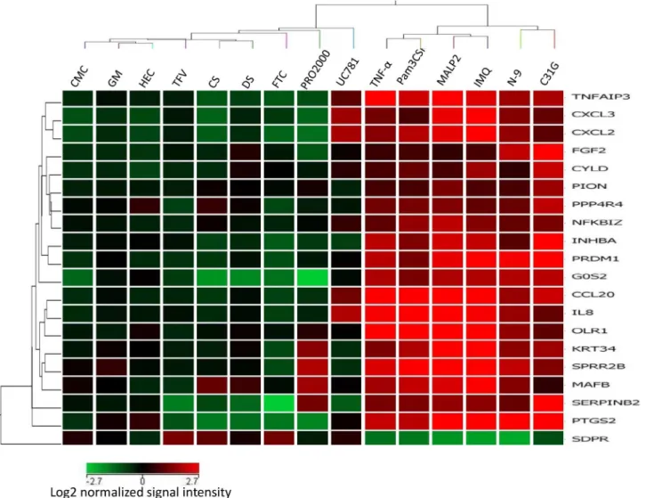
We used a panel of PIC-DG to test their discriminatory power and to evaluate the pro-inflam-matory potential of several microbicides /active ingredients: non-ionic detergent C31G (an ac-tive ingredient of Savvy), non-ionic polymers—PRO2000, cellulose sulfate (CS), dextran sulfate (DS), reverse transcriptase inhibitors (RTI)—nucleoside/nucleotide RTI (NRTI)—tenofovir (TFV) and emtricitabine (FTC), and non-NRTI (NNRTI)—UC781. The heatmap of cluster analysis of microarray data demonstrates that C31G clusters together with PICs, while the other candidate microbicides proved to be not inflammatory by this gene expression pro-file (Fig 6).
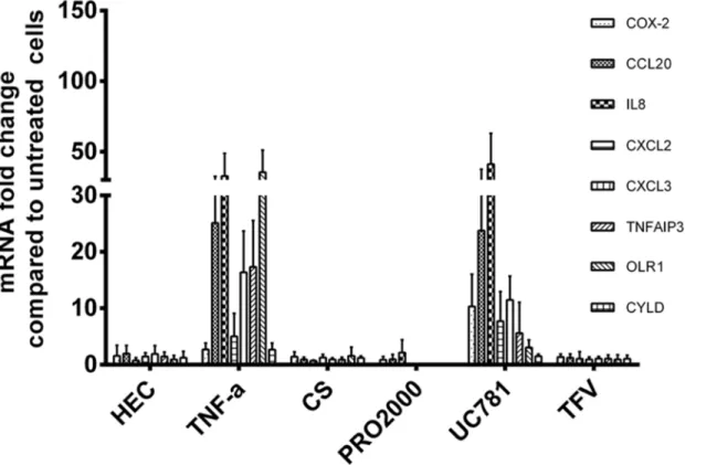
It should be noted, however, that although UC781 clustered with the NIC group, it caused upregulation of several of the PIC-DGs belonging to inflammation/immune response category which was confirmed by real-time RT-PCR analysis (Fig 7).
Panel of selected PIC-DGs clearly discriminates normal vaginal
flora-from BV-related bacteria in the Vk2 cell test model
Earlier we demonstrated thatP.bivia, bacteria associated with BV caused significant NFkB acti-vation and increase in IL-8 level in Vk2 cells [41]. In the study reported herein, vaginal epitheli-al expression of seven selected PIC-DGs,PTGS2,CCL20,IL8,CXCL2,CXCL3,TNFAIP3, OLR1, following exposure toP.biviaand beneficial commensalLactobacillus (L.) gasseriwas compared using qPCR. Strong induction of all seven PIC-DG byP.bivia(3 to 75 fold increase),
Table 4. Functional categories of the PIC/NIC discriminatory genesa.
Gene Inflammatory/ immune response
Immune Cell Trafficking
Cellular movement
Cellular Growth & Proliferation
Cell Death Cancer
P value 4.62E-07 - 3.19E-03 2.24E-07 - 3.19E-03 2.79E08 -3.19E-03
6.92E-07 - 3.19E-03 1.17E06 -3.19E-03
1.43E05 -3.14E-03
PTGS2 x x x x x x
CXCL2 x x x x x x
CXCL3 x x x x x x
FGF2 x x x x x x
IL8 x x x x x x
INHBA x x x x x x
TNFAIP3 x x x x x
NFKBIZ x x x x x
OLR1 x x x x x
CCL20 x x x x
CYLD x x x x
PRDM1 x x x x
MAFB x x x x x
SERPINB2 x x x x x
SPRR2A x
SDPR x
G0S2 x
# Molecules
14 10 12 12 14 13
aClassification is based on IPA functional analysis and published literature. P values are estimated by IPA
but not byL.gasseri, was observed (Fig 8), thus validating the discriminatory power of these genes in pathologically relevant conditions of the cervicovaginal tract.
Discussion
The molecular and cellular mechanisms underpinning HIV-1 vaginal transmission are not yet fully understood. Numerous studies indicate that the initial targets of HIV-1 infection could primarily be CD4+ T cells, with dendritic cells (DC), Langerhans cells (LC), and/or macro-phages also playing a role (reviewed in [18,65]). Inflammation and immune activation of cervi-covaginal mucosa are considered factors that increase susceptibility to HIV infection due to recruitment and activation of HIV-target immune cells and increased production of
Fig 3. Top network of Vk2 PIC/NIC discriminatory genes generated by Ingenuity Pathway Analysis.Red/pink color indicates upregulation of the genes (microarray data). Connections of NFkB complex with other genes is shown in blue color.
doi:10.1371/journal.pone.0128557.g003
Fig 4. NFκB activation in Vk2 cells in response to PIC treatments.Fast degradation of IκB-αin the cytoplasm and translocation of p65/NFkB to the
nucleus following Vk2 cells exposure to PICs was detected in cytoplasmic and nuclear fractions using corresponding antibodies.
immunostimulatory factors that paradoxically intensify HIV replication at the sites of viral ex-posure. [13–18]. In addition, cervicovaginal inflammation may cause disruption of the epitheli-al barrier, thus providing a portepitheli-al for virepitheli-al entry [66,67]. Therefore, it is critical to screen candidate microbicides for potential mucosal immunomodulatory/inflammatory effects. The goal of this study was to develop anin vitromethod for preclinical evaluation of pro-inflamma-tory/immunomodulatory potential of new candidate microbicides and identify new biomarkers of mucosal alteration. To this end, we compared gene expression profiles of Vk2 cells treated with PICs and NICs in order to get a panel of genes that are consistently altered by PICs com-pared to NICs, thus distinguishing between these two groups.
To make the discriminatory genes more encompassing we selected PICs that belong to dif-ferent molecular and mechanistic groups. TNF-αis a well characterized pleiotropic proiflam-matory cytokine [68,69]. N-9, a nonionic cell membrane disrupting surfactant, is a microbicide that increased the probability of HIV-1 transmission in clinical trials, supposedly, due to vagi-nal mucosa irritation and inflammation [70,71]. Furthermore, cervicovaginal lavages collected from women who had used N-9 for three days enhanced HIV replicationin vitro[40]. The other pro-inflammatory group included different TLR-ligands (TLR-L) (Table 1). TLRs are sig-naling transmembrane proteins that recognize and rapidly respond to various conserved mi-crobial pathogen-associated molecular patterns (PAMPs). The interaction of TLRs with their corresponding PAMPs leads to initiation of innate immune response and triggers pro-inflam-matory pathways, in which activation of NFκB plays a central role (see reviews [58,59,61,62].
Fig 5. Real-time qPCR validation of changes in expression of eight selected genes observed in microarray analysis.Bars represent the mean±SD of fold change relative to growth medium control. At least 3 independent experiments were performed for each treatment. All genes were normalized to GAPDH. Asterisks placed vertically denote p values for each PIC treatment relative to HEC used as a reference. (***p<0.0005,**p<0.005,*p<0.05; Student t-test)
There are 10 TLRs identified in the human thus far. The female lower genital tract mucosa was found to express most of them [37,72–75]. There is strong evidence supporting the link be-tween the presence of infectious conditions in the lower genital tract, including sexually trans-mitted infections (STI) and BV, and increased risk of HIV-1 acquisition [19–21]. It is possible that stimulation of innate immune and inflammatory signaling by the PAMP-activated cervi-covaginal TLRs through cytokine and chemokine induction increases availability of HIV-1 tar-get cells in the mucosal epithelium, facilitating primary HIV-1 infection.
In this study we identified a set of 20 genes of which 19 genes are consistently activated and one gene is downregulated in human vaginal epithelial cells exposed to various compounds that shared proinflammatory/immunomodulatory activity. Our selection of the stimuli was confirmed by the functional analysis of these discriminatory genes using the IPA program and published data, which revealed that out of the 20 selected genes, 14 genes are known to be in-volved in inflammatory and immune responses. For most of our inflammation-related genes, their role in inflammatory responses is largely understood, albeit primarily in tissues other
Fig 6. Transcription profile of the 20 discriminatory genes expression in Vk2 cells exposed to candidate microbicides and selected PICs and NICs. Columns represent treatments, rows represent genes. Gene expression levels are indicated by color: red is for upregulation and green is for downregulation. Expression data are averages from at least six experiments/microarrays for each treatment. Clustering based on 20 PIC/NIC discriminatory genes places C31G (known as causing inflammatory response) to the PIC category, while dextran sulfate (DS) and cellulose sulfate (CS)—into the NIC group.
than the cervicovaginal mucosa. At the same time, novel features that might link them to HIV susceptibility have recently emerged (discussed below).
Althoughin vivomodels have obvious limitations compared to thein vitroones, the set of genes or biomarkers reported herein can be tested early in the process of drug discovery or product development to identify compounds with properties that may result in undesirable
Fig 7. Real-time qPCR validation of changes in expression of eight selected genes observed in the microarray analysis of microbicide candidates. Experimental details are as inFig 5. HEC and TNF-αare added as references.
doi:10.1371/journal.pone.0128557.g007
Fig 8. PIC-DG expression following bacterial colonization of Vk2 cells as revealed by quantitative real time RT-PCR.P. bivia (right) induced strong upregulation of all seven PIC-DEGs, while L. gasseri (left) did not cause any changes. Results are presented as mean±SD of three experiments.
mucosal effects. In an attempt to validate this set of biomarkers, we tested microbicide candi-dates previously characterized in clinical studies with known cervicovaginal mucosal effects [2] and we were able to properly qualify those compounds. In clinical trials, CS, PRO2000 and DS appeared to be safe and did not cause adverse mucosal effects/epithelial disruption, while C31G might have caused vaginal alterations comparable to the effect observed with N-9 [2,76–
80]. Clustering analysis of the microarray data classified C31G as PIC, while CS, DS, PRO2000 aligned with the NIC group in good correlation with the results from clinical trials. Tenofovir, currently completing a phase III confirmatory trial in the form of 1% vaginal gel, did not show a pro-inflammatory profile. This is in agreement with results from numerous safety studies in-cluding the Phase IIb trial, CAPRISA 004 [81–83]. Interestingly, although clustering with NICs, UC781 showed upregulation of certain PIC-associated genes. Product development of UC781 as a vaginal microbicide, which had reached clinical stage, was discontinued due to pharmaceutical and safety issues. High concentrations of UC781 revealed significant changes in the mucosa of macaques and rabbits [84].
In addition, the discriminatory power of seven selected PIC-DGs was confirmed in the bac-terial exposure experiment using commensal and pathogenic microorganisms. We found strong induction of these selected PIC-DGs in Vk2 cells byP.bivia(3 to 75 fold increase), bac-teria associated with BV [47–49]. By contrast,L gasseri, one of the dominant beneficial Lacto-bacillusspecies in the vagina of healthy women [43,44,46,85] did not cause any changes (Fig 8). Upregulation of inflammation-related genes by the BV-related bacteria may have pathogenic implications and provide further insight on the mechanisms underpinning increased rates of HIV-1 transmission in women with BV [86,87].
Clearly these new biomarkers do not identify every potential adverse effect on the cervicova-ginal mucosa or environment. CS, which failed to protect women from HIV and may have even increased their susceptibility, did not show inflammatory effects, neither in this model nor in clinical studies. However, it has been reported that it might downregulate epithelial junctional proteins or alter the vaginal microbiome facilitating HIV infection [9,88].
In addition to generating a group of new biomarkers of vaginal mucosal alteration, the re-ported discriminatory genes may also provide clues to better understanding of the mucosal changes that coexist with and may facilitate HIV transmission. For instance, these genes in-clude several widely known chemokines. IL8, is one of the major mediators of the inflammato-ry response [89,90]. This chemokine, also known as CXCL8, attracts T cells and neutrophils, stimulates adhesion of monocytes to endothelial cells via interaction with its receptors CXCR1 and CXCR2 [91]. High vaginal IL-8 levels were observed at BV [92] and in response to some pro-inflammatory compounds [29]. IL-8 was found to stimulate HIV-1 replication in T cells and macrophages and increase HIV-1 transmission in cervical explants tissues [93,94]. Much less is known about CXCL2 (MIP-2α, GRO-2, GRO-β) and CXCL3 (MIP-2β, GRO-3, GRO-γ) that are structurally related to IL-8. Like IL-8, they also interact with CXCR2 and are involved in inflammatory and immune responses by attracting and activating immune cells [95].
Another chemokine which is significantly upregulated by PICs isCCL20, or macrophage in-flammatory protein 3α(MIP-3α). CCL20 is the only chemokine that interacts with CCR6 re-ceptor [96] which is expressed by Th17 lymphocytes and by LCs. Several studies indicate that the CD4+Th17 cells are early HIV/SIV preferential targets and are implicated in HIV patho-genesis [97–101]. Recruitment of CCR6-expressing Th17 cells through CCL20-CCR6 interac-tions was demonstrated in diverse tissues [102–104]. In the vaginal epithelium these
are able to migrate from the mucosa to the lymph nodes where virions can be transmitted to lymphocytes for productive infection. Inin vivoandex vivostudies, involvement of LCs in HIV sexual infection has been clearly demonstrated [65,106–110]. Inex vivoexperiments, in-flammatory stimuli, TNF-αand Pam3CSK4 (a ligand for TLR1/TLR2 heterodimer) strongly increased HIV-1 transmission by LCs [107]. Conversely, suppression of CCL20 production and possible prevention of LCs and Th17 attraction and activation correlated with restriction of mucosal transmission of SIV, as demonstrated by glycerol monolaureate studies in non-human primates [13].
Upregulation ofPTGS2by all PICs observed in this study is a strong assertion of the proin-flammatory potential of the selected PICs. The protein encoded byPTGS2, most often called COX-2 (cyclooxygenase-2), is an inducible enzyme that is expressed in response to various pathophysiological stimuli. It is one of the key enzymes involved in inflammation. It catalyzes the first steps of conversion of arachidonic acid into prostaglandins (PG) that play an impor-tant role in inflammatory and immunomodulatory processes [111–113]. One of the major COX-2 products, PGE2 was shown to directly enhance HIV-1 long terminal repeat (LTR) tran-scription in human T cells [114]. We have earlier demonstrated that PGE2 levels were elevated in the vaginal epithelial cells following COX-2 induction in response to diverse pro-inflamma-tory/immunomodulatory stimuli [33,34]. Activation ofPTGS2gene resulting in PGE2 eleva-tion could be a factor contributing to HIV sexual transmission in inflammatory condieleva-tions.
TNFAIP3(tumor necrosis factor, alpha-induced protein 3) is another gene that plays an im-portant role in regulation of inflammation and immunity and regulates NFκB. We also ob-served strong upregulation ofOLR1, a gene for oxidized low density lipoprotein (lectin-like) receptor 1—cell surface protein belonging to the C-type lectin family.OLR1can be rapidly acti-vated by a wide range of stimuli including pro-inflammatory and tissue damaging ones. Its ac-tivation triggers several signaling pathways including NFκB [115,116].
NFκB signaling pathway is the central regulator of inflammation and immune activation [60]. We demonstrate here that NFκB is activated in vaginal cells in response to all PIC treat-ments used in this study (Fig 4). The network of PIC discriminatory gene interactions illus-trates the central role of NFκB complex in their regulation (Fig 3). Importantly, NFkB binding elements are present in enhancer located in HIV-1 LTR and provide signal-specific activation of HIV expression in response to NFkB stimuli [117,118]. It might be possible that PIC treat-ments activate NFkB pathway in the TLR-expressing HIV-1-target cells present in the vaginal epithelium. Multiple genes activated by PICs (INHBA, FGF2, PRDM1, MafB, and SERPINB2) are involved in signal transduction or regulation of immune response [54,55,119–121], which can also promote HIV-1 transcription and replication [122].
Conclusion
cervicovaginal immunoinflammatory and mucosal altering processes that facilitate or limit HIV transmission having important implications for the design of novel prevention strategies.
Supporting Information
S1 Table. List of primers.
(DOCX)
S2 Table. Probesets significantly deregulated in Vk2 cells by TNF-α.
(XLSX)
S3 Table. Probesets significantly deregulated in Vk2 cells by Pam3CSK4.
(XLSX)
S4 Table. Probesets significantly deregulated in Vk2 cells by MALP2.
(XLSX)
S5 Table. Probesets significantly deregulated in Vk2 cells by imiquimod.
(XLSX)
S6 Table. Probesets significantly deregulated in Vk2 cells by N-9.
(XLSX)
S7 Table. Probesets significantly deregulated in Vk2 cells by HEC.
(XLSX)
S8 Table. Probesets significantly deregulated in Vk2 cells by CMC
(XLSX)
S9 Table. Significantly deregulated probesets common to all TLR ligands.
(XLSX)
S10 Table. Probesets significantly deregulated in Vk2 cells that are common to all treat-ments with proinflammatory-immunomodulatory compounds.
(DOCX)
Author Contributions
Conceived and designed the experiments: IAZ GFD. Performed the experiments: TJ NY IAZ SSJ TF HSY RNF. Analyzed the data: IAZ RS JB. Wrote the paper: IAZ GFD. Editorial com-ments/critiques: RS RNF.
References
1. Shattock RJ, Moore JP (2003) Inhibiting sexual transmission of HIV-1 infection. Nat Rev Microbiol 1: 25–34. PMID:15040177
2. Romano JW, Robbiani M, Doncel GF, Moench T (2012) Non-specific microbicide product develop-ment: then and now. Curr HIV Res 10: 9–18. PMID:22264041
3. Abdool Karim SS, Baxter C (2012) Overview of microbicides for the prevention of human immunodefi-ciency virus. Best Pract Res Clin Obstet Gynaecol 26: 427–439. doi:10.1016/j.bpobgyn.2012.01.010 PMID:22386823
4. Obiero J, Mwethera PG, Hussey GD, Wiysonge CS (2012) Vaginal microbicides for reducing the risk of sexual acquisition of HIV infection in women: systematic review and meta-analysis. BMC Infect Dis 12: 289. doi:10.1186/1471-2334-12-289PMID:23130761
6. Abdool Karim Q, Abdool Karim SS, Frohlich JA, Grobler AC, Baxter C, et al. (2010) Effectiveness and safety of tenofovir gel, an antiretroviral microbicide, for the prevention of HIV infection in women. Sci-ence 329: 1168–1174. doi:10.1126/science.1193748PMID:20643915
7. Van Damme L, Govinden R, Mirembe FM, Guedou F, Solomon S, et al. (2008) Lack of effectiveness of cellulose sulfate gel for the prevention of vaginal HIV transmission. N Engl J Med 359: 463–472. doi:10.1056/NEJMoa0707957PMID:18669425
8. Feldblum PJ, Adeiga A, Bakare R, Wevill S, Lendvay A, et al. (2008) SAVVY vaginal gel (C31G) for prevention of HIV infection: a randomized controlled trial in Nigeria. PLoS ONE 3: e1474. doi:10. 1371/journal.pone.0001474PMID:18213382
9. Mesquita PM, Cheshenko N, Wilson SS, Mhatre M, Guzman E, et al. (2009) Disruption of tight junc-tions by cellulose sulfate facilitates HIV infection: model of microbicide safety. J Infect Dis 200: 599– 608. doi:10.1086/600867PMID:19586414
10. Stone AB, Harrison PF, Lusti-Narasimhan M (2013) Microbicides from a regulatory perspective. AIDS 27: 2261–2269. doi:10.1097/QAD.0b013e32836239b4PMID:23612007
11. Gray RH, Wawer MJ (2012) Probability of heterosexual HIV-1 transmission per coital act in sub-Saha-ran Africa. J Infect Dis 205: 351–352. doi:10.1093/infdis/jir751PMID:22241799
12. Hughes JP, Baeten JM, Lingappa JR, Magaret AS, Wald A, et al. (2012) Determinants of per-coital-act HIV-1 infectivity among African HIV-1-serodiscordant couples. J Infect Dis 205: 358–365. doi:10. 1093/infdis/jir747PMID:22241800
13. Li Q, Estes JD, Schlievert PM, Duan L, Brosnahan AJ, et al. (2009) Glycerol monolaurate prevents mucosal SIV transmission. Nature 458: 1034–1038. doi:10.1038/nature07831PMID:19262509
14. Royce RA, Sena A, Cates W Jr, Cohen MS (1997) Sexual transmission of HIV. N Engl J Med 336: 1072–1078. PMID:9091805
15. Appay V, Sauce D (2008) Immune activation and inflammation in HIV-1 infection: causes and conse-quences. J Pathol 214: 231–241. PMID:18161758
16. Borrow P, Shattock RJ, Vyakarnam A (2010) Innate immunity against HIV: a priority target for HIV pre-vention research. Retrovirology 7: 84. doi:10.1186/1742-4690-7-84PMID:20937128
17. Shacklett BL (2010) Immune responses to HIV and SIV in mucosal tissues: 'location, location, loca-tion'. Curr Opin HIV AIDS 5: 128–134. doi:10.1097/COH.0b013e328335c178PMID:20543589
18. Haase AT (2010) Targeting early infection to prevent HIV-1 mucosal transmission. Nature 464: 217– 223. doi:10.1038/nature08757PMID:20220840
19. Galvin SR, Cohen MS (2004) The role of sexually transmitted diseases in HIV transmission. Nat Rev Microbiol 2: 33–42. PMID:15035007
20. Thurman AR, Doncel GF (2011) Innate immunity and inflammatory response to Trichomonas vagina-lis and bacterial vaginosis: relationship to HIV acquisition. Am J Reprod Immunol 65: 89–98. doi:10. 1111/j.1600-0897.2010.00902.xPMID:20678168
21. Ward H, Ronn M (2010) Contribution of sexually transmitted infections to the sexual transmission of HIV. Curr Opin HIV AIDS 5: 305–310. doi:10.1097/COH.0b013e32833a8844PMID:20543605
22. Jespers V, Millwood IY, Poynten IM, Van Damme L, Kaldor JM (2013) The evolving design and meth-ods for trials evaluating the safety of candidate vaginal microbicides: a systematic review. Sex Transm Dis 40: 729–736. doi:10.1097/01.olq.0000431070.38601.03PMID:23945427
23. Hladik F, Doncel GF (2010) Preventing mucosal HIV transmission with topical microbicides: chal-lenges and opportunities. Antiviral Res 88 Suppl 1: S3–9. doi:10.1016/j.antiviral.2010.09.011PMID: 21109065
24. Pudney J, Quayle AJ, Anderson DJ (2005) Immunological microenvironments in the human vagina and cervix: mediators of cellular immunity are concentrated in the cervical transformation zone. Biol Reprod 73: 1253–1263. PMID:16093359
25. Doncel GF, Chandra N, Fichorova RN (2004) Preclinical assessment of the proinflammatory potential of microbicide candidates. J Acquir Immune Defic Syndr 37 Suppl 3: S174–180. PMID:16419269
26. Fichorova RN (2004) Guiding the vaginal microbicide trials with biomarkers of inflammation. J Acquir Immune Defic Syndr 37 Suppl 3: S184–193. PMID:16419271
27. Zhong M, He B, Yang J, Bao R, Zhang Y, et al. (2012) L-selectin and P-selectin are novel biomarkers of cervicovaginal inflammation for preclinical mucosal safety assessment of anti-HIV-1 microbicide. Antimicrob Agents Chemother 56: 3121–3132. doi:10.1128/AAC.05950-11PMID:22391529
29. Fichorova RN, Bajpai M, Chandra N, Hsiu JG, Spangler M, et al. (2004) Interleukin (IL)-1, 6, and IL-8 predict mucosal toxicity of vaginal microbicidal contraceptives. Biol Reprod 71: 761–769. PMID: 15128598
30. Cummins JE Jr, Doncel GF (2009) Biomarkers of cervicovaginal inflammation for the assessment of microbicide safety. Sex Transm Dis 36: S84–91. doi:10.1097/OLQ.0b013e3181994191PMID: 19218890
31. Doncel GF, Clark MR (2010) Preclinical evaluation of anti-HIV microbicide products: New models and biomarkers. Antiviral Res 88 Suppl 1: S10–18. doi:10.1016/j.antiviral.2010.09.018PMID:21109063
32. Fields S, Song B, Rasoul B, Fong J, Works MG, et al. (2014) New candidate biomarkers in the female genital tract to evaluate microbicide toxicity. PLoS One 9: e110980. doi:10.1371/journal.pone. 0110980PMID:25333937
33. Joseph T, Zalenskaya IA, Yousefieh N, Schriver SD, Cote LC, et al. (2012) Induction of Cyclooxygen-ase (COX)-2 in Human Vaginal Epithelial Cells in Response to TLR ligands and TNF-alpha. Am J Reprod Immunol 67: 482–490 doi:10.1111/j.1600-0897.2011.01099.xPMID:22235849
34. Zalenskaya IA, Cerocchi OG, Joseph T, Donaghay MA, Schriver SD, et al. (2011) Increased COX-2 expression in human vaginal epithelial cells exposed to nonoxynol-9, a vaginal contraceptive microbi-cide that failed to protect women from HIV-1 infection. Am J Reprod Immunol 65: 569–577. doi:10. 1111/j.1600-0897.2010.00964.xPMID:21241401
35. Fichorova RN, Rheinwald JG, Anderson DJ (1997) Generation of papillomavirus-immortalized cell lines from normal human ectocervical, endocervical, and vaginal epithelium that maintain expression of tissue-specific differentiation proteins. Biol Reprod 57: 847–855. PMID:9314589
36. Fichorova RN, Desai PJ, Gibson FC III, Genco CA (2001) Distinct proinflammatory host responses to Neisseria gonorrhoeae infection in immortalized human cervical and vaginal epithelial cells. Infect Immun 69: 5840–5848. PMID:11500462
37. Fichorova RN, Cronin AO, Lien E, Anderson DJ, Ingalls RR (2002) Response to Neisseria gonor-rhoeae by cervicovaginal epithelial cells occurs in the absence of toll-like receptor 4-mediated signal-ing. J Immunol 168: 2424–2432. PMID:11859134
38. Fichorova RN, Lee Y, Yamamoto HS, Takagi Y, Hayes GR, et al. (2012) Endobiont viruses sensed by the human host—beyond conventional antiparasitic therapy. PLoS One 7: e48418. doi:10.1371/ journal.pone.0048418PMID:23144878
39. Fichorova RN, Anderson DJ (1999) Differential expression of immunobiological mediators by immor-talized human cervical and vaginal epithelial cells. Biol Reprod 60: 508–514. PMID:9916021
40. Fichorova RN, Tucker LD, Anderson DJ (2001) The molecular basis of nonoxynol-9-induced vaginal inflammation and its possible relevance to human immunodeficiency virus type 1 transmission. J In-fect Dis 184: 418–428. PMID:11471099
41. Fichorova RN, Yamamoto HS, Delaney ML, Onderdonk AB, Doncel GF (2011) Novel vaginal microflo-ra colonization model providing new insight into microbicide mechanism of action. MBio 2: e00168– 00111. doi:10.1128/mBio.00168-11PMID:22027006
42. Onderdonk AB, Lee ML, Lieberman E, Delaney ML, Tuomala RE (2003) Quantitative microbiologic models for preterm delivery. J Clin Microbiol 41: 1073–1079. PMID:12624032
43. Antonio MA, Hawes SE, Hillier SL (1999) The identification of vaginal Lactobacillus species and the demographic and microbiologic characteristics of women colonized by these species. J Infect Dis 180: 1950–1956. PMID:10558952
44. Pavlova SI, Kilic AO, Kilic SS, So JS, Nader-Macias ME, et al. (2002) Genetic diversity of vaginal lac-tobacilli from women in different countries based on 16S rRNA gene sequences. J Appl Microbiol 92: 451–459. PMID:11872120
45. Redondo-Lopez V, Cook RL, Sobel JD (1990) Emerging role of lactobacilli in the control and mainte-nance of the vaginal bacterial microflora. Rev Infect Dis 12: 856–872. PMID:2237129
46. Pendharkar S, Magopane T, Larsson PG, de Bruyn G, Gray GE, et al. (2013) Identification and char-acterisation of vaginal lactobacilli from South African women. BMC Infect Dis 13: 43. doi:10.1186/ 1471-2334-13-43PMID:23351177
47. Hill GB (1993) The microbiology of bacterial vaginosis. Am J Obstet Gynecol 169: 450–454. PMID: 8357043
48. Ling Z, Kong J, Liu F, Zhu H, Chen X, et al. (2010) Molecular analysis of the diversity of vaginal micro-biota associated with bacterial vaginosis. BMC Genomics 11: 488. doi:10.1186/1471-2164-11-488 PMID:20819230
50. Irizarry RA, Bolstad BM, Collin F, Cope LM, Hobbs B, et al. (2003) Summaries of Affymetrix GeneChip probe level data. Nucleic Acids Res 31: e15. PMID:12582260
51. Benjamini Y, Hochberg Y (1995) Controlling the false discovery rate: A practical and powerful ap-proach to multiple testing. Journal of the Royal Statistical Society Series B 57: 289–300.
52. Livak KJ, Schmittgen TD (2001) Analysis of relative gene expression data using real-time quantitative PCR and the 2(-Delta Delta C(T)) Method. Methods 25: 402–408. PMID:11846609
53. Zalenskaya IA, Bradbury EM, Zalensky AO (2000) Chromatin structure of telomere domain in human sperm. Biochem Biophys Res Commun 279: 213–218. PMID:11112441
54. Kim H, Seed B (2010) The transcription factor MafB antagonizes antiviral responses by blocking re-cruitment of coactivators to the transcription factor IRF3. Nat Immunol 11: 743–750. doi:10.1038/ni. 1897PMID:20581830
55. Schroder WA, Major L, Suhrbier A (2011) The role of SerpinB2 in immunity. Crit Rev Immunol 31: 15– 30. PMID:21395508
56. Turini ME, DuBois RN (2002) Cyclooxygenase-2: a therapeutic target. Annu Rev Med 53: 35–57. PMID:11818462
57. Williams CS, Mann M, DuBois RN (1999) The role of cyclooxygenases in inflammation, cancer, and development. Oncogene 18: 7908–7916. PMID:10630643
58. Doyle SL, O'Neill LA (2006) Toll-like receptors: from the discovery of NFkappaB to new insights into transcriptional regulations in innate immunity. Biochem Pharmacol 72: 1102–1113. PMID:16930560
59. Drexler SK, Foxwell BM (2010) The role of toll-like receptors in chronic inflammation. Int J Biochem Cell Biol 42: 506–518. doi:10.1016/j.biocel.2009.10.009PMID:19837184
60. Hoffmann A, Baltimore D (2006) Circuitry of nuclear factor kappaB signaling. Immunol Rev 210: 171– 186. PMID:16623771
61. Medzhitov R, Janeway C Jr (2000) The Toll receptor family and microbial recognition. Trends Micro-biol 8: 452–456. PMID:11044679
62. Sabroe I, Parker LC, Dower SK, Whyte MK (2008) The role of TLR activation in inflammation. J Pathol 214: 126–135. PMID:18161748
63. Szameit S, Vierlinger K, Farmer L, Tuschl H, Noehammer C (2008) Microarray-based in vitro test sys-tem for the discrimination of contact allergens and irritants: identification of potential marker genes. Clin Chem 54: 525–533. doi:10.1373/clinchem.2007.097386PMID:18202158
64. Tureci O, Bian H, Nestle FO, Raddrizzani L, Rosinski JA, et al. (2003) Cascades of transcriptional in-duction during dendritic cell maturation revealed by genome-wide expression analysis. FASEB J 17: 836–847. PMID:12724343
65. Hladik F, Sakchalathorn P, Ballweber L, Lentz G, Fialkow M, et al. (2007) Initial events in establishing vaginal entry and infection by human immunodeficiency virus type-1. Immunity 26: 257–270. PMID: 17306567
66. Coombs RW, Reichelderfer PS, Landay AL (2003) Recent observations on HIV type-1 infection in the genital tract of men and women. AIDS 17: 455–480. PMID:12598766
67. Weiler AM, Li Q, Duan L, Kaizu M, Weisgrau KL, et al. (2008) Genital ulcers facilitate rapid viral entry and dissemination following intravaginal inoculation with cell-associated simian immunodeficiency virus SIVmac239. J Virol 82: 4154–4158. doi:10.1128/JVI.01947-07PMID:18272571
68. Beutler B, Cerami A (1989) The biology of cachectin/TNF—a primary mediator of the host response. Annu Rev Immunol 7: 625–655. PMID:2540776
69. Warren JS (1990) Interleukins and tumor necrosis factor in inflammation. Crit Rev Clin Lab Sci 28: 37–59. PMID:2121159
70. Niruthisard S, Roddy RE, Chutivongse S (1991) The effects of frequent nonoxynol-9 use on the vagi-nal and cervical mucosa. Sex Transm Dis 18: 176–179. PMID:1658953
71. Van Damme L, Ramjee G, Alary M, Vuylsteke B, Chandeying V, et al. (2002) Effectiveness of COL-1492, a nonoxynol-9 vaginal gel, on HIV-1 transmission in female sex workers: a randomised con-trolled trial. Lancet 360: 971–977. PMID:12383665
72. Fazeli A, Bruce C, Anumba DO (2005) Characterization of Toll-like receptors in the female reproduc-tive tract in humans. Hum Reprod 20: 1372–1378. PMID:15695310
73. Herbst-Kralovetz MM, Quayle AJ, Ficarra M, Greene S, Rose WA Jr, et al. (2008) Quantification and comparison of toll-like receptor expression and responsiveness in primary and immortalized human female lower genital tract epithelia. Am J Reprod Immunol 59: 212–224. doi:10.1111/j.1600-0897. 2007.00566.xPMID:18201283
75. Yu L, Wang L, Chen S (2009) Toll-like receptors, inflammation and tumor in the human female repro-ductive tract. Am J Reprod Immunol 62: 1–8. doi:10.1111/j.1600-0897.2009.00712.xPMID: 19527227
76. Peterson L, Nanda K, Opoku BK, Ampofo WK, Owusu-Amoako M, et al. (2007) SAVVY (C31G) gel for prevention of HIV infection in women: a Phase 3, double-blind, randomized, placebo-controlled trial in Ghana. PLoS One 2: e1312. PMID:18091987
77. Adams JL, Kashuba AD (2012) Formulation, pharmacokinetics and pharmacodynamics of topical mi-crobicides. Best Pract Res Clin Obstet Gynaecol.
78. Cone RA, Hoen T, Wong X, Abusuwwa R, Anderson DJ, et al. (2006) Vaginal microbicides: detecting toxicities in vivo that paradoxically increase pathogen transmission. BMC Infect Dis 6: 90. PMID: 16740164
79. Van Damme L (2004) Clinical microbicide research: an overview. Trop Med Int Health 9: 1290–1296. PMID:15598260
80. Ballagh SA, Baker JM, Henry DM, Archer DF (2002) Safety of single daily use for one week of C31G HEC gel in women. Contraception 66: 369–375. PMID:12443969
81. Sokal DC, Karim QA, Sibeko S, Yende-Zuma N, Mansoor LE, et al. (2013) Safety of tenofovir gel, a vaginal microbicide, in South African women: results of the CAPRISA 004 Trial. Antivir Ther 18: 301– 310. doi:10.3851/IMP2311PMID:22914267
82. Gengiah TN, Baxter C, Mansoor LE, Kharsany AB, Abdool Karim SS (2012) A drug evaluation of 1% tenofovir gel and tenofovir disoproxil fumarate tablets for the prevention of HIV infection. Expert Opin Investig Drugs 21: 695–715. doi:10.1517/13543784.2012.667072PMID:22394224
83. Johnson TJ, Clark MR, Albright TH, Nebeker JS, Tuitupou AL, et al. (2012) A 90-day tenofovir reser-voir intravaginal ring for mucosal HIV prophylaxis. Antimicrob Agents Chemother 56: 6272–6283. doi: 10.1128/AAC.01431-12PMID:23006751
84. Patton DL, Sweeney YT, Balkus JE, Rohan LC, Moncla BJ, et al. (2007) Preclinical safety assess-ments of UC781 anti-human immunodeficiency virus topical microbicide formulations. Antimicrob Agents Chemother 51: 1608–1615. PMID:17353240
85. Anderson A, Sanunu M, Schneider C, Clad A, Karygianni L, et al. (2014) Rapid species-level identifi-cation of vaginal and oral lactobacilli using MALDI-TOF MS analysis and 16S rDNA sequencing. BMC Microbiol 14: 312. doi:10.1186/s12866-014-0312-5PMID:25495549
86. Atashili J, Poole C, Ndumbe PM, Adimora AA, Smith JS (2008) Bacterial vaginosis and HIV acquisi-tion: a meta-analysis of published studies. AIDS 22: 1493–1501. doi:10.1097/QAD.
0b013e3283021a37PMID:18614873
87. Buve A, Jespers V, Crucitti T, Fichorova RN (2014) The vaginal microbiota and susceptibility to HIV. AIDS 28: 2333–2344. PMID:25389548
88. Ravel J, Gajer P, Fu L, Mauck CK, Koenig SS, et al. (2012) Twice-daily application of HIV microbi-cides alter the vaginal microbiota. MBio 3.
89. Harada A, Sekido N, Akahoshi T, Wada T, Mukaida N, et al. (1994) Essential involvement of interleu-kin-8 (IL-8) in acute inflammation. J Leukoc Biol 56: 559–564. PMID:7964163
90. Hoffmann E, Dittrich-Breiholz O, Holtmann H, Kracht M (2002) Multiple control of interleukin-8 gene expression. J Leukoc Biol 72: 847–855. PMID:12429706
91. Baggiolini M, Moser B (1997) Blocking chemokine receptors. J Exp Med 186: 1189–1191. PMID: 9379143
92. Spandorfer SD, Neuer A, Giraldo PC, Rosenwaks Z, Witkin SS (2001) Relationship of abnormal vagi-nal flora, proinflammatory cytokines and idiopathic infertility in women undergoing IVF. J Reprod Med 46: 806–810. PMID:11584481
93. Lane BR, Lore K, Bock PJ, Andersson J, Coffey MJ, et al. (2001) Interleukin-8 stimulates human immunodeficiency virus type 1 replication and is a potential new target for antiretroviral therapy. J Virol 75: 8195–8202. PMID:11483765
94. Narimatsu R, Wolday D, Patterson BK (2005) IL-8 increases transmission of HIV type 1 in cervical ex-plant tissue. AIDS Res Hum Retroviruses 21: 228–233. PMID:15795529
95. Vandercappellen J, Van Damme J, Struyf S (2008) The role of CXC chemokines and their receptors in cancer. Cancer Lett 267: 226–244. doi:10.1016/j.canlet.2008.04.050PMID:18579287
96. Schutyser E, Struyf S, Van Damme J (2003) The CC chemokine CCL20 and its receptor CCR6. Cyto-kine Growth Factor Rev 14: 409–426. PMID:12948524
98. Kader M, Wang X, Piatak M, Lifson J, Roederer M, et al. (2009) Alpha4(+)beta7(hi)CD4(+) memory T cells harbor most Th-17 cells and are preferentially infected during acute SIV infection. Mucosal Immunol 2: 439–449. doi:10.1038/mi.2009.90PMID:19571800
99. McKinnon LR, Nyanga B, Chege D, Izulla P, Kimani M, et al. (2011) Characterization of a human cer-vical CD4+ T cell subset coexpressing multiple markers of HIV susceptibility. J Immunol 187: 6032– 6042. doi:10.4049/jimmunol.1101836PMID:22048765
100. Monteiro P, Gosselin A, Wacleche VS, El-Far M, Said EA, et al. (2011) Memory CCR6+CD4+ T cells are preferential targets for productive HIV type 1 infection regardless of their expression of integrin beta7. J Immunol 186: 4618–4630. doi:10.4049/jimmunol.1004151PMID:21398606
101. Prendergast A, Prado JG, Kang YH, Chen F, Riddell LA, et al. (2010) HIV-1 infection is characterized by profound depletion of CD161+ Th17 cells and gradual decline in regulatory T cells. AIDS 24: 491– 502. doi:10.1097/QAD.0b013e3283344895PMID:20071976
102. Hirata T, Osuga Y, Takamura M, Kodama A, Hirota Y, et al. (2010) Recruitment of CCR6-expressing Th17 cells by CCL 20 secreted from IL-1 beta-, TNF-alpha-, and IL-17A-stimulated endometriotic stro-mal cells. Endocrinology 151: 5468–5476. doi:10.1210/en.2010-0398PMID:20881253
103. Hirota K, Yoshitomi H, Hashimoto M, Maeda S, Teradaira S, et al. (2007) Preferential recruitment of CCR6-expressing Th17 cells to inflamed joints via CCL20 in rheumatoid arthritis and its animal model. J Exp Med 204: 2803–2812. PMID:18025126
104. Miossec P (2009) IL-17 and Th17 cells in human inflammatory diseases. Microbes Infect 11: 625– 630. doi:10.1016/j.micinf.2009.04.003PMID:19371791
105. Cremel M, Berlier W, Hamzeh H, Cognasse F, Lawrence P, et al. (2005) Characterization of CCL20 secretion by human epithelial vaginal cells: involvement in Langerhans cell precursor attraction. J Leukoc Biol 78: 158–166. PMID:15831560
106. Blauvelt A, Glushakova S, Margolis LB (2000) HIV-infected human Langerhans cells transmit infec-tion to human lymphoid tissue ex vivo. AIDS 14: 647–651. PMID:10807187
107. de Jong MA, de Witte L, Bolmstedt A, van Kooyk Y, Geijtenbeek TB (2008) Dendritic cells mediate herpes simplex virus infection and transmission through the C-type lectin DC-SIGN. J Gen Virol 89: 2398–2409. doi:10.1099/vir.0.2008/003129-0PMID:18796707
108. Fahrbach KM, Barry SM, Ayehunie S, Lamore S, Klausner M, et al. (2007) Activated CD34-derived Langerhans cells mediate transinfection with human immunodeficiency virus. J Virol 81: 6858–6868. PMID:17442711
109. Sugaya M, Lore K, Koup RA, Douek DC, Blauvelt A (2004) HIV-infected Langerhans cells preferential-ly transmit virus to proliferating autologous CD4+ memory T cells located within Langerhans cell-T cell clusters. J Immunol 172: 2219–2224. PMID:14764689
110. Ballweber L, Robinson B, Kreger A, Fialkow M, Lentz G, et al. (2011) Vaginal langerhans cells nonpro-ductively transporting HIV-1 mediate infection of T cells. J Virol 85: 13443–13447. doi:10.1128/JVI. 05615-11PMID:21976645
111. Hata AN, Breyer RM (2004) Pharmacology and signaling of prostaglandin receptors: multiple roles in inflammation and immune modulation. Pharmacol Ther 103: 147–166. PMID:15369681
112. Harizi H, Grosset C, Gualde N (2003) Prostaglandin E2 modulates dendritic cell function via EP2 and EP4 receptor subtypes. J Leukoc Biol 73: 756–763. PMID:12773508
113. Kabashima K, Sakata D, Nagamachi M, Miyachi Y, Inaba K, et al. (2003) Prostaglandin E2-EP4 sig-naling initiates skin immune responses by promoting migration and maturation of Langerhans cells. Nat Med 9: 744–749. PMID:12740571
114. Dumais N, Barbeau B, Olivier M, Tremblay MJ (1998) Prostaglandin E2 Up-regulates HIV-1 long ter-minal repeat-driven gene activity in T cells via NF-kappaB-dependent and-independent signaling pathways. J Biol Chem 273: 27306–27314. PMID:9765256
115. Khaidakov M, Mitra S, Kang BY, Wang X, Kadlubar S, et al. (2011) Oxidized LDL receptor 1 (OLR1) as a possible link between obesity, dyslipidemia and cancer. PLoS One 6: e20277. doi:10.1371/ journal.pone.0020277PMID:21637860
116. Li D, Mehta JL (2000) Antisense to LOX-1 inhibits oxidized LDL-mediated upregulation of monocyte chemoattractant protein-1 and monocyte adhesion to human coronary artery endothelial cells. Circu-lation 101: 2889–2895. PMID:10869259
117. Jones KA, Peterlin BM (1994) Control of RNA initiation and elongation at the HIV-1 promoter. Annu Rev Biochem 63: 717–743. PMID:7979253
118. Nolan GP, Baltimore D (1992) The inhibitory ankyrin and activator Rel proteins. Curr Opin Genet Dev 2: 211–220. PMID:1386268
120. Keller AD, Maniatis T (1991) Identification and characterization of a novel repressor of beta-interferon gene expression. Genes Dev 5: 868–879. PMID:1851123
121. Verstrepen L, Verhelst K, van Loo G, Carpentier I, Ley SC, et al. (2010) Expression, biological activi-ties and mechanisms of action of A20 (TNFAIP3). Biochem Pharmacol 80: 2009–2020. doi:10.1016/ j.bcp.2010.06.044PMID:20599425