Location-Dependent Excitatory Synaptic Interactions in
Pyramidal Neuron Dendrites
Bardia F. Behabadi1*, Alon Polsky2, Monika Jadi3, Jackie Schiller4, Bartlett W. Mel1,5
1Department of Biomedical Engineering, University of Southern California, Los Angeles, California, United States of America,2Synaptic Physiology Section, National Institute of Neurological Disorders and Stroke, National Institutes of Health, Bethesda, Maryland, United States of America,3Computational Neurobiology Laboratory, Salk Institute for Biological Studies, La Jolla, California, United States of America,4Department of Physiology, Technion Medical School, Bat-Galim, Haifa, Israel,5Neuroscience Graduate Program, University of Southern California, Los Angeles, California, United States of America
Abstract
Neocortical pyramidal neurons (PNs) receive thousands of excitatory synaptic contacts on their basal dendrites. Some act as classical driver inputs while others are thought to modulate PN responses based on sensory or behavioral context, but the biophysical mechanisms that mediate classical-contextual interactions in these dendrites remain poorly understood. We hypothesized that if two excitatory pathways bias their synaptic projections towards proximal vs. distal ends of the basal branches, the very different local spike thresholds and attenuation factors for inputs near and far from the soma might provide the basis for a classical-contextual functional asymmetry. Supporting this possibility, we found both in compartmental models and electrophysiological recordings in brain slices that the responses of basal dendrites to spatially separated inputs are indeed strongly asymmetric. Distal excitation lowers the local spike threshold for more proximal inputs, while having little effect on peak responses at the soma. In contrast, proximal excitation lowers the threshold, but also substantially increases the gain of distally-driven responses. Our findings support the view that PN basal dendrites possess significant analog computing capabilities, and suggest that the diverse forms of nonlinear response modulation seen in the neocortex, including uni-modal, cross-modal, and attentional effects, could depend in part on pathway-specific biases in the spatial distribution of excitatory synaptic contacts onto PN basal dendritic arbors.
Citation:Behabadi BF, Polsky A, Jadi M, Schiller J, Mel BW (2012) Location-Dependent Excitatory Synaptic Interactions in Pyramidal Neuron Dendrites. PLoS Comput Biol 8(7): e1002599. doi:10.1371/journal.pcbi.1002599
Editor:Boris S. Gutkin, E´cole Normale Supe´rieure, College de France, CNRS, France ReceivedJanuary 26, 2012;AcceptedMay 21, 2012;PublishedJuly 19, 2012
Copyright:ß2012 Behabadi et al. This is an open-access article distributed under the terms of the Creative Commons Attribution License, which permits unrestricted use, distribution, and reproduction in any medium, provided the original author and source are credited.
Funding:This research was supported by NIMH grant#MH065918-01 and Israel-US BSF grant#2009341. The funders had no role in study design, data collection and analysis, decision to publish, or preparation of the manuscript.
Competing Interests:The authors have declared that no competing interests exist. * E-mail: behabadi@qualcomm.com
Introduction
Pyramidal neurons, the principal cells of the neocortex, receive at least two broad classes of excitatory inputs. Classical driver inputs, which give rise to the neuron’s basic receptive field properties, are generally associated with vertical connections from other cortical layers [1–3]. Non-classical excitatory inputs mod-ulate neural responses based on sensory [4,5], attentional [6,7], cross-modal [8], and other ‘‘contextual’’ information [9,10], and are thought to be carried by the dense network of horizontal connections within a cortical area, and feedback connections from other areas [3,5,11–13]. Conceptually, excitatory forms of modulation include pure threshold-lowering effects which left-shift a neuronal (or dendritic) input-output curve without changing its gain (Figure 1A), pure gain-boosting effects that multiplicatively scale input-output curves without changing their thresholds (Figure 1B), as well as a spectrum of mixed effects that include both threshold and gain changes (Figure 1C) [for review see 14]. Previous studies have identified a variety of mechanisms that could allow one excitatory pathway to boost a cell’s responsiveness to another. Some have involved direct modulation of the soma [15–17], while others have focused on signal interactions through the main apical trunk, such as the coupling of apical and somatic spike-generating mechanisms [18–20] or the gating of distally evoked responses through the apical trunk to the soma [21–23]. In
contrast to these relatively long range interactions that affect the entire apical tree or the cell as a whole, other studies have focused on excitatory interactions operating on a more local scale – within individual thin dendrites [24–33]. Among these earlier studies, however, a mechanism with the flexibility to produce a broad spectrum of excitatory classical-contextual interactions has not so far been identified.
Results
Assessing the location dependence of the NMDA/AMPA peak conductance ratio
Excitatory inputs to pyramidal neuron basal dendrites can trigger local spikes mediated primarily by N-methyl-D-aspartate receptor (NMDAR) channels [30,39,40,43–45]. The location-dependence of NMDA spike properties evoked by stimulation at different distances from the soma was recently demonstrated using UV laser uncaging of glutamate onto basal dendrites of layer 5 pyramidal neurons in acute slices [44], and was further quantified herein order to set the location-dependence (or lack thereof) of the NMDA-AMPA peak conductance ratio in our compartmental model (Figure 2). Though more proximal sites generate larger somatic responses and have higher spike thresholds as expected from passive cable theory [36], we found no significant difference in a measure of the local spike-thresholding nonlinearity as a function of input location (Figure 2A–D). Specifically, the ‘‘nonlinearity relative to the linear extrapolation’’ (NRLE) was quantified at each stimulated site by finding the point along that
site’s input-output curve that maximized the ratio of the actual to the predicted voltage response based on a linear fit to all preceding data points (Figure 2B, see Materials and Methods for further details). Intuitively, the maximum NRLE value occurred at the largest/sharpest upturn in the input-output curve. A comparison of NRLE values is shown for proximal and distal sites in Figure 2D (red columns), with the proximal-distal cutoff at 100mm. The
difference was not significant (proximal NRLE = 3.1261.37, N = 15 cells, 35 locations, distal NRLE = 3.2161.55, N = 10 cells, 18 locations; p = 0.84). When NMDARs, but not AMPARs (Alpha-amino-3-hydroxy-5-methyl-4-isoxazole propionic acid) were blocked with 50mM APV (2-amino-5-phosphonovaleric acid) and 100mM MK-801 ((+)-5-methyl-10,11-dihydro-5H -di-benzo[a,d]cyclohepten-5,10-imine maleate), a complete collapse of the dendritic spike nonlinearity resulted, and NRLE scores dropped to values below 1 that were equivalent for both proximal and distal sites (proximal NRLE = 0.7660.16, N = 10; distal NRLE = 0.8460.37, N = 5; p = 0.71).We tuned our compartmen-tal model to produce similarly uniform NRLE values (Figure 2E– G), which we found could be achieved by setting a spatially uniform NMDA-AMPA peak conductance ratio along the length of the branch (ratio was 2.38:1, corresponding to the red dashed line in Figure 2H; red points show NRLE values at different distances from the soma, corresponding to the red bars in Figure 2G). In general, the choice of NMDA-AMPA ratio had a straightforward effect on NRLE scores, as shown in Figure 2H for cases with higher (magenta) or lower (cyan) uniform ratios, and cases with linearly increasing (green) or decreasing (blue) ratios. The use of a uniform NMDA/AMPA ratio in our simulations meant that any location-dependent effects produced by the model arose from synaptic interactions and nonuniform cable properties rather than differences in the synapses themselves.
Mapping out two-input summation in the compartmental model
Using the uniform NMDA-AMPA ratio that resulted from the above fitting procedure, we used the compartmental model to map out the summation ‘‘arithmetic’’ for two inputs delivered simultaneously to a basal dendrite. Figure 3A shows somatic voltage responses to varying combinations of proximal and distal inputs (at 80mm, 120mm, and 160mm from the soma), over a range of stimulus intensities (0 to 40 activated synapses). For convenience, numbers below each pair of schematic electrodes indicate spatial separations between the stimulus sites.
When the two stimuli were co-localized (3 subplots on main diagonal), the branch input-output function could be described by
Figure 1. A spectrum of possible excitatory driver-modulator (classical-contextual) interactions.Conceptual curve families illustrate:A,
pure threshold-lowering,B,pure gain-boosting, andC,mixed modulatory effects. doi:10.1371/journal.pcbi.1002599.g001
Author Summary
a sigmoidal (s-shaped) nonlinearity of a stereotyped form (Figure 3B). The finding of a sigmoidal input-output function was consistent with previous descriptions of synaptic integration in pyramidal neuron thin dendrites [28,31,39,40,43–45], as was the horizontal and vertical scaling of the input-output function depending on distance (Figure 3C) [see also 44].
The pattern of responses grew markedly more complex when the two inputs were spatially separated (3 off-diagonal plots in Figure 3A). In the lower right panel of Figure 3A,for example, a 2-D sigmoidal structure was still apparent, but the proximal sigmoid (corresponding to voltage values running along the x-axis and red curves in Figure 3B, C), and the distal sigmoid (voltage responses along the y-axis and blue curves in Figure 3B, C), were now very different. This difference gave rise to an asymmetric 2-D sigmoidal function with curved, irregular contours. A representative case for stimuli at 90 and 150mm (Figure 4A) is shown in 3-D format in Figure 4B.To determine whether this asymmetric pattern of 2-input summation depended on the detailed time courses of the AMPA and NMDA conductances, voltage-dependent Na+
and K+
currents, capacitive effects, or any other temporal dynamics in the full compartmental model, we tested whether the effects could be reproduced by a 2-compartment model containing only 5 time-invariant conductances, thus lacking any temporal dynamics at all (Figure 4C). The responses of the 2-compartment model were nearly indistinguishable from those produced by the full compart-mental model (compare Figures 4B and 4D), suggesting that the proximal-distal interactions we observed depend on the voltage-dependence of the NMDA channels, and the asymmetric placement of the two stimulus sites relative to the low input-resistance soma, but not on detailed aspects of synaptic timing or other membrane dynamics (see Figure S1 in Text S1 for details and analysis).
Experimental test of proximal-distal summation asymmetry
Using methods analogous to those in Figures 3 and 4, we carried out two-input summation experiments in brain slices. Proximal and distal sites were activated separately and together over a range of stimulus intensities until an NMDA spike was generated at each stimulus site. In some experiments (i.e., when needed), CNQX was applied in the bath or TTX was applied at the soma to prevent somatic spiking, which would have otherwise occluded the underlying synaptic summation effects. Example traces are shown in Figure 5A for increasing stimulus intensity at the distal electrode, including a distally-evoked dendritic spike. A similar progression is shown for the proximal site in Figure 5B, as well as for the same progression of distal inputs in the presence of a constant proximal stimulus (Figure 5C; the proximal input level in this case corresponded to the asterisked curve in Figure 5B).Summary plots for this cell are shown in Figure 5D and E where the peak somatic depolarization is plotted as a function of the distal or proximal driver stimulus intensity, respectively. The curve
families in Figure 5D and E are analogous to slices through the 3-D plots of Figure 4, where different input-output curves correspond to increasing levels of modulation at the second site (as in Figure 1).Note that the assignment of driver and modulator labels to the two sites was arbitrary, and simply reflected the direction in which the data was sliced and plotted. The set of modulation levels used in Figure 5D corresponds to the lowest (dashed) curve in 5E, and vice versa. Triangles indicate the point where a distal spike alone was generated (with zero proximal input), while the transition from square to pentagon indicates the proximal spike alone (with zero distal input). The circle marks the just-suprathreshold response for the distal stimulus when the proximal bias was just-subthreshold for its own spike.
Figure 5F and G .show 294 2-input summation cases from 6 cells (the same data is broken down by cell in Figure S2 in Text S1).The substantial variation seen in local spike thresholds and response magnitudes, as indicated by the scatter of like symbols, could be attributed to differences in the locations of the stimulating electrodes, differences in the relative efficiency of electrical stimulation vs. laser uncaging, and substantial cell-to-cell variation in branch diameters. To facilitate comparison of the data to model predictions, which were generated with fixed electrode locations chosen to match the average NMDA-spike responses at proximal and distal locations in our experiments, we normalized the data using fiducial points for each cell corresponding to the four symbols in Figure 5D, E(see Materials and Methods). Data with and without pharmacological blockers was combined given that a case-by-case analysis revealed no systematic difference in the shapes of the input-output curves under the different conditions. Individual curves (thin lines) in the normalized data were color-coded to indicate coarse levels of modulation intensity, from low (black) to high (blue) (Figure 6). Average curves within in each color-coded set are shown as bold lines to facilitate comparison between model and data.
The model and experimental data sets were very similar in form (compare top and bottom rows of Figure 6). Confirming the pattern evident in Figure 5D, the proximal input, when viewed as the modulator, both lowered the threshold and increased the magnitude of the distally-driven input-output curve (Figure 6A, C, see progression from black to green to red curves). Slicing the same data in the orthogonal direction as was done in Figure 5E, the distal input when viewed as the modulator initially lowered the threshold of the proximally driven response (i.e. left-shifted the input-output curve) without increasing its magnitude, eventually leading to a flattening/linearization of the proximal input-output curve at high levels of modulation.
Proximal-distal summation in the spike-rate regime: model predictions
The close match between experimental data and modeling results in Figure 6 indicates that the models capture key features of
Figure 2. Location independence of the dendritic spike-threshold nonlinearity. A, Experimental setup. Whole-cell recordings were performed from the soma of a layer 5 pyramidal neuron. The cell was loaded with OGB-1 (200mM) and was visualized using fluorescence confocal
microscopy. Purple ‘‘clouds’’ denote sites of glutamate uncaging.B,Somatic responses to increasing stimulus intensity using UV laser focal uncaging of glutamate 60mm from the soma. Black dashed trace shows extrapolated response based on linear fit to series of subthreshold response peaks.
Ratio of actual to extrapolated response at local spike threshold defines the ‘‘Nonlinearity Relative to Linear Extrapolation’’ (NRLE) ratio (see Materials and Methods).C,Same as (B), but for stimulus site 160mm from soma.D,NRLE values at proximal and distal sites were equivalent (,3) under control conditions, and were reduced to equivalent values (,1) by NMDA channel blockers APV and MK-801. Bars indicate mean6SD.E,Model responses at soma to increasing stimulus intensity (#of synapses) at 70mm.F,Same as (E) but for stimulus at 160mm.G,As in the experimental data, model NRLE
values under control and NMDA block conditions were nearly constant along the proximal-distal axis. Error bars indicate SD across four different dendritic branches in the model, highlighted in the inset in (E).H,Red data are same as in (G). When the NMDA-AMPA ratio is made uniformly higher or lower over the length of the dendrite (magenta and cyan dashed lines, respectively), the NRLE measure roughly follows suit (magenta and cyan points). Similarly, if the NMDA-AMPA ratio increases or decreases linearly along the length of the dendrite (diagonal green and blue dashed lines, respectively), the NRLE ratio also varies roughly linearly (green and blue points).
proximal-distal summation under conditions where the neuron remains subthreshold for somatic spike generation. We ran additional simulations to examine the cell’s input-output behavior under more realisticin vivo-like conditions where the neuron was driven to fire action potentials. 50 Hz independent Poisson spike trains were delivered to two groups of synapses at 90 and 190mm
from the soma (similar to the location pairs in Figures 4C and 6A,B), and output spike rates were recorded at the soma over a
500 ms period. Spike timing was asynchronous both within and between synapse groups. To enable a single dendrite to drive output spikes, the soma was biased with a noisy current injection that produced a,1 Hz background firing rate (see Materials and
Methods).Somatic responses are shown for separate activation of the distal (Figure 7A) and proximal (Figure 7B) sites at 3 stimulus intensities. Lower plots show the effect of increasing proximal modulation on distal input-output curves (Figure 7C) and vice
Figure 3. Proximal-distal interactions: predictions of the detailed compartmental model.A,Location pairs are indexed on outer x and y axes, and depicted by electrode icons in insets (number shown under electrodes is separation distance). Proximal and distal stimulus intensity is indexed on inner x and y axes, respectively, in each subplot. Striped lines in 3 subplots on main diagonal are shown in (B).B,Dendritic spike threshold and amplitude recorded at the soma increased markedly as electrodes approached soma. C, Superposition of curves normalized to first suprathreshold point shows nearly invariant basic shape of input-output curve. The normalization was a x,y-scaling of each curve such that the first suprathreshold data point was placed at the middle of the plot.
doi:10.1371/journal.pcbi.1002599.g003
versa (Figure 7D). Colored squares correspond to traces in Figure 7A, B.
Despite the much more complex dynamics in these spiking simulations compared to the subthreshold case, the results were overall very similar. The proximal input when viewed as the
modulator both lowered the threshold and increased the gain of the distally-driven input-output curve (see black bars in Figure 7C), whereas a distal input when viewed as the modulator only lowered the threshold but did not increase the gain of the proximal input’s input-output curve (Figure 7D).
Figure 4. Proximal-distal interactions in a time-invariant 2-compartmental model are nearly indistinguishable from those produced by the detailed compartmental model.A,Schematic proximal and distal ‘stimulating electrodes’ are shown activating one highlighted terminal basal dendrite.B,Peak somatic responses for inputs at 90 and 150mm as illustrated in (A). Plot shares color bar with Figure 3A.C,2-compartment
circuit diagram with proximal and distal NMDA conductances.D,Time-invariant responses for 2-compartment model. Parameters were hand tuned to resemble (C): Axial, distal leak and proximal leak conductances were 2.5,0.25, and 4 A.U., respectively. NMDA peak conductance was 0.5 A.U. per synapse. Overall peak response in 2-compartment model was scaled to match overall peak in (C) (Vsoma= 15.2 mV). For details on 2-compartment
To verify that the nonlinear proximal-distal interaction shown in Figure 7C, D reflected abona fide within-dendrite effect, we ran control simulations in which proximal and distal inputs were delivered to two different dendrites. In contrast to the nonlinear within-branch interactions, firing rates generated by the separate branches combined nearly linearly at the soma (Figure 7E, F), consistent with linear between-branch summa-tion reported in previous studies [28,30]. We also found that nonlinear within-branch interactions remained very similar when proximal and distal inputs were distributed ‘‘regionally’’ across multiple branches of a dendritic subtree rather than limited to a single branch (Figure S3 in Text S1). Overall, the pattern of asymmetric summation between proximal and distal sites found under simulated in vivo-like conditions closely paralleled the subthreshold results.
Evidence for proximal-distal segregation of input pathways on pyramidal neuron dendrites
Pending the availability of anatomical ‘‘connectome’’ data that establishes whether pathway-specific biases exist in the spatial targeting of excitatory synapses onto PN basal dendrites, a pathway’s tendency to terminate proximally vs. distally can potentially be distinguished by electrophysiological measures. In particular, cable theory predicts that somatic EPSPs generated by proximal synapses will have faster rise times and narrower half widths than similar synapses activated distally [37,46]. In a possible example of this effect, data of Yoshimura et al. [47] from kitten visual cortex suggests that vertical inputs from layer 4 onto layer 2–3 pyramidal neurons, and long-range horizontal (LH) connections between layer 2–3 PNs, may terminate at different distances from the soma. Unitary EPSPs evoked by LH axons connecting layer 2–3 pyramidal cells at separations of 350 to 1000 um had significantly shorter half widths (34.5619.9 vs. 53.0628.1 ms, p,0.05, t-test) and faster rise times (3.962.5 ms vs. 5.062.5 ms, p,0.04, Wilcoxon rank-sum test) than unitary EPSPs evoked by stimulation of vertical inputs from layer 4. Consistent with the predictions of cable theory, in compartmental simulations designed to replicate the Yoshimura et al. data (see Materials and Methods), we found that the shorter half widths and faster rise times for LH connections, compared to longer half-widths and slower rise-times for vertical inputs from layer 4,suggests that as a population, the LH connections in primary visual cortex target more proximal dendritic locations than do vertical inputs (Figure 8). In light of our findings here (Figure 7), these LH connections, which are generally thought to carry contextual information, would be expected to exert a gain-boosting effect on cell responses driven by the vertical inputs from layer 4. This is consistent with reports of multiplicative boosting of classical receptive field responses in visual cortex by horizontally offset contextual cues [4,48].
Discussion
Using a combined modeling and experimental approach we have identified a new biophysical mechanism tied to synapse location on basal dendrites that could provide a basis for asymmetrical interactions between classical and contextual inputs to neocortical pyramidal neurons. The mechanism depends on the increase in input resistance and voltage attenuation for stimulus sites at increasing distances from the soma, and on the voltage-dependence of NMDA channels, but does not depend critically on synaptic time courses, cable delays, or other aspects of membrane dynamics. A variety of differences other than synapse location may also contribute to nonlinear classical-contextual interactions in the neocortex, including different neurotransmitter receptor subtypes [49,50], different short-term synaptic dynamics [16,51], or different presynaptic firing patterns [52]. In the location-dependent mech-anism we have identified here however, even if synaptic properties and activation patterns are everywhere the same, the differential biasing of synaptic projections along the proximal-distal axis of PN basal dendrites provides a simple and flexible means for excitatory inputs to PNs to exert a spectrum of classical-contextual interactions (Figure 7). Modulatory pathways designed to lower the threshold of classical RF responses would either co-terminate with, or target more distal sites than their associated driver inputs (Figure S4A in Text S1). Modulatory pathways designed to boost response gain, including attentional inputs [7,53], contextual cues in the extra-classical receptive field [4,48,54,55], and extrasensory inputs underlying gain field effects [9,10], would, according to this view, preferentially target more proximal sites than their corresponding classical driver inputs (Figure S4B in Text S1).
Relations to previous work
Previous studies in layer 5 and CA1 pyramidal neurons have mostly focused on nonlinear interactions between proximal and distal inputs to the apical dendritic tree [18–20,22,41]. Though these studies like ours are concerned with nonlinear synaptic interactions in PN dendrites, our findings and conclusions can be distinguished from earlier work in two major respects. First, the biophysical mechanisms we describe differ from previously reported mechanisms. Oakley et al. [41] explored the interaction of glutamate-evoked calcium plateau potentials (.500 ms) evoked at different points along the apical trunk of a layer 5 pyramidal cell; the main proximal-distal effect reported was that the response to a combined proximal and distal input was dominated by the proximal response, whereas the distally-evoked response was largely occluded. Other studies have focused on the coupling between sodium and/or calcium spike-generating mechanisms in the distal apical tuft region and the cell’s main firing mechanism at the soma [18–20,22]. The Larkum et al. [19] and Jarsky et al. [22] studies focus on the gating of distally-generated sodium spikes travelling to the soma, where the gating was controlled by a
Figure 5. Proximal-distal interactions: experimental results.A,Somatic responses evoked by 50 Hz double pulse stimulation with bipolar theta electrode at distal site (210mm from the soma), including a clearly visible dendritic spike.B,Somatic responses to proximal input alone at
120mm, evoked by laser flash photolysis of caged glutamate.C,Distally evoked responses in the presence of constant proximal modulation activated
simultaneously; modulatory input alone is indicated by asterisked trace in (B).D,Summary plot: each successive curve corresponds to a higher proximal modulation. Modulator-alone peaks are given by y-intercepts. Triangle indicates just-suprathreshold response to distal input, also shown in (E). Circle marks just-suprathreshold response for the distal stimulus when the proximal bias was simultaneously just-subthreshold for its own spike.
E,Same cell as (D), with proximal and distal roles reversed. Square and pentagon correspond to just sub- and just supra-threshold response peaks, respectively, also shown in (D).F, G,Combined results of 294 stimulus pairs in 6 cells (cell-by-cell results shown in Figure S2 in Text S1). Inset, stimulus sites are indicated by black triangles (electrical stimulation) and purple clouds (laser uncaging), dendrite length in ball-and-stick cartoon is 275mm.
Blue case is same as in (A–E). Red case included TTX (1mM) perfused from an electrode near the soma to prevent somatic spiking which would have
masked the subthreshold integration process being studied. Grey and orange cases used electrical stimulation at proximal site instead of uncaging, and included CNQX (10mM) in the bath to block AMPAR responses in order to prevent somatic spiking due to fast AMPA currents.
depolarizing input that ‘‘rescues’’ the forward-propagating spike at a point on the path to the soma where it would otherwise fail. Other work [18,20] has examined the modulatory role played by dendritic calcium spikes. When a somatic action potential back-propagates into the apical tree and combines with a distal depolarizing current injection to trigger a dendritic calcium (BAC) spike, the resulting current then flows ‘‘back’’ to the soma to produce a burst of somatic action potentials. The number of spikes in that burst, vs. the single spike that triggered it, can be thought of as the multiplier that accounts for the cell’s overall gain increase [20]. Remondes and Schuman [21] and Takashashi and Magee [23] showed similar coupling effects involving mixed fast and slow spikes evoked by temporoammonic input to the distal apical trees of CA1 pyramidal cells, where the coupling of these distal regenerative events to somatic firing was enabled by NMDA currents activated more proximally in the stratum radiatum [see also 56].Even more complex apical-somatic coupling effects involving timing and inhibitory circuits have also been reported [57,58].
In contrast to these previously described mechanisms involving inputs to the main apical trunk, and/or coupling of distal Na+
and Ca2+ spikes to the soma through the main apical trunk, the
location-dependent summation nonlinearity we report here depends on (1) the voltage-dependence of synaptically activated NMDA channels, previously shown to be the major regenerative current carriers in PN thin dendrites [32,39,40,44,52], and (2) the asymmetric cable properties that result when a thin dendrite connects to a larger trunk or soma [36]. Interestingly, Branco et al. [33] have recently shown that a PN’s ability to distinguish excitatory stimulus sweeps towards or away from the cell body on a basal dendrite also depends on an interaction between NMDA currents and spatially-varying cable properties. This suggests that similar biophysical building blocks may contribute to very different forms of nonlinear synaptic integration.
The second major respect in which the location-dependent summation mechanism we have described differs from previously reported integrative mechanisms in PNs is the level of spatial resolution. Synaptic interactions mediated through the main apical trunk, especially when they involve coupling of distal apical and somatic spiking mechanisms [18–23] are acting on a relatively global scale within the dendritic arbor. In contrast, the mechanism we have identified operates within the confines of individual thindendrites [see also 33] – and could potentially account for modulatory effects that operate on a finer than receptive field scale [4,59,60]. This biophysical capability, if exploited by PNs, could explain how attentional [59,60] and contextual [4] influences can selectively alter the responsiveness of a single receptive field subunit within a multi-subunit ‘‘complex’’ cell in the cortex [4,59,60]. In order for neurons to take advantage of subunit-specific modulation effects, driver inputs representing different stimulus variants – different receptive field positions, different color channels, etc. – would need to be segregated onto different dendritic branches [26,27,61] so that they could be separately targeted by modulatory pathways. A recent report that usedin vivo optical recording of Ca2+
signals to study inputs to the dendrites of orientation-tuned neurons in mouse visual cortex came close to addressing this issue [62], but did not reach the question as to whether the different dendrites of an orientation-tuned neuron differ in some feature other than orientation, such as different
receptive field locations. Some evidence has been found for spatial segregation within the dendritic trees of sensory neurons [63], though direct evidence that such segregation occurs in the neocortex, and on what spatial scale, is currently lacking.
Driver-modulator segregation on pyramidal neuron basal dendrites
Though our study has focused on 2-input summation effects within a single dendrite, this does not imply that each dendrite necessarily processes unique classical and/or contextual signals: the same classical and contextual pathways might project to multiple basal dendrites or the tree as a whole, while maintaining their segregation in the radial dimension. Direct evidence for excitatory pathway segregation even at this coarser level of resolution is also lacking, but has a strong precedent: the targeting of different synaptic pathways to different dendritic zones is the rule rather than the exception in CNS organization [35,64,65], a rule that certainly applies to pyramidal neurons in other respects: apical tuft dendrites are innervated by different axons than basal dendrites both in the neocortex [66–70] and hippocampus [71], and within the basal arbor itself different classes of interneurons are known to selectively target somatic-perisomatic vs. distal sites [72–74], just as we propose here for excitation. Furthermore, a spatial segregation of classical and contextual excitatory inputs to basal dendrites would likely depend on location-dependent neural plasticity mechanisms. In keeping with this, Gordon et al. [75] recently showed that the rules for synaptic long-term potentiation are different at proximal vs. distal sites on pyramidal neuron basal dendrites [see also 76,77]. Confirmation or refutation of the modulation-by-location hypothesis will require high-resolution anatomical and physiological mapping techniques capable of identifying the major sources of excitatory synapses onto PN basal dendrites, including long-range horizontal and cortico-cortical connections [65,78–81], in conjunction with physiolog-ical recordings of somatic and dendritic potentials under varying states of response modulationin vivo[82,83]. To the extent that excitatory projection biases onto PN basal dendrites are found, the present framework will be of help in interpreting the functional consequences of such biases for cortical circuit computations.
Implications for dendrites with normalized synapses
The nonuniform cable properties of thin dendrites connected to main trunks or the soma mean that synapses at more proximal sites experience lower input resistances than their distal counter-parts, so that a larger number of (equivalent) synapses is needed to push the membrane at a proximal site into the NMDA voltage-dependent regenerative range compared to a distal site (Figure 4B,D). It is thus interesting to note that on apical oblique dendrites of CA1 pyramidal cells, recent evidence indicates that spine volumes and PSD areas are largest near the proximal ends of the branches and grow systematically smaller moving distally, suggesting that excitatory synaptic conductances are at least partially normalized to the local input resistance [84]. Such a scheme would help equalize the stimulus intensity requirements for pathways projecting selectively to proximal vs. distal sites on these branches, though it is not currently known whether this form of pathway segregation occurs in CA1.
horizontally using fiducial points for each cell (triangle, square, pentagon, circle; see Materials and Methods) to allow comparison to model results despite different stimulus locations, efficacy, branch input resistances, etc.
Similarity of excitatory and inhibitory location effects
The location-dependent excitatory effects reported here are intriguingly similar in form, though opposite in direction, to location-dependent inhibitory modulation effects we have recently described in these same dendrites [85]. In that related study, we found that inhibitory inputs to PN basal dendrites also differently affect a dendrite’s sigmoidal input-output curve depending on their location: a distal inhibitory input increases the threshold for an NMDA spike triggered by a more proximal input, that is, it right-shifts the proximal input’s sigmoidal response curve. In contrast, a proximal inhibitory input both increases the threshold and lowers the gain of the sigmoidal response to a more distal input, analogous, but opposite, to the combined threshold and gain effects associated with proximal excitatory modulation. The very similar form of these excitatory and inhibitory modulation effects strengthens the case that PN thin dendrites, by virtue of their voltage-dependent NMDA currents and asymmetric cable properties, possess significant nonlinear analog processing capa-bilities tied to synapse location [33,39,44]. These include the
ability for excitatory and inhibitory modulatory pathways to bi-directionally manipulate the thresholds and gains of dendritic input-output curves through biases in the spatial distribution of their synaptic influences along the proximal-distal axis of perisomatic thin dendrites. In the case of excitation, biases would be set up in the direct excitatory projections onto PN dendrites. In the case of inhibition, biases would be established indirectly by manipulating a pathway’s relative activation of dendrite vs. soma-targeting interneurons.
‘‘Dark computation’’ in the neocortex?
If in future experiments systematic variations in excitatory synapse distributions on PN thin dendrites are determined to play a significant role in mediating classical-contextual interactions, it is understandable how such a location-based computing mechanism could have escaped notice up to this point. Unlike other modulation mechanisms in which driver and modulator synapses are distinguishable based on measurable physical characteristics, such as synapse size or post-synaptic receptor type [35],
Figure 7. Model predictions of spike rate responses also show strong proximal-distal asymmetry.A,B,Somatic responses to 50 Hz independent Poisson inputs delivered to 3 (blue), 6 (green), and 9 (magenta) distal synapses centered at 190mm in (A) and 17 (blue), 21 (green), and
25 (magenta) proximal synapses centered at 90mm in (B).C,Mean firing rates for distal drive with proximal modulation increasing from curve to curve (averages of 20 runs). Slope changes are accentuated by black bars centered at point of maximum slope. Colored squares correspond to traces in (A–B).D,Same as (C), but for proximal drive with distal modulation. Black bars accentuate left shifting of i-o curve.E, F,Similar input configuration to (C–D), but with proximal and distal inputs (same distances) on two different dendrites. Modulatory effect from both perspectives is linear, as evidenced by the nearly constant additive (vertical shifting) effect of either proximal or distal cross-branch modulation acting on the driver’s input-output curves.G,Diagram illustrates driver-modulator interaction shown in (C). Proximal synapses when viewed as contextual modulators (left) lower the thresholdhand increase the gainaof the dendritic sigmoid nonlinearity. Distal synapses viewed as modulators (right) exert a left-shifting (threshold lowering) effect. Note diagrams are schematic representations of the modeling results; absolute and relative positions of the driver and modulator inputs in the schematics should not be given a literal spatial interpretation.
doi:10.1371/journal.pcbi.1002599.g007
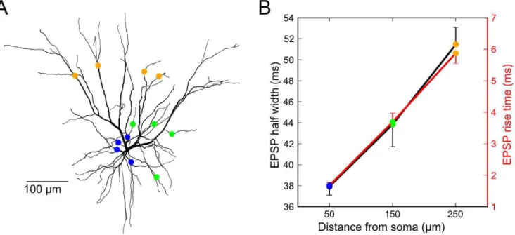
Figure 8. EPSP time-course analysis of L3 model neuron suggests that ‘‘modulatory’’ long-distance horizontal connections terminate proximally and vertical L4 ‘‘driver’’ inputs terminate distally.A,L3 model morphology [89, see Materials and Methods for details] with colored markers indicating one set of the locations of the 4 synapses evoking the responses shown in (B).B,Four synaptic inputs were placed on 100 sets of 4 randomly selected dendrites 50, 150, and 250mm from the soma, evoking 4.6, 3.6, and 2.9 mV EPSPs on average. EPSP half width and
risetime grew with distance from the soma. Error bars indicate s.d. of the mean across random dendritic sets. Compare to Table 1 from Yoshimura et al. [47] showing that EPSP half widths between modulatory long-distance horizontal and vertical L4 ‘‘driver’’ inputs increased (34.5619.9 vs. 53.0628.1ms, p,0.05, t-test) as did EPSP rise times (3.962.5ms vs. 562.5ms, p,0.04, Wilcoxon rank-sum test, Figure 2B rise time data from Yoshimura et al. [47] was digitized with Engauge Digitizer for statistical analysis in Matlab).
modulation-by-location in its pure form would be locally invisible (i.e. ‘‘dark’’), in the sense that under the microscope, dendrites would appear to be lined with an undifferentiated population of excitatory synapses. Only when the remote source of each synaptic contact has been traced, could the nature – or even the existence – of the location-based computation be inferred. The possibility that analog location-dependent computations do routinely occur within the dendrites of neocortical PNs, that contribute to the modulation of PN response by a multitude of attentional, contextual, and cross-modal influences, highlights the continuing need for multi-disciplinary approaches in analyzing neocortical circuits.
Materials and Methods
Multi-compartment modeling
All simulations were performed using the NEURON modeling package (version 7.0 r276) [86]. Unless otherwise indicated, all simulation studies utilized a 3D reconstructed layer 5 pyramidal cell morphology (see Figures 2E inset and 4A) that was a smoothed version of the ‘‘j4’’ morphology [87,88], to which a myelinated axon was added to model axonal spike initiation [89]. Ion channel models and distributions were constrained by a variety of data [28,30,38,90,91]. Parameters are shown in Table 1. NEURON files are available upon request.
Excitation was delivered at varying distances from the soma through combined NMDAR/AMPAR type synapses. The AMPA component in each synapse had a fixed peak conductance while the NMDA peak conductance doubled (2.23 to 4.46 nS) from the first to second pulse in 50 Hz double-pulse stimulation experi-ments (Figures 3–6).Values were fit based on measured physio-logical summation nonlinearities for single and double pulse stimulation experiments [30], were in keeping with increases in NMDA conductance upon repeated stimulation [92], and non-saturation of the NMDA receptor [93]. Both AMPA and NMDA conductances were modeled as difference-of-exponential functions with kinetics appropriate for 35uC (see Table 1). The NMDA channel model included an instantaneous voltage-dependent Mg-block of the form B(V) = 1/(1+e2(V+12)/10
). Hodgkin-Huxley style sodium and potassium conductances were included in the axon, soma and dendrites, with the sodium conductance decreasing linearly to zero at a distance of 200mm from the soma [38]. For
single-pulse simulations mimicking single pulse UV glutamate uncaging in Figure 2, NMDA peak conductance was set to 3.56 nS to match the NRLE of thein vitrodata.
Synapse clusters were centered at specified locations with 0.5mm spacing [34]. Terminal dendrites were corrected for the membrane area contribution of unmodeled spines by increasing membrane capacitance and conductance by a factor of 2.0 [34]. In simulations with NMDAR block, the NMDA channel peak conductance was set to 0.
The axon, soma, and all dendritic subtrees containing activated synapses were divided into electrical compartments, or ‘‘segments’’ of length no greater than one tenth of the section’s length constant at 100 Hz [94], or 10mm - whichever was smaller. In other
dendrites, 3 segments were used per section without loss of simulation accuracy.
Suprathreshold (spike rate) results used the same model as above except for the excitation which was in the form of unsynchronized 50 Hz Poisson trains, and the peak NMDA conductance was fixed to 3.9 nS. To achieve a low background firing rate (,1 Hz), the
axo-somatic spike generating mechanism the soma was biased with a noisy current injection (0.7561 nA) updated every integration time step (0.1 ms).Spike rates were averaged over the 500 ms stimulus period.
NRLE measure
For each data point (xi, yi) beginning with the second point on each input-output curve, a line was fit to all preceding data points, and extrapolated to the point (xi+1, yextrap). The ratio of the actual y value to the linearly extrapolated y value yi+1/yextrap was computed, and the maximum of this ratio along a given input-output curve was taken as the NRLE for that curve.
EPSP time-course analysis
The EPSP study in Figure 8 utilized a published L3 model [89] with the following changes: 1) Spine correction was changed to be the same as described above, which does not distort the morphology, enabling our analysis of EPSP properties versus distance, and 2) Rmand Cmwere increased by a factor of 1.6 so that the EPSP half-width ranges in the model were similar to those in Yoshimura et al. [47]. Synapses were AMPA-type only and were modeled as difference-of-exponential functions with trise,fall= 0.2,2 ms and 2 nS peak conductance. EPSP properties were similar when synapses contained mixed NMDA/AMPA conductances similar to those used elsewhere in the paper (data not shown).
2-compartment circuit analysis
Time-invariant voltage responses were calculated using methods described elsewhere [85], but with the addition of a second NMDA conductance (see Figure 4C). The Kirchhoff’s current law equations were as follows:
IdistzIprox~0,
Table 1.Model parameters.
Property Value References
Passive Properties
Rm dendrites: 10 kVcm2 [89]
axon nodes: 50Vcm2 [89]
other: 20 kVcm2 [89]
Cm dendrites: 2mF/cm2 [89]
myelinated axon: 0.05mF/cm2 [89]
other: 1mF/cm2 [89]
Ra 100Vcm [89]
Eleak 270 mV
Active Properties
g
gNa dendrites: 0.006 S/cm2 [38]
non-myelinated axon: 5 S/cm2 [38]
myelinated axon: 0.006 S/cm2 [38]
soma: 0.25 S/cm2 [38]
g
gK dendrites: 0.0003 S/cm2 [29]
non-myelinated axon: 0.05 S/cm2 [29]
soma: 0.03 S/cm2 [29]
ENa +60 mV
EK 290 mV
Synapses AMPAR gmax= 1.5 nS [38,90,91]
trise,fall= 0.05, 0.5 ms [38,90,91]
NMDAR gmax= 3.56, or 3.9 nS [30,92,93,95]
trise,fall= 2.1, 18.8 ms [30,92,93,95]
EAMPA/NMDAR 0 mV
doi:10.1371/journal.pcbi.1002599.t001
where
Idist~(Vdist{ENMDA)Nsyn dist
g
gNMDAB(Vdist)z(Vdist{Eleak)gleak dist
and
Iprox~(Vprox{ENMDA)Nsyn prox
g
gNMDAB(Vprox)z(Vprox{Eleak)gleak prox ð1Þ
By exploiting the relationship between Vproxand Vdist:
Vprox~VdistzIdist=ga ð2Þ
to eliminate the dependence on Vprox in Eq. (1) the resulting equation was solved numerically for Vdist, then Vprox was computed using Eq. (2). Here, B(V) = 1/(1+e2(V+22)/12
), slightly ‘softer’ than the magnesium block term used in the multi-compartment model. This was done to account for the ‘‘dilution’’ of the NMDA voltage-dependent non-linearity by co-activated AMPA channels, which were not explicitly included in the 2-compartment model. In Figure S1 in Text S1, CTO, the ‘current-to-overcome’, was defined as –(Isoma-INMDA_Drive), corresponding to the ‘net leak’ in the control and modulation conditions.
Slice preparation and electrophysiological recording
Neocortical brain slices 300–350mm thick were prepared from
18- to 28-day-old Wistar rats. All experimental procedures were in accordance with guidelines of the Technion Institutional Animal Care and Use Committee. Extracellular solution contained 125 mM NaCl, 25 mM NaHCO3, 25 mM glucose, 3 mM KCl, 1.25 mM NaH2PO4, 2 mM CaCl2 and 1 mM MgCl2 (pH 7.4) at 35–36uC. Intracellular solution contained 115 mM K+-gluconate,
20 mM KCl, 2 mM Mg-ATP, 2 mM ATP, 10 mM Na2-phosphocreatine, 0.3 mM GTP, 10 mM HEPES and 0.15 mM Calcium Green-1 (CG-1) or 0.2 mM Oregon Green 488 Bapta-1 (OGB-1), pH 7.2. GABAA receptor blocker bicuculline methio-dide (BCC; 1–20mM) was added to the extracellular solution in some experiments. Whole-cell patch-clamp recordings were made from visually identified layer-5 pyramidal neurons using infrared-differential interference contrast optics. Electrophysiological re-cordings were performed using Multi-Clamp 700A (Axon Instru-ments, Foster City, CA), and the data were acquired and analyzed using Pclamp 8.2 (Axon Instruments), Igor (Wavemetrics, Lake Oswego, OR), and in-house software. All statistical analyses used the Student’s t-test.
Focal stimulation
The neurons were filled with a calcium-sensitive dye (CG-1 or OGB-1) and the basal dendritic tree was imaged with a confocal imaging system (Olympus Fluoview) mounted on an upright BX51WI Olympus microscope (Tokyo, Japan) equipped with a 606(0.9 n.a.; Olympus) water objective. The theta stimulating
electrodes were filled with Alexa Fluor 647. Full images were obtained with a temporal resolution of 1 Hz and in the line scan mode with a temporal resolution of 512 Hz. Images were analyzed using Tiempo (Olympus), Igor (Wavemetrics), and in-house software. Focal synaptic stimulation was performed with a theta patch pipette located in close proximity to the selected basal dendritic segment, as guided by the fluorescent image of the dendrite. We limited ourselves to dendritic regions that were more
distal than the initial 50-mm segment of the basal dendrites, as we
could not obtain focal synaptic activation in those regions.
Glutamate uncaging
For the uncaging experiments, caged glutamate (4-methoxy-7-nitroindolinyl(MNI)-glutamate; Tocris, San Diego, CA) was photolyzed with a 361 nm UV-laser beam (Enterprise 2; Coherent, Palo Alto, CA) using point scan mode. The caged glutamate (5–10 mM) was delivered locally to a branch using pressure ejection (5–10 mbar) from an MNI-glutamate-containing electrode (2mm diameter).
Normalization of two-input data using a fiducial point template
The four fiducial points indicated by shapes in Figure 5D,E were used to normalize the data from each of the 6 cells (Figure 5F,G). The normalization results are shown in Figure 6 C,D. The square and pentagon indicate subthreshold and just-suprathreshold responses for the proximal spike alone, the triangle indicates the just-suprathreshold response for the distal spike alone, and the circle was just-suprathreshold for the distal stimulus when the proximal bias was simultaneously just-subthreshold for its own spike. We noted that over the data set: (1) the proximal spike was more than twice the height of the distal spike (compare height of pentagon and triangle); (2) the proximal just-subthreshold response was about 2/3 the height of the distal spike response (compare y-coordinates of square and triangle); and (3) the threshold for spike generation by a distal input was roughly cut in half when boosted by a just-subthreshold proximal bias (compare x-coordinates of circle and triangle. Given these observations, we created a template set of fiducial points based on the average ratios found in the experiments: triangle = (1, 1); square = (0, 0.6); pentagon = (0, 2.4); circle = (0.6, 2.2).For any given cell, the 2-D data (distal drive, proximal modulation) was scaled using the horizontal and vertical scaling factors that minimized the MSE between actual and template fiducial points. Note that only 2 scaling factors found through MSE minimization for each cell were used to scale all 29–56 data points for that cell. Overfitting was thus avoided. It is worth noting that we previously attempted a more ‘intuitive’ normalization procedure based on only the two fiducial points corresponding to the proximal and distal spikes, but because that normalization scheme did not capture threshold lowering and spike boosting affects of ‘medium’ strength modulators, that approach resulted in a poorer match between the data and the model. Thus, we used the additional fiducial points which capture more of the relevant features of each experimental data set.
The data viewed from the orthogonal perspective (proximal driver, distal modulation) was more uniform, so that only a single fiducial point was needed for normalization: each plot was rigidly scaled to place the pentagon at the point (1, 2.4) (Figure 6D).
Supporting Information
Text S1 Supplementary Figures S1-S4. (PDF)
Acknowledgments
Author Contributions
Conceived and designed the experiments: BFB AP JS BWM. Performed the experiments: AP JS. Analyzed the data: BFB AP. Contributed reagents/materials/analysis tools: MJ. Wrote the paper: BFB BWM.
References
1. Mountcastle VB (1997) The columnar organization of the neocortex. Brain 120 (Pt 4): 701–722.
2. Lu¨bke J, Roth A, Feldmeyer D, Sakmann B (2003) Morphometric analysis of the columnar innervation domain of neurons connecting layer 4 and layer 2/3 of juvenile rat barrel cortex. Cereb Cortex 13: 1051–1063.
3. Binzegger T, Douglas RJ, Martin KAC (2004) A quantitative map of the circuit of cat primary visual cortex. J Neurosci 24: 8441–8453.
4. Kapadia MK, Ito M, Gilbert CD, Westheimer G (1995) Improvement in visual sensitivity by changes in local context: parallel studies in human observers and in V1 of alert monkeys. Neuron 15: 843–856.
5. Angelucci A, Bressloff PC (2006) Contribution of feedforward, lateral and feedback connections to the classical receptive field center and extra-classical receptive field surround of primate V1 neurons. Prog Brain Res 154: 93–120. 6. McAdams CJ, Maunsell JH (2000) Attention to both space and feature modulates
neuronal responses in macaque area V4. J Neurophysiol 83: 1751–1755. 7. Reynolds JH, Heeger DJ (2009) The normalization model of attention. Neuron
61: 168–185.
8. Barraclough NE, Xiao D, Baker CI, Oram MW, Perrett DI (2005) Integration of visual and auditory information by superior temporal sulcus neurons responsive to the sight of actions. J Cogn Neurosci 17: 377–391.
9. Zipser D, Andersen RA (1988) A back-propagation programmed network that simulates response properties of a subset of posterior parietal neurons. Nature 331: 679–684.
10. Salinas E, Sejnowski TJ (2001) Gain modulation in the central nervous system: where behavior, neurophysiology, and computation meet. Neuroscientist 7: 430– 440.
11. Rockland KS, Lund JS (1983) Intrinsic laminar lattice connections in primate visual cortex. J Comp Neurol 216: 303–318.
12. Gilbert CD, Wiesel TN (1989) Columnar specificity of intrinsic horizontal and corticocortical connections in cat visual cortex. J Neurosci 9: 2432–2442. 13. Boucsein C, Nawrot MP, Schnepel P, Aertsen A (2011) Beyond the cortical
column: abundance and physiology of horizontal connections imply a strong role for inputs from the surround. Front Neurosci 5: 32.
14. Silver RA (2010) Neuronal arithmetic. Nat Rev Neurosci 11: 474–489. 15. Chance FS, Abbott LF, Reyes AD (2002) Gain modulation from background
synaptic input. Neuron 35: 773–782.
16. Abbott LF, Chance FS (2005) Drivers and modulators from push-pull and balanced synaptic input. Prog Brain Res 149: 147–155.
17. Haider B, McCormick DA (2009) Rapid neocortical dynamics: cellular and network mechanisms. Neuron 62: 171–189.
18. Larkum ME, Zhu JJ, Sakmann B (1999) A new cellular mechanism for coupling inputs arriving at different cortical layers. Nature 398: 338–341.
19. Larkum ME, Zhu JJ, Sakmann B (2001) Dendritic mechanisms underlying the coupling of the dendritic with the axonal action potential initiation zone of adult rat layer 5 pyramidal neurons. J Physiol (Lond) 533: 447–466.
20. Larkum ME, Senn W, Lu¨scher H-R (2004) Top-down dendritic input increases the gain of layer 5 pyramidal neurons. Cereb Cortex 14: 1059–1070. 21. Remondes M, Schuman EM (2002) Direct cortical input modulates plasticity
and spiking in CA1 pyramidal neurons. Nature 416: 736–740.
22. Jarsky T, Roxin A, Kath WL, Spruston N (2005) Conditional dendritic spike propagation following distal synaptic activation of hippocampal CA1 pyramidal neurons. Nat Neurosci 8: 1667–1676.
23. Takahashi H, Magee JC (2009) Pathway interactions and synaptic plasticity in the dendritic tuft regions of CA1 pyramidal neurons. Neuron 62: 102–111. 24. Mel BW (1992) NMDA-based pattern discrimination in a modeled cortical
neuron. Neural Comput 4: 502–517.
25. Mel BW (1993) Synaptic integration in an excitable dendritic tree. J Neurophysiol 70: 1086–1101.
26. Mel BW, Ruderman DL, Archie KA (1998) Translation-invariant orientation tuning in visual ‘‘complex’’ cells could derive from intradendritic computations. J Neurosci 18: 4325–4334.
27. Archie KA, Mel BW (2000) A model for intradendritic computation of binocular disparity. Nat Neurosci 3: 54–63.
28. Poirazi P, Brannon T, Mel BW (2003) Arithmetic of subthreshold synaptic summation in a model CA1 pyramidal cell. Neuron 37: 977–987.
29. Poirazi P, Brannon T, Mel BW (2003) Pyramidal neuron as two-layer neural network. Neuron 37: 989–999.
30. Polsky A, Mel BW, Schiller J (2004) Computational subunits in thin dendrites of pyramidal cells. Nat Neurosci 7: 621–627.
31. Losonczy A, Magee JC (2006) Integrative properties of radial oblique dendrites in hippocampal CA1 pyramidal neurons. Neuron 50: 291–307.
32. Larkum ME, Nevian T, Sandler M, Polsky A, Schiller J (2009) Synaptic integration in tuft dendrites of layer 5 pyramidal neurons: a new unifying principle. Science 325: 756–760.
33. Branco T, Clark BA, Ha¨usser M (2010) Dendritic discrimination of temporal input sequences in cortical neurons. Science 329: 1671–1675.
34. Larkman AU (1991) Dendritic morphology of pyramidal neurones of the visual cortex of the rat: III. Spine distributions. J Comp Neurol 306: 332–343. 35. Sherman SM, Guillery RW (1998) On the actions that one nerve cell can have
on another: distinguishing ‘‘drivers’’ from ‘‘modulators.’’ Proc Natl Acad Sci U S A 95: 7121–7126.
36. Rall W, Rinzel J (1973) Branch input resistance and steady attenuation for input to one branch of a dendritic neuron model. Biophys J 13: 648–687. 37. Zador AM, Agmon-Snir H, Segev I (1995) The morphoelectrotonic transform: a
graphical approach to dendritic function. J Neurosci 15: 1669–1682. 38. Nevian T, Larkum ME, Polsky A, Schiller J (2007) Properties of basal dendrites
of layer 5 pyramidal neurons: a direct patch-clamp recording study. Nat Neurosci 10: 206–214.
39. Branco T, Ha¨usser M (2011) Synaptic integration gradients in single cortical pyramidal cell dendrites. Neuron 69: 885–892.
40. Schiller J, Major G, Koester HJ, Schiller Y (2000) NMDA spikes in basal dendrites of cortical pyramidal neurons. Nature 404: 285–289.
41. Oakley JC, Schwindt PC, Crill WE (2001) Dendritic calcium spikes in layer 5 pyramidal neurons amplify and limit transmission of ligand-gated dendritic current to soma. J Neurophysiol 86: 514–527.
42. Milojkovic BA, Zhou W-L, Antic SD (2007) Voltage and calcium transients in basal dendrites of the rat prefrontal cortex. J Physiol (Lond) 585: 447–468. 43. Rhodes P (2006) The properties and implications of NMDA spikes in neocortical
pyramidal cells. J Neurosci 26: 6704–6715.
44. Major G, Polsky A, Denk W, Schiller J, Tank DW (2008) Spatiotemporally graded NMDA spike/plateau potentials in basal dendrites of neocortical pyramidal neurons. J Neurophysiol 99: 2584–2601.
45. Antic SD, Zhou W-L, Moore AR, Short SM, Ikonomu KD (2010) The decade of the dendritic NMDA spike. J Neurosci Res 88: 2991–3001.
46. Stuart G, Spruston N, Ha¨usser M, editors (2007) Dendrites. 2nd edition. New York: Oxford University Press. 560 p.
47. Yoshimura Y, Sato H, Imamura K, Watanabe Y (2000) Properties of horizontal and vertical inputs to pyramidal cells in the superficial layers of the cat visual cortex. J Neurosci 20: 1931–1940.
48. Kapadia MK, Westheimer G, Gilbert CD (2000) Spatial distribution of contextual interactions in primary visual cortex and in visual perception. J Neurophysiol 84: 2048–2062.
49. Fleidervish IA, Binshtok AM, Gutnick MJ (1998) Functionally distinct NMDA receptors mediate horizontal connectivity within layer 4 of mouse barrel cortex. Neuron 21: 1055–1065.
50. Reichova I, Sherman SM (2004) Somatosensory corticothalamic projections: distinguishing drivers from modulators. J Neurophysiol 92: 2185–2197. 51. Li J, Guido W, Bickford ME (2003) Two distinct types of corticothalamic EPSPs
and their contribution to short-term synaptic plasticity. J Neurophysiol 90: 3429–3440.
52. Polsky A, Mel BW, Schiller J (2009) Encoding and decoding bursts by NMDA spikes in basal dendrites of layer 5 pyramidal neurons. J Neurosci 29: 11891– 11903.
53. McAdams CJ, Maunsell JHR (1999) Effects of attention on orientation-tuning functions of single neurons in macaque cortical area V4. J Neurosci 19: 431–441. 54. Nelson JI, Frost BJ (1985) Intracortical facilitation among co-oriented, co-axially
aligned simple cells in cat striate cortex. Exp Brain Res 61: 54–61.
55. Polat U, Mizobe K, Pettet MW, Kasamatsu T, Norcia AM (1998) Collinear stimuli regulate visual responses depending on cell’s contrast threshold. Nature 391: 580–584.
56. Dudman JT, Tsay D, Siegelbaum SA (2007) A role for synaptic inputs at distal dendrites: instructive signals for hippocampal long-term plasticity. Neuron 56: 866–879.
57. Dvorak-Carbone H, Schuman EM (1999) Patterned activity in stratum lacunosum moleculare inhibits CA1 pyramidal neuron firing. J Neurophysiol 82: 3213–3222.
58. Ang CW, Carlson GC, Coulter DA (2005) Hippocampal CA1 circuitry dynamically gates direct cortical inputs preferentially at theta frequencies. J Neurosci 25: 9567–9580.
59. Moran J, Desimone R (1985) Selective attention gates visual processing in the extrastriate cortex. Science 229: 782–784.
60. Reynolds JH, Chelazzi L, Desimone R (1999) Competitive mechanisms subserve attention in macaque areas V2 and V4. J Neurosci 19: 1736–1753. 61. Morita K (2008) Possible role of dendritic compartmentalization in the spatial
working memory circuit. J Neurosci 28: 7699–7724.
62. Jia H, Rochefort NL, Chen X, Konnerth A (2010) Dendritic organization of sensory input to cortical neurons in vivo. Nature 464: 1307–1312.
63. Bollmann JH, Engert F (2009) Subcellular topography of visually driven dendritic activity in the vertebrate visual system. Neuron 61: 895–905.
64. Shepherd GM (2003) The Synaptic Organization of the Brain. 5th edition. USA: Oxford University Press.
65. Petreanu L, Mao T, Sternson SM, Svoboda K (2009) The subcellular organization of neocortical excitatory connections. Nature 457: 1142–1145. 66. Felleman DJ, Van Essen DC (1991) Distributed hierarchical processing in the
primate cerebral cortex. Cereb Cortex 1: 1–47.
67. Barbas H, Rempel-Clower N (1997) Cortical structure predicts the pattern of corticocortical connections. Cereb Cortex 7: 635–646.
68. Ichinohe N, Fujiyama F, Kaneko T, Rockland KS (2003) Honeycomb-like mosaic at the border of layers 1 and 2 in the cerebral cortex. J Neurosci 23: 1372–1382.
69. Kuramoto E, Furuta T, Nakamura KC, Unzai T, Hioki H, et al. (2009) Two types of thalamocortical projections from the motor thalamic nuclei of the rat: a single neuron-tracing study using viral vectors. Cereb Cortex 19: 2065– 2077.
70. Rubio-Garrido P, Pe´rez-de-Manzo F, Porrero C, Galazo MJ, Clasca´ F (2009) Thalamic input to distal apical dendrites in neocortical layer 1 is massive and highly convergent. Cereb Cortex 19: 2380–2395.
71. Andersen P, Morris R, Amaral D, Bliss T, O’Keefe J (2006) The Hippocampus Book. 1st ed. Oxford University Press, USA.
72. Somogyi P, Tama´s G, Lujan R, Buhl EH (1998) Salient features of synaptic organisation in the cerebral cortex. Brain Res Brain Res Rev 26: 113–135. 73. McBain CJ, Fisahn A (2001) Interneurons unbound. Nat Rev Neurosci 2: 11–
23.
74. Karube F, Kubota Y, Kawaguchi Y (2004) Axon branching and synaptic bouton phenotypes in GABAergic nonpyramidal cell subtypes. J Neurosci 24: 2853– 2865.
75. Gordon U, Polsky A, Schiller J (2006) Plasticity compartments in basal dendrites of neocortical pyramidal neurons. J Neurosci 26: 12717–12726.
76. Golding NL, Staff NP, Spruston N (2002) Dendritic spikes as a mechanism for cooperative long-term potentiation. Nature 418: 326–331.
77. Froemke RC, Poo M-M, Dan Y (2005) Spike-timing-dependent synaptic plasticity depends on dendritic location. Nature 434: 221–225.
78. Denk W, Horstmann H (2004) Serial block-face scanning electron microscopy to reconstruct three-dimensional tissue nanostructure. PLoS Biol 2: e329. 79. Hayworth K, Kasthuri N, Schalek R, Lichtman J (2006) Automating the
collection of ultrathin serial sections for large volume TEM reconstructions. Microsc Microanal 12: 86–87.
80. Callaway EM (2008) Transneuronal circuit tracing with neurotropic viruses. Curr Opin Neurobiol 18: 617–623.
81. Seung HS (2009) Reading the book of memory: sparse sampling versus dense mapping of connectomes. Neuron 62: 17–29.
82. Go¨bel W, Helmchen F (2007) New angles on neuronal dendrites in vivo. J Neurophysiol 98: 3770–3779.
83. Nagayama S, Zeng S, Xiong W, Fletcher ML, Masurkar AV, et al. (2007) In vivo simultaneous tracing and Ca(2+) imaging of local neuronal circuits. Neuron 53: 789–803.
84. Katz Y, Menon V, Nicholson DA, Geinisman Y, Kath WL, et al. (2009) Synapse distribution suggests a two-stage model of dendritic integration in CA1 pyramidal neurons. Neuron 63: 171–177.
85. Jadi M, Polsky A, Schiller J, Mel BW (2012) Location-Dependent effects of inhibition on local spiking in pyramidal neuron dendrites. PLoS Comput Biol: In press.
86. Carnevale NT, Hines ML (2006) The NEURON Book. Cambridge, UK: Cambridge University Press.
87. Douglas RJ, Martin KA, Whitteridge D (1991) An intracellular analysis of the visual responses of neurones in cat visual cortex. J Physiol 440: 659–696. 88. Behabadi BF, Mel BW (2007) J4 at sweet 16: a new wrinkle? Neural Comput 19:
2865–2870.
89. Mainen ZF, Sejnowski TJ (1996) Influence of dendritic structure on firing pattern in model neocortical neurons. Nature 382: 363–366.
90. Forti L, Bossi M, Bergamaschi A, Villa A, Malgaroli A (1997) Loose-patch recordings of single quanta at individual hippocampal synapses. Nature 388: 874–878.
91. Smith MA, Ellis-Davies GC, Magee JC (2003) Mechanism of the distance-dependent scaling of Schaffer collateral synapses in rat CA1 pyramidal neurons. J Physiol 548: 245–258.
92. Popescu G, Robert A, Howe JR, Auerbach A (2004) Reaction mechanism determines NMDA receptor response to repetitive stimulation. Nature 430: 790– 793.
93. Mainen ZF, Malinow R, Svoboda K (1999) Synaptic calcium transients in single spines indicate that NMDA receptors are not saturated. Nature 399: 151–155. 94. Hines ML, Carnevale NT (2001) NEURON: a tool for neuroscientists.
Neuroscientist 7: 123–135.