593
CHALLENGING CLINICAL CASES
Key words:
Urolithiasis; Urinary Diversion; Ureteral Calculi; Endoscopy
Int Braz J Urol. 2013; 39: 593-6
__________________ Submitted for publication: February 19, 2012
__________________ Accepted after revision: May 27, 2013
Lithiasis after urinary diversion is an uncommon condition that poses therapeutic chal-lenges. The authors report the case of a patient submitted to cystectomy and ureterosig-moidostomy 35 years ago due to bladder endometriosis. The patient presented with a ureteral stone and was treated by retrograde endoscopic extraction.
INTRODUCTION
Ureterosigmoidostomy has been used as a form of urinary diversion for either benign or malignant conditions. Urinary lithiasis has been reported as ureterosigmoidostomy complication in 3-10% of the cases in recent series (1,2). Bac-terial colonization and diversion-associated me-tabolic derangements are main risk factors (3) in this situation.
Treatment of urinary stones depends on clinical, anatomic and stone factors and there are many therapeutic options that might be conside-red when evaluating uncommon cases. Neverthe-less, the choice for the best approach depends on
the availability of equipment and expertise of the surgeon.
CASE REPORT
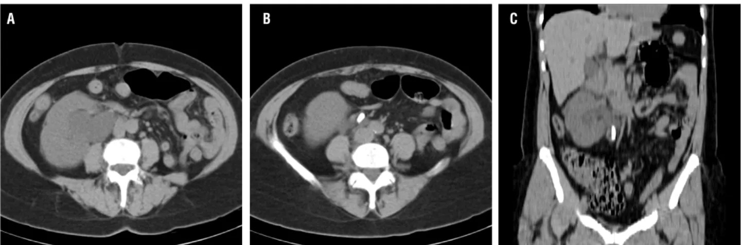
We report the case of a 62 years old woman submitted to cystectomy and ureterosigmodos-tomy 35 years ago due to bladder endometriosis. Potassium citrate was used to prevent hyperchlo-remic metabolic acidosis. The follow-up was un-remarkable until the patient’s current presentation with acute right flank pain. A CT scan showed left kidney agenesis and right kidney hydronephrosis. A 2 cm stone with 580HU was located in the pro-ximal right ureter (Figure-1).
Endoscopic management of ureteral calculus in a patient
with ureterosigmoidostomy diversion
_______________________________________________
Leonardo de Albuquerque dos Santos Abreu, Celso Lara, Marco Antonio Dionísio, Alexandre Dias
Pelosi, Fátima Aparecida Ferreira Figueiredo
University Gama Filho (LASA); Hospital Federal dos Servidores do Estado (LASA); Department of Urology - State University of Rio de Janeiro (CL, MAD, FAFF); Rede D’Or (ADP, FAFF) and Inca - Instituto Nacional de Câncer (ADP), Rio de Janeiro, Brazil
ABSTRACT
ARTICLE
INFO
_________________________________________________________ ___________________
Vol. 39 (4): 593-596, July - August, 2013
IBJU |ENDOSCOPIC MANAGEMENT OF URETERAL CALCULUS
594
An abdominal radiograph could not show the stone confirming it as radiolucent. Serum creati-nine was 1.6 mg/ dL. A percutaneous nephrostomy--ultrasound guided was emergently performed to relieve obstruction. While waiting for the definitive treatment the patient lost the nephrostomy and un-derwent a new percutaneous nephrostomy.
The initial treatment plan was to perform an anterograde holmium LASER flexible ureterolito-tripsy. Under fluoroscopic guidance a middle calyx was punctured which allowed adequate access to the ureteropelvic junction (UPJ). Dilation was carried out with axial ureteral dilators over a 0.035” guidewire and a 9.5Fr access sheath was introduced until close to the UPJ entry. When the flexible ureteroscope was introduced distal migration of the stone was noticed. Because of ureteral tortuosity we were unable to per-form LASER lithotripsy and retrieve the stone (Figu-re-2). In this procedure a retrograde access was tried, however, we were unable to catheterize the ostium.
A retrograde approach with a side view endoscope was then carried. During sigmoidosco-py, right ureteral ostium was located at a distance of approximately 15 cm from the anal verge. The ureteral ostium was catheterized with a cholangio-pancreatography catheter (Figure-3). A retrograde pyelogram confirmed the 2 cm stone in the distal ureter. A 0.035’’ hydrophilic guide wire was inser-ted and dilation of the uretero-sigmoid junction was carried with a balloon catheter (Figure-4). Af-ter dilation it was possible to see the calculus pro-truding through the ostium and a biliary balloon catheter was used to pull it down (Figure-5). The
Figure 1 - Pre-operative CT scan.
A B C
Figure 2 - Ureteroscope deflection in distal ureter.
IBJU |ENDOSCOPIC MANAGEMENT OF URETERAL CALCULUS
595
calculus had a soft appearance, however, we were unable to recover it for further composition analy-sis. A CT scan confirmed that the patient was sto-ne free postoperatively and the sto-nephrostomy was removed. Renal function returned to baseline after treatment and the patient was well until the last follow-up 1 year after the procedure.
DISCUSSION
Treatment options for ureteral stones in patients with urinary diversion range from mini-mally invasive procedures such as extracorporeal shock-wave lithotripsy (ESWL) to conventional open surgery.
Open surgery may be a definitive option, however, morbidity associated with a reoperation and the risks of complications must be considered. ESWL may be a good option for proximal ureteral stones but for radiolucent or distal stones the re-sults may not be optimal (4,5).
Chemolytic dissolution of stones or residu-al stone fragments is a useful adjunct to conventio-nal treatment. Although uric acid stones have good response to chemolytic therapy, oral chemolysis may take several days for complete dissolution (6). Moreover, urinary alcalinization may be difficult in patients with chronic metabolic acidosis. Direct chemolysis can also be done through the nephros-tomy tube. Nevertheless, the risks of infection and
metabolic complications may be increased in this particular situation.
Choosing a retrograde or anterograde ap-proach will depend on location of the stone. For stones located in medium or proximal ureter an an-terograde approach is feasible. Renal access should be carried out in medium or upper calix to allow passage of the flexible ureteroscope without angu-lation as described by Lackmichi et al. (7). However, urologists must be aware that more than one flexi-ble ureteroscope may be necessary to accomplish the procedure as was emphasized by the authors. In the reported case anterograde ureterolithotripsy was unsuccessfully because of distal migration of the stone and tortuosity of the ureter.
For stones located in the distal ureter re-trograde approach may be necessary. Fitzgerald et al. (8) described ureterolithotripsy with a semirigid ureteroscope of a large ureteral stone. A guide wire was passed through a previously placed nephros-tomy down the sigmoid/rectum which helped to locate and dilate the ureterointestinal junction. Mosler et al. (9) were the first to report the use of a side view duodenoscope for retrograde approa-ch. Insufflation with air during endoscopy helped to locate the ureterointestinal junction allowing dilation of the anastomosis and removal of cal-culus with basket without lithotripsy. Hyams et al. (10) reported successful retrograde access in 21 of 28 attempts without the aid of previous renal
IBJU |ENDOSCOPIC MANAGEMENT OF URETERAL CALCULUS
596
percutaneous access. A flexible ureteroscope was also used to retrieve ureteral stones. In the reported case a side view duodenoscope was used for retro-grade access and we also found that air insufflation helped location and dilation of the ureterointestinal junction. Lesion of ureterosigmoid junction during dilation for retrograde access might be of concern. Nevertheless, complications were not reported on Hyams’ series, as well as in our case.
In the reported case lithotripsy was unne-cessary. Nevertheless, flexible or semirigid urete-roscopes with the aid of LASER or other lithotrip-ters may be used whenever necessary.
CONCLUSIONS
Endoscopic management of ureteral sto-nes after urinary diversion is feasible and can be achieved with little morbidity. The use of retrogra-de or anterograretrogra-de approach for ureteral stones af-ter urinary diversions should combine endoscopic and endourologic technics in a multidisciplinary environment.
CONFLICT OF INTEREST
None declared.
REFERENCES
1. Tollefson MK, Elliott DS, Zincke H, Frank I: Long-term out-come of ureterosigmoidostomy: an analysis of patients with >10 years of follow-up. BJU Int. 2010; 105: 860-3.
2. Nitkunan T, Leaver R, Patel HR, Woodhouse CR: Modified ure-terosigmoidostomy (Mainz II): a long-term follow-up. BJU Int. 2004; 93: 1043-7.
3. Okhunov Z, Duty B, Smith AD, Okeke Z: Management of uroli-thiasis in patients after urinary diversions. BJU Int. 2011; 108: 330-6.
4. Maker V, Layke J: Gastrointestinal injury secondary to extra-corporeal shock wave lithotripsy: a review of the literature since its inception. J Am Coll Surg. 2004; 198: 128-35. 5. Cardinaux C, Tawadros T, Praz V, Wisard M, Treuthardt C,
Ji-chlinski P: Ischemic cecal perforation secondary to ESWL of junctional stone in ureterosigmoidostomy (Mainz Pouch II). Urology. 2009; 73: 1423.e1-2.
6. Bernardo NO, Smith AD: Chemolysis of urinary calculi. Urol Clin North Am. 2000; 27: 355-65.
7. Lackmichi MA, Niang L, Labou I, Thibault F, Ravery V, Gat-tegno B, et al.: Anterograde flexible ureteroscopy for stones of the uretero-sigmoid junction of a Mainz II pouch. Prog Urol. 2006; 16: 505-7.
8. Fitzgerald KB, Aslan P, Preminger GM: Endourological man-agement of a large distal ureteral calculus in a patient with ureterosigmoidostomy diversion. J Urol. 1998; 159: 2081-2. 9. Mosler P, Kiesslich R, Stein R, Galle PR, Thüroff JW, Kanzler
S: Use of a duodenoscope in the management of a ureteral calculus in patient with ureterosigmoidostomy (Mainz pouch II;rectosigmoid pouch). Endoscopy. 2003; 35: 1086-7. 10. Hyams ES, Winer AG, Shah O: Retrograde ureteral and renal
ac-cess in patients with urinary diversion. Urology. 2009; 74: 47-50.
_____________________
Correspondence address: