Structure of Importin-
α
from a Filamentous
Fungus in Complex with a Classical Nuclear
Localization Signal
Natalia E. Bernardes1☯, Agnes A. S. Takeda1☯, Thiago R. Dreyer1, Fernanda Z. Freitas2, Maria Célia Bertolini2, Marcos R. M. Fontes1
*
1Departamento de Física e Biofísica, Instituto de Biociências, Universidade Estadual Paulista, UNESP, Botucatu, SP, Brazil,2Departamento de Bioquímica e Tecnologia Química, Instituto de Química, Universidade Estadual Paulista, UNESP, Araraquara, SP, Brazil
☯These authors contributed equally to this work. *[email protected]
Abstract
Neurospora crassais a filamentous fungus that has been extensively studied as a model or-ganism for eukaryotic biology, providing fundamental insights into cellular processes such as cell signaling, growth and differentiation. To advance in the study of this multicellular or-ganism, an understanding of the specific mechanisms for protein transport into the cell nu-cleus is essential. Importin-α(Imp-α) is the receptor for cargo proteins that contain specific
nuclear localization signals (NLSs) that play a key role in the classical nuclear import path-way. Structures of Imp-αfrom different organisms (yeast, rice, mouse, and human) have
been determined, revealing that this receptor possesses a conserved structural scaffold. However, recent studies have demonstrated that the Impαmechanism of action may vary
significantly for different organisms or for different isoforms from the same organism. There-fore, structural, functional, and biophysical characterization of different Impαproteins is
nec-essary to understand the selectivity of nuclear transport. Here, we determined the first crystal structure of an Impαfrom a filamentous fungus which is also the highest resolution
Impαstructure already solved to date (1.75Å). In addition, we performed calorimetric
analy-sis to determine the affinity and thermodynamic parameters of the interaction between
Imp-αand the classical SV40 NLS peptide. The comparison of these data with previous studies
on Impαproteins led us to demonstrate thatN. crassaImp-αpossess specific features that
are distinct from mammalian Imp-αbut exhibit important similarities to rice Imp-α,
particular-ly at the minor NLS binding site.
Introduction
The filamentous fungusNeurospora crassahas been studied by classical and molecular genet-ics, providing several insights into cellular processes, which include cell signaling, growth and differentiation, secondary metabolism, circadian rhythm and genome defense [1]. Together
a11111
OPEN ACCESS
Citation:Bernardes NE, Takeda AAS, Dreyer TR, Freitas FZ, Bertolini MC, Fontes MRM (2015)
Structure of Importin-αfrom a Filamentous Fungus in
Complex with a Classical Nuclear Localization Signal. PLoS ONE 10(6): e0128687. doi:10.1371/journal. pone.0128687
Academic Editor:Beata G Vertessy, Institute of Enzymology of the Hungarian Academy of Science, HUNGARY
Received:February 19, 2015
Accepted:April 29, 2015
Published:June 19, 2015
Copyright:© 2015 Bernardes et al. This is an open access article distributed under the terms of the
Creative Commons Attribution License, which permits unrestricted use, distribution, and reproduction in any medium, provided the original author and source are credited.
Data Availability Statement:The coordinates and structure factors from the structure (impotin-alpha from N. crassa-SV40NLS) have been deposited in
the protein data bank data base (www.pdb.org) under
accession code 4RXH.
Funding:This work was supported by Fundação de
Amparo a Pesquisa do Estado de São Paulo (www.
fapesp.br: MRMF, MCB, NEB), Conselho Nacional de
Desenvolvimento Científico e Tecnológico (www.
with the yeastsSaccharomyces cerevisiaeandSchizosaccharomyces pombe,N. crassahas largely been used as a model organism to study fundamental aspects of eukaryotic biology. Neverthe-less, compared to yeast,N. crassahas developed greater morphological and developmental complexity due its multicellular characteristics, and althoughN. crassais not known to be a pathogen, similarities exist between the saprotrophic Neurospora and pathogenic fungi [1].
The genomic sequence ofN. crassaexhibits an apparent lack of functional gene duplication [1], which has allowed several studies to better understand the metabolic pathways of this ex-perimentally tractable organism. We have usedN. crassaas a model organism to understand the regulation of basic cellular processes, such as glycogen metabolism. Many proteins and transcription factors have been identified that likely play a role in this process [2,3]. Some of these proteins and all transcription factors possess putative nuclear localization signals (NLSs), either monopartite or bipartite, suggesting that they can be transported into the nucleus via the classical nuclear pathway [3], which is mediated by importins (importin-α/importin-β) [4,5].
The classical NLSs (cNLSs) are the best characterized targeting signals and are typically di-vided into one (monopartite) or two (bipartite) clusters of basic residues [6], whose consensus sequences correspond to K[K/R]X[K/R] for monopartite cNLSs and [K/R][K/R]X10_12[K/R]3/5
for bipartite cNLSs, where [K/R]3/5corresponds to at least three of either lysine or arginine
res-idues of five consecutive amino acids [6–8]. Structural studies have shown that both classes are recognized by the nuclear import receptor importin-α(Impα) [7,9,10]. Impαis composed of tandem armadillo (Arm) repeats that generate a curved and elongated shape. Impαalso con-tains conserved residues that form two NLS binding sites known as major (Arms 1–4) and minor (Arms 4–8) NLS binding sites [9,10]. Monopartite NLSs primarily interact with the major binding site, whereas bipartite NLSs interact with both minor and major NLS binding sites [10–12].
The modulation of differential expression is a key feature for understanding the metabolism and growth of an organism [13,14], and an understanding of the nuclear import mechanism of fungi may provide insights into these pathways. Although, Impαstructures have been eluci-dated, an understanding of the specificity of NLS recognition and regulation remains limited [15]. To better understand NLS recognition and gain insights into theN. crassanuclear import process, we report the first crystal structure of Impαfrom a filamentous fungus (NcImpα) in complex with a classical monopartite NLS (SV40 large T antigen NLS). This is also the highest resolution Impαstructure solved to date (1.75Å). Isothermal titration calorimetric (ITC)
stud-ies were used to determine the affinity and thermodynamic parameters of the interaction be-tween Impαand the classical SV40 NLS peptide. Both crystallographic and calorimetric analyses confirmed NcImpαrecognition of cNLS via both major and minor binding sites. Moreover, we observed interactions in the minor binding site that are more similar to those in Impαfrom rice (Oryza sativa) [16] than those in Impαfrom yeast [9]. This study highlights the importance of the analysis of the interactions between NLSs and Impαproteins from vari-ous organisms. Particularly, it may aid in the identification of specific NLSs fromN. crassa, providing insights into the nuclear import pathway in fungi and enabling future biotechnologi-cal applications.
Materials and Methods
Protein expression and purification
The recombinant Impαprotein fromN. crassa(NcImpα) was expressed as a truncated hexa-His fusion protein, consisting of residues 75–529, using theEscherichia colihost strain Roset-taTM (DE3) pLysS (Novagen), as previously described [17]. The protein was purified using nickel affinity chromatography and eluted in a 0.15–3.0 M imidazole linear gradient, followed
Aperfeiçoamento de Pessoal de Nível Superior (www.
capes.br: AAST).
by dialysis. NcImpαwas stored under cryogenic temperatures in a buffer composed of 20 mM Tris-HCl, pH 8, and 100 mM NaCl.
Synthesis of NLS peptides
The peptide corresponding to SV40 NLS (125PPKKKRKV132) was synthesized by Proteimax (Brazil) with a purity of higher than 99%. The peptides contained additional residues at the N-and C-termini compared with the minimally identified NLS [18].
Isothermal titration calorimetry
ITC measurements were performed using a MicroCal iTC200 microcalorimeter (GE Health-care) calibrated according to the manufacturer0s instructions. NcImpαand the SV40 NLS pep-tide were prepared and dialyzed in buffer (20 mM Tris-HCl, pH 8.0, and 100 mM NaCl). The sample cell was loaded with 50μM NcImpαthat was titrated with the SV40 NLS peptide at a concentration of 1 mM (protein:peptide molar ratio of 1:20). Titrations were conducted at 10°C and consisted of 20 injections of 2.0μL in an interval of 240 s with a 1000 rpm homogeni-zation speed. The heat of dilution was determined by titration of the peptide sample into the protein sample buffer (20mM Tris-HCl, pH 8.0, and 100 mM NaCl) in separate control assays and was subtracted from the corresponding titrations. The assays temperature was chosen to avoid protein aggregation displayed for higher temperatures and to permit direct comparison to previously Impα/SV40NLS ITC studies performed in the same condition [19]. The data were processed using MicroCal Origin Software to obtain values for stoichiometry (N), dissoci-ation constants (Kd), enthalpy (ΔH), and binding-type input parameters were adjusted to ob-tain the best fitting model. The values ofKdandΔH were used to calculate free energy (ΔG) and entropy (ΔS) values.
Crystallization, X-ray data collection and structure determination
NcImpαwas concentrated to 12 mg/ml using an Amicon 30 kDa cutoff filter unit (Millipore) and stored at -20°C. Crystals of NcImpαin complex with SV40 NLS were obtained at a 1:8 pro-tein:peptide molar ratio in 20 mM Bicine, pH 8.5, and 20% (w/v) polyethylene glycol 6000 at 4°C [20] using MRC2 Well Crystallization Plates and an Orix4 system (Douglas Instruments).
X-ray diffraction data were collected from a single crystal of NcImpα/SV40NLS at a wave-length of 1.0Åusing a synchrotron radiation source (X25 beamline, National Synchrotron Light Source, NSLS, Upton, NY, USA) and a PILATUS detector. A crystal was mounted in a nylon loop and flash-cooled in a stream of nitrogen at -173.15°C using 20% (v/v) glycerol as the cryoprotectant. The crystal-to-detector distance was 270 mm with an oscillation range of 0.5°, resulting in the collection of a total of 720 images. The data were processed using the HKL2000 suite [21]. The crystal belonged to the space groupP212121(Table 1) and was
iso-morphous to previously obtained crystals [20].
PDB accession code
Coordinates and structure factors from NcImpα/SV40NLS have been deposited in the PDB under accession code 4RXH.
Results
Structure of NcImp
α
in complex with SV40NLS
NcImpαwas expressed as an N-terminal truncation lacking residues 1–74 (which corresponds to the IBB domain; [30]) that are responsible for autoinhibition. Furthermore, crystallization was performed in the presence of an NLS ligand to stabilize the truncated protein, as previously reported [17,20], and to investigate the protein-NLS binding features.
X-ray diffraction data collection and refinement statistics of the NcImpα/SV40 NLS com-plex are summarized inTable 1. The crystals are not isomorphous to other Impα/SV40
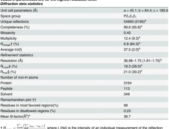
Table 1. X-ray data-collection and refinement statistics for NcImpα/SV40NLS structure.
Values in parentheses are for the highest resolution shell.
Diffraction data statistics
Unit cell parameters (Å) a = 45.1; b = 64.4; c = 185.6
Space group P212121
Unique reflections 54993 (5195)a
Completeness (%) 99.6 (95.8)a
Mosaicity 0.40
Multiplicity 12.4 (9.3)a
Rmerge†(%) 6.8 (84.3)a
Average I/σ(I) 37.5 (2.0)a
Refinement statistics
Resolution (Å) 36.98–1.75 (1.81–1.75)a
Rcryst‡(%) 18.3 (28.5)a
Rfree§ (%) 21.0 (30.2)a
Number of non-H atoms
Protein 3164
Peptide 113
Solvent 349
Ramachandran plot††
Residues in most favored regions(%) 99
Residues in disallowed regions (%) 0.23
Mean B-factor(Å2)e 36.7
†Rmerge¼
P hkl
P
iðjIhkl;i <Ihkl>jÞ
P
hkl;i<Ihkl> , whereIi(hkl) is the intensity of an individual measurement of the reflection
with Miller indices hkl and hI(hkl)i is the mean intensity of this reflection. Calculated for I>-3σ(I) [21].
‡Rcryst¼
P
hklðjjFobshklj jFcalchkljÞ
jFobshklj , wherejFobsjandjFcalcjare the observed and calculated structure-factor
amplitudes, respectively.
§Rfreeis equivalent toRcrystbut was calculated with reflections (5%) omitted from the refinement process.
Calculated based on the Luzzati plot with the program SFCHECK [31].
† †Calculated with the program PROCHECK [31].
aValues in parentheses are for the highest-resolution shell.
complexes [10] and diffracted to high resolution (1.75Å), which is the highest resolution ob-tained for an Impαto date.
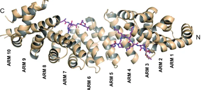
The final model of the NcImpα/SV40NLS complex consists of 428 residues of NcImpα(79–
507), the peptide ligands bound to the major (seven residues) and minor (four residues) sites, and 349 water molecules (Table 1). The structure exhibits an elongated and curved shape com-posed of ten tandem Arm repeats, each containing threeα-helices (H1, H2 and H3;), as ob-served in other Impαstructures [9,10,16,30,32] (Fig 1). The loop containing residues 461–
465 could not be modeled due an absence of electron density. The concave surface of the pro-tein maintains the conserved array of Trp and Asn residues and negatively charged residues that interact with positively charged residues from the NLS ligands.
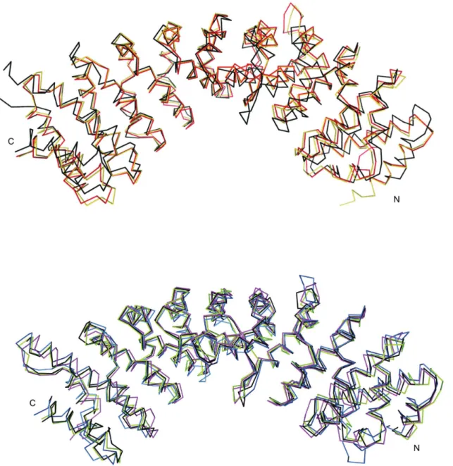
Superposition of the Cαatoms of NcImpα(residues 83–505) with other importin-α struc-tures results in a root-mean-square deviation (r.m.s.d.) of 1.78Åfor ImpαfromM. musculus (MmImpα; 3UL1; [33]), 1.77Åfor human Impα5 (HsImpα5; 4WV6; [34]), 1.22Åfor human Impα1 (HsImpα1; 2JDQ; [35]), 1.21Åfor rice Impα(OsImpα; [36]) and 1.13Åfor yeast Impα(ScImpα; [9]).
Binding of the SV40 NLS peptide at the NcImp
α
major binding site
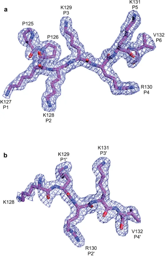
The SV40 NLS peptide binds the major binding site of NcImpαin an extended conformation with an orientation that is antiparallel to the Arm repeats. The electron density for the peptide in the major binding site is well defined, allowing for the unambiguous modeling of the eight peptide residues (125PPKKKRKV132,Fig 2a). The SV40 NLS peptide exhibits a conserved bind-ing mode at the major bindbind-ing site, which is analogous to mouse (MmImpα; [10]), yeast (ScImpα; [9]) and rice (OsImpα; [16]) Impαproteins. The N-terminal residue (P125) also ex-hibits a conformation that is similar to that observed in the structures of OsImpα/SV40 NLS and MmImpαin complex with an extended SV40 NLS peptide (G110-G132, referred as
Fig 1. Overall structure of the NcImpαin complex with the SV40 NLS peptide.NcImpαstructure represented in cartoon exhibits an elongated and curved shape composed of ten tandem Arm repeats. The SV40NLS peptides, represented in magenta sticks, are bound to the major (Arms 1–4) and minor (Arms 4–8) binding sites of NcImpα.
Fig 2. Electron-density map (coefficients 2jFobsj−jFcalcj) of the NcImpα/SV40NLS structure in the area corresponding to SV40NLS peptides (contoured at 2.0 s.d.) at major (A) and minor (B) binding sites.
CN-SV40NLS) [37]. ScImpα/SV40 NLS and MmImpα/SV40 NLS were crystallized using trun-cated version of the SV40 NLS peptide (P126-V132).
The average B-factor of the peptide at the major binding site (33.2Å2) is lower than the av-erage B-factor for the protein (37.7Å2), indicating stability for the interaction of the peptide at this site. The conserved asparagines N150, N192 and N235 of NcImpαstabilize the backbone of the SV40 NLS peptide via hydrogen bonds, whereas the residues W146, W188 and W231 form pockets for the side chains of K129 and K131 at theP3andP5positions of the SV40NLS
peptide, respectively. The K127 of SV40NLS participates in hydrophobic interactions with G195 of NcImpαat theP1binding site. K128 of SV40 NLS forms hydrogen bonds with G154,
A152 and T159 and a salt bridge with D196 of NcImpα, as observed for lysine residues occupy-ing theP2position in previous structures [9,10]. In theP4site, the side chain of R130 interacts
with L109 and K111 of NcImpαvia hydrogen bonds and hydrophobic interactions with P115 and S153. Finally, K131 (positionP5) forms hydrogen bonds with Q185 and hydrophobic
in-teractions with F142, and V132 (positionP6) forms a hydrogen bond with S110 of NcImpα
(Fig 3a,S1 Fig).
Interestingly, the N-terminal residue P125 of the SV40NLS peptide, that is not in theP1−
P6positions, forms hydrogen bonds and hydrophobic interactions with R238 and D270 of
NcImpα. P125 aids in the stabilization of interactions between the protein and a peptide NLS at the major binding site, as observed previously for MmImpα/CN-SV40NLS and OsImpα/ SV40NLS structures [16,37].
Binding of the SV40 NLS peptide at the NcImp
α
minor binding site
The electron density for the SV40 NLS peptide at the minor binding site is also well defined and allows the unambiguous modeling of four residues of the peptide (128KRKV132,Fig 2b). As observed for the major binding site, asparagines residues (N319 and N361 in this case) of NcImpαdefine and guide the backbone of the peptide. The average B-factor for the SV40 NLS peptide bound at the minor binding site of NcImpαis higher than the average B-factor for the protein (39.3 and 37.7Å2, respectively), indicating a lower stability for the peptide at this site compared with the major site.
The interactions between the SV40NLS peptide and NcImpαin the minor site exhibit great-er similarities with the intgreat-eractions between this peptide and OsImpα[16](Table 2) than those with MmImpαor ScImpα[9,10]. In the NcImpα/SV40NLS structure, K129 at theP1’position
forms hydrogen bonds with G323, V321 and T328 of NcImpα. R130, which occupies theP2’
position, is accommodated between the hydrophobic side chains of W357 and W399 and inter-acts with E396 via salt bridges and with S360 via hydrogen bonds, which results in the lowest B-factor value (36.2Å2) compared to the other residues of the peptide. K131 at theP3’position
is stabilized by helix dipoles, negatively charged residues and hydrogen bonds with N283, G281, and T322. Finally, V132 atP4’interacts with D280, R315 and N319 of NcImpαvia
hy-drophobic interactions (Fig 3b,S2 Fig).
Affinity of the SV40 NLS peptide for NcImp
α
Fig 3. Schematic diagram of the interactions between SV40NLS and NcImpαfor major (A) and minor (B) binding sites.The SV40NLS peptide main
and side-chains are drawn in magenta. NcImpαresidues interacting with the peptide are indicated by yellow (side-chain) and orange (main-chain). Polar contacts are shown as dashed lines and arcs with radiating spokes indicate hydrophobic contacts.
but with higher affinity for one site. These results are consistent with previous structural and functional results that indicate the presence of the major and minor binding sites in Impαin complex with the SV40NLS peptide [9,10] and are comparable with ITC experiments per-formed with MmImpα/SV40NLS complex [19]. Furthermore, the negative contribution for the enthalpy (ΔH) and positive value for the entropy (ΔS) suggest that both hydrogen bonds and hydrophobic interactions play a role in this interaction, whereas conformational changes are unfavorable. These data corroborate with the structural information obtained in the present study.
Discussion
Comparison of NcImp
α
with other Imp
α
structures
The crystal structure of NcImpαresembles other Impαstructures [9,10,16] but exhibits a con-formation that is more concave compared with MmImpα(Fig 5a). Its superposition (residues 83–504) with other Impαproteins reveals a higher r.m.s.d. for Cαatoms for MmImpα (resi-dues 78–496; PDB ID: 3UL1; r.m.s.d.: 1.78Å) and HsImpα1 (residues 78–493; PDB ID: 4WV6; r.m.s.d.: 1.77Å) (Fig 5a) compared with ScImpα(residues 89–507; PDB ID: 1BK5; r.m.s.d.: 1.13Å), OsImpα(residues 74–493; PDB ID: 4BQK; r.m.s.d.: 1.21Å) and HsImpα5 (residues 84–502; PDB ID: 2JDQ; r.m.s.d.: 1.22Å) (Fig 5b). The primary sequence identities between
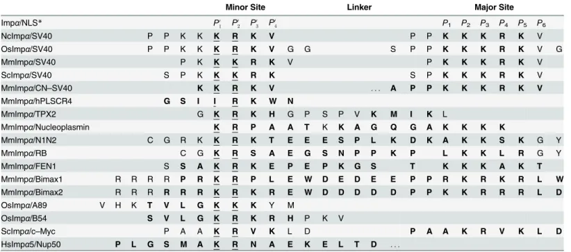
Table 2. Binding to specific pockets of Impα/NLS structures from different organisms.
Minor Site Linker Major Site
Impα/NLS* P0
1 P
0
2 P
0
3 P
0
4 P1 P2 P3 P4 P5 P6
NcImpα/SV40 P P K K K R K V P P K K K R K V
OsImpα/SV40 P P K K K R K V G G S P P K K K R K V G
MmImpα/SV40 P K K K R K V P K K K R K V
ScImpα/SV40 S P K K K R K S P K K K R K V
MmImpα/CN–SV40 K K R K V . . . A P P K K K R K V
MmImpα/hPLSCR4 G S I I R K W N
MmImpα/TPX2 G K R K H G P S P V K M I K L
MmImpα/Nucleoplasmin K R P A A T K K A G Q G A K K K K
MmImpα/N1N2 C G R K K R K T E E E S P L K D K A K K S K G Y
MmImpα/RB C G K R S A E G S N P P K P L K K L R G Y
MmImpα/FEN1 S S A K R K E P E P K G S T K K K A K T
MmImpα/Bimax1 R R R R P R K R P L E W D E D E E P P R K R K R L W
MmImpα/Bimax2 R R R R R R K R K R E W D D D D D P P K K R R R L D
OsImpα/A89 V H K T V L G K K K Y M
OsImpα/B54 S V L G K R K R H P K V
ScImpα/c–Myc P A A K R V K L D P A A K R V K L D
HsImpα5/Nup50 P L G S M A K R N A E K E L T D . . .
*Binding to specific binding pockets of Impαbased on structural data are shown in bold. The NLSs are aligned as observed to bind to the NLS-binding sites (P0
1–P
0
4, minor binding site;P1—P6, major binding site, as defined in [11]). The suspension points corresponds to the extension of the NLS peptides that not bind into the linker region. NcImpα/SV40NLS; OsImpα/SV40NLS [16]; MmImpα/SV40NLS [10]; ScImpα/SV40NLS [9];MmImpα/CN-SV40NLS [37]; MmImpα/hPLSCR4 [38]; MmImpα/TPX2 [39]; MmImpα/Nucleoplasmin [10]; MmImpα/N1N2 [7]; MmImpα/RB [7]; MmImpα/FEN1 [40]; MmImpα/Bimax1 [41]; MmImpα/Bimax2 [41]; OsImpα/A89 [16]; OsImpα/B54 [16]; ScImpα/c-Myc [11]; HsImpα5/Nup50 [42].
NcImpαand other Impαproteins directly reflect the r.m.s.d. values for Cαatoms: ScImpα
(64.69% identity; r.m.s.d.: 1.13Å), OsImpα(63.16% identity; r.m.s.d.: 1.21Å), HsImpα5 (60.22% identity; r.m.s.d.: 1.22Å), MmImpα(46.26% identity; r.m.s.d.: 1.78Å), HsImpα1 (45.01%; r.m.s.d.: 1.77Å). Furthermore, ScImpα, OsImpαand HsImpα5 belong to the same phylogenetic family as NcImpα(α1 family; [43,44]), whereas MmImpαand HsImpα1 belongs to a different family (α2 family). These sequence and structural comparisons raise an impor-tant question concerning Impαfrom different organisms or isoforms from the same organism. How different is the binding mode of an NLS ligand to a particular Impα?
Several studies have shown that Impαproteins from different families exhibit preferences for specific NLSs [45–48]. Thus, examining the binding mode of the SV40NLS peptide, which has been crystallized with various Impαproteins, may provide insights into the binding speci-ficities of these proteins. Crystal structures of Impα/SV40NLS complexes have been
Fig 4. Isothermal calorimetric data for SV40NLS peptide binding to NcImpα. (a)Raw power output (μcal/ s) per unit time (min) of replicate titrations(b)Integrated data (kcal.mol−1of injectant versus molar ratio of SV40NLS to NcImpα). These data were obtained from the raw power output as the area underneath each peak, which is then corrected for baseline heat injections and SV40NLS dilution heat and mixing. The solid line represents the best fit of the data.
doi:10.1371/journal.pone.0128687.g004
Table 3. Isothermal titration calorimetry data for SV40NLS binding to NcImpα.
Major site Minor Site
N 0.89±0.02 0.86±0.02
Kd(μM) 1.23±0.22 1.69±0.46
ΔH (kcal/mol) -2.47±0.06 -3.21±0.12
ΔS (cal/mol/deg) 18.3 15.1
determined for ScImpα[9], MmImpα(in complex with SV40NLS and with an extended SV40NLS peptide, referred to as CN-SV40—[10,37]), OsImpα[16] and NcImpα(the present study). In all of these structures, the SV40NLS peptide binds strongly to the major site via sev-eral interactions, resulting in well-defined electron density and B-factor values that are similar to or lower than the average B-factor value for the protein. These results are consistent with the determined affinities for the MmImpα/SV40NLS [19] and NcImpα/SV40NLS complexes using ITC, in which both studies demonstrated the presence of two binding sites for the peptide
Fig 5. Comparison among Impαcrystal structures from different organisms.The Impαproteins were superimposed using the Cαatoms of each protein (residues 83 -505)(a)NcImpα, MmImpα(PDB ID: 3UL1) and HsImpα1 (PDB ID: 4WV6) are represented in black, red and orange, respectively.(b)NcImpα, OsImpα(PDB ID: 4BQK), ScImpα(PDB ID: 1BK5) and HsImpα5 (PDB ID: 2JDQ) are represented in black, blue, green and pink, respectively.
exhibiting with different affinities. In all of these structures, SV40NLS binding to the major site is essentially identical, in which all six positions (P1−P6) of the peptide (127KKKRKV132,
Table 2) are occupying similar regions at the protein. The main differences are related to the number of peptide residues bound before theP1position (OsImpαexhibits 4 residues before
P1, NcImpαexhibits 2 residues, MmImpαexhibits 1 residue and MmImpα/CN-SV40NLS
ex-hibits 4 residues). However, these differences result from the length of the SV40NLS peptide used in these structural studies rather than differences in Impαproteins from different organisms.
The role of the minor binding site for NcImp
α
specificity
The major site was usually associated with the binding of monopartite NLSs, whereas the minor site was associated with a secondary binding site for bipartite NLSs [7,10,40]. Recently, studies have demonstrated that some monopartite NLSs may bind to only the minor site [16, 38], indicating the importance of this binding site for Impαspecificity for cargo proteins.
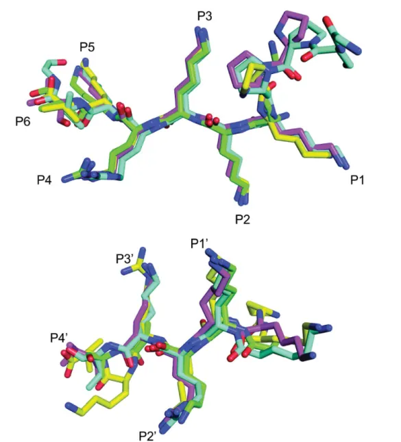
In contrast to the binding of SV40NLS to the major site (Fig 6a), the binding mode of the peptide at the minor binding site is different among different Impαproteins. As shown in Table 2andFig 6b, SV40NLS binds to ScImpαand MmImpαvia Lys and Arg residues at the P1’andP2’positions, whereas the peptide binds OsImpαand NcImpαvia Lys residues at both
P1’andP2’positions, i.e., the binding of the peptide is shifted one position in the OsImpαand
NcImpαstructures.
Interestingly, Lys and Arg residues at theP1’andP2’positions, respectively, are observed for
the majority of monopartite (hPLSCR1, hPLSCR4, c-Myc, and TPX2 [11,38,39,49]) and bi-partite NLS complexes (nucleoplasmin, N1N2, RB and FEN1 [10,37,40]), and it is also the predominant binding mode for MmImpα/CN-SV40NLS [37]. Furthermore, the authors of MmImpα/SV40NLS structure [10] also observed staggering of peptide one position N-termi-nally which may lead to an alternative binding mode with Lys and Arg residues at theP1’and
P2’positions. The binding of KK residues at theP1’andP2’positions for SV40NLS in MmImpα
and ScImpαseems to be exceptions in these particular cases.
An additional interesting characteristic of SV40NLS binding to Impαis the presence of ad-ditional interactions between both the N- and the C-termini of the peptide and MmImpαin the minor site: two positions beforeP1’(P126, N-terminus) and in theP5’position (V132).
These interactions are observed in only the mammalian receptor despite all SV40NLS peptides used for crystallization with Impαproteins have these same residues. Structural studies on OsImpαhave indicated that the plant-specific NLS peptide preferentially binds at the minor site in OsImpαand at the major site in MmImpα[16]. The similarity of SV40NLS binding at the minor site between OsImpαand NcImpαraises questions concerning the importance of this NLS binding site for the import mechanism ofN. crassa.
preventing the binding of the plant-specific NLS peptide to MmImpα[16]. However, these substitutions may also prevent the binding of K127 of SV40NLS to NcImpαand OsImpα, ex-plaining the presence of this interaction in MmImpα, which belongs to theα2 family, and the absence of this interaction in NcImpα, which belongs to theα1 family.
The majority of monopartite (hPLSCR4, c-Myc, and TPX2 [11,38,39]) and bipartite NLS complexes (nucleoplasmin, N1N2, RB and FEN1 [7,10,40]) presented KR residues at theP0
1
andP0
2positions which seems to be the most favorable residues to occupy these positions. The
binding of KK residues attheP0
1andP
0
2positions for SV40NLS in MmImpαseems to be an
ex-ception which probably occurs because it is also favorable [10] to bind additional residues at
Fig 6. Comparison of SV40NLS peptides binding to the major (a) and minor (b) binding sites for Impαstructures from different organisms.
SV40NLS peptides in complex with NcImpα(magenta), ScImpα(green), OsImpα(blue) and MmImpα(yellow) were superimposed using the Cαatoms of the peptides. Positions binding corresponding to the major (P1-P5) and minor (P10-P40) sites are identified along the chains (binding pocket labels were based on peptide binding with NcImpα).
Fig 7. Non-conserved residues between Impαproteins near to the minor NLS binding region.(a)Partial alignment of amino acid sequences of Impα
proteins from different organisms. Conserved residues are shown in black. The residues S402, K497 and E493 for NcImpαand their equivalent residues for Impαproteins are shown in red.(b)Superposition between Cαof NcImpαand MmImpαstructures (performed as described inFig 5) highlights the residues S402, K497 and E493 of NcImpα(magenta) and the residues T402, S483 and A487 of MmImpα(green). These particular residues may be associated to NLS binding specificity to the minor NLS binding site. E493 side-chain is shown in a hypothetical conformation in this figure because this residue presents lack of electron density in this structure due to its high flexibility.
the N- and C-terminal regions of the peptide for some Impαproteins. For OsImpα/SV40NLS and NcImpα/SV40NLS complexes, KR binding at theP0
1andP
0
2positions is more favorable
be-cause fewer interactions occur with the N- and C-terminal regions of the peptide. This observa-tion accounts for the shift in the binding of SV40NLS to OsImpαand NcImpαcompared with MmImpα.
Affinity assays between NLS peptides and Impαproteins have been performed based in in-direct affinity measurements [16,40,49–54], using Surface Plasmon Resonance (SPR) [32], and ITC [19,38,55]. Indirect affinity measurements between NLSs and Impαproteins re-ported dissociation constants in the nM range and permitted the comparison among these molecules. SPR experiments was only able to estimate the peptide-protein affinity in theμM range which was lately confirmed by ITC experiments. The comparison between ITC analysis of MmImpα/SV40NLS [19] and NcImpα/SV40NLS is consistent with the features of the minor binding site observed in the structure. The higher affinity of the peptide at the minor binding site for MmImpα(Kd= 0.98±0.08μM) compared with NcImpα(Kd= 1.69±0.46μM) may be associated with the presence of additional interactions at N- and C-termini in the MmImpα/ SV40NLS complex. The crystal structure of this complex [10] has revealed that the residues K131 and V132 of the SV40 NLS peptide form salt bridges with the conserved E354 and R315 of MmImpα, respectively. Additionally, a hydrogen bond between V132 and R135 aids in the stabilization of the backbone of the SV40 NLS C-terminus. The importance of additional inter-actions N- and C-termini has been also observed with ITC assays for a phosphorylated NLS peptide which enhanced by 10-fold its affinity to MmImpαcompared to unphosphorylated version [54]. Furthermore, it has been observed that the full length nucleoplasmin protein binds to Impα/Impβcomplex with a 2-fold increase in affinity compared to just nucleoplamin NLS peptide [53]. Interesting, ITC studies with the hPLSCR4 NLS peptide (273GSIIRKWN280) which binds only the minor site of MmImpα[38] showed lower affinity (Kd= 48.7±6.5μM) compared with that of the SV40NLS peptide. This fact may be mainly attributed to the absence of a positively charged residue in the hPLSCR4 peptide at theP1’position.
In conclusion, despite the structural similarities among Impαproteins, this study and other recent studies with this receptor from different organisms or different isoforms from the same organism clearly demonstrated differences in the binding specificities for cargo proteins. The differences between NcImpαand MmImpαmay result from the phylogenetic distance among the proteins and the functions of each protein family in organism development, which results in differences in affinities for NLSs. The elucidation of NcImpαin complex with specific NLSs peptides from fungi may provide an explanation for the differences between these proteins.
Supporting Information
S1 Fig. Schematic diagram of the interactions between the SV40NLS peptide (purple) and
NcImpα(orange) at the major binding site.Polar contacts are shown with dashed lines, and
hydrophobic contacts are indicated bay arcs with radiating spokes. Carbon, nitrogen and oxy-gen atoms are shown in black, blue and red, respectively. Generated with the program LIG-PLOT [26].
(EPS)
S2 Fig. Schematic diagram of the interactions between the SV40NLS peptide (purple) and
NcImpα(orange) at the minor binding site.Polar contacts are shown with dashed lines, and
hydrophobic contacts are indicated bay arcs with radiating spokes. Carbon, nitrogen and oxy-gen atoms are shown in black, blue and red, respectively. Generated with the program LIG-PLOT [26].
Acknowledgments
This work was financially supported by FAPESP (Fundação de Amparo à Pesquisa do Estado de São Paulo, Brazil), CNPq (Conselho Nacional de Desenvolvimento Científico e Tecnológico, Brazil), CAPES (Coordenação de Aperfeiçoamento de Pessoal de Nível Superior, Brazil) and PNPD/ CAPES providing research grants and fellowships.
Author Contributions
Conceived and designed the experiments: MRMF AAST MCB. Performed the experiments: NEB TRD FZF AAST. Analyzed the data: MRMF AAST MCB NEB TRD FZF. Contributed re-agents/materials/analysis tools: MRMF MCB. Wrote the paper: MRMF AAST MCB NEB TRD FZF.
References
1. Galagan JE, Calvo SE, Borkovich KA, Selker EU, Read ND, Jaffe D, et al. The genome sequence of the filamentous fungus Neurospora crassa. Nature. 2003 Apr; 422(6934):859–868. PMID:12712197 2. Freitas FZ, Chapeaurouge A, Perales J, Bertolini MC. A systematic approach to identify STRE-binding
proteins of the gsn glycogen synthase gene promoter in Neurospora crassa. Proteomics. 2008 May; 8 (10):2052–2061. doi:10.1002/pmic.200700921PMID:18425733
3. Gonçalves RD, Cupertino FB, Freitas FZ, Luchessi AD, Bertolini MC. A genome-wide screen for Neu-rospora crassa transcription factors regulating glycogen metabolism. Mol Cell Proteomics. 2011 Nov; 10(11):M111.007963. doi:10.1074/mcp.M111.007963PMID:21768394
4. Stewart M. Molecular mechanism of the nuclear protein import cycle. Nat Rev Mol Cell Biol. 2007 Mar; 8(3):195–208. doi:10.1038/nrm2114PMID:17287812
5. Chook YM, Blobel G. Structure of the nuclear transport complex karyopherin-beta2-Ran x GppNHp. Nature. 1999 May; 399(6733):230–237. doi:10.1038/20375PMID:10353245
6. Dingwall C, Laskey RA. Nuclear targeting sequences-a consensus? Trends Biochem Sci. 1991 Dec; 16(12):478–481. doi:10.1016/0968-0004(91)90184-W
7. Fontes MRM, Teh T, Jans D, Brinkworth RI, Kobe B. Structural basis for the specificity of bipartite nu-clear localization sequence binding by importin-alpha. J Biol Chem. 2003 Jul; 278(30):27981–27987. doi:10.1074/jbc.M303275200PMID:12695505
8. Kosugi S, Hasebe M, Matsumura N, Takashima H, Miyamoto-Sato E, Tomita M, et al. Six classes of nu-clear localization signals specific to different binding grooves of importin alpha. J Biol Chem. 2009 Jan; 284(1):478–485. doi:10.1074/jbc.M807017200PMID:19001369
9. Conti E, Uy M, Leighton L, Blobel G, Kuriyan J. Crystallographic analysis of the recognition of a nuclear localization signal by the nuclear import factor karyopherin alpha. Cell. 1998 Jul; 94(2):193–204. doi: 10.1016/S0092-8674(00)81419-1PMID:9695948
10. Fontes MR, Teh T, Kobe B. Structural basis of recognition of monopartite and bipartite nuclear localiza-tion sequences by mammalian importin-alpha. J Mol Biol. 2000 Apr; 297(5):1183–1194. doi:10.1006/ jmbi.2000.3642PMID:10764582
11. Conti E, Kuriyan J. Crystallographic analysis of the specific yet versatile recognition of distinct nuclear localization signals by karyopherin alpha. Structure. 2000 Mar; 8(3):329–338. doi: 10.1016/S0969-2126(00)00107-6PMID:10745017
12. Rexach M, Blobel G. Protein import into nuclei: association and dissociation reactions involving trans-port substrate, transtrans-port factors, and nucleoporins. Cell. 1995 Dec; 83(5):683–692. doi: 10.1016/0092-8674(95)90181-7PMID:8521485
13. Stinnett SM, Espeso EA, Cobeño L, Araújo-Bazán L, Calvo AM. Aspergillus nidulans VeA subcellular localization is dependent on the importin alpha carrier and on light. Mol Microbiol. 2007 Jan; 63(1):242–
255. doi:10.1111/j.1365-2958.2006.05506.xPMID:17163983
14. Cupertino FB, Freitas FZ, de Paula RM, Bertolini MC. Ambient pH controls glycogen levels by regulat-ing glycogen synthase gene expression in Neurospora crassa. New insights into the pH signalregulat-ing path-way. PLoS One. 2012; 7(8):e44258. doi:10.1371/journal.pone.0044258PMID:22952943
16. Chang CW, Couñago RLM, Williams SJ, Bodén M, Kobe B. Crystal structure of rice importin-αand structural basis of its interaction with plant-specific nuclear localization signals. Plant Cell. 2012 Dec; 24 (12):5074–5088. doi:10.1105/tpc.112.104422PMID:23250448
17. Takeda AAS, Freitas FZ, Magro AJ, Bernardes NE, Fernandes CAH, Gonçalves RD, et al. Biophysical characterization of the recombinant importin-αfrom Neurospora crassa. Protein Pept Lett. 2013 Jan; 20(1):8–16. doi:10.2174/09298665130103PMID:22789101
18. Knudsen NØ, Andersen SD, Lützen A, Nielsen FC, Rasmussen LJ. Nuclear translocation contributes to regulation of DNA excision repair activities. DNA Repair (Amst). 2009 Jun; 8(6):682–689. doi:10. 1016/j.dnarep.2009.03.005
19. Cutress ML, Whitaker HC, Mills IG, Stewart M, Neal DE. Structural basis for the nuclear import of the human androgen receptor. J Cell Sci. 2008 Apr; 121(Pt 7):957–968.
20. Bernardes NE, Takeda AAS, Freitas FZ, Bertolini MC, Fontes MRM. Crystallization and preliminary X-ray crystallographic analysis of importin-αfrom Neurospora crassa. Acta Crystallogr F Struct Biol Com-mun. 2014 Apr; 70(Pt 4):501–504. doi:10.1107/S2053230X14005068PMID:24699749
21. Otwinowski Z, Minor W. Processing of X-ray diffraction data collected in oscillation mode. In: Charles W Carter J, editor. Macromolecular Crystallography Part A. vol. 276 of Methods in Enzymology. Academ-ic Press; 1997. p. 307–326.
22. McCoy AJ, Grosse-Kunstleve RW, Storoni LC, Read RJ. Likelihood-enhanced fast translation func-tions. Acta Crystallogr D Biol Crystallogr. 2005 Apr; 61(Pt 4):458–464. doi:10.1107/
S0907444905001617PMID:15805601
23. Emsley P, Cowtan K. Coot: model-building tools for molecular graphics. Acta Crystallogr D Biol Crystal-logr. 2004 Dec; 60(Pt 12 Pt 1):2126–2132. doi:10.1107/S0907444904019158PMID:15572765 24. Adams PD, Afonine PV, Bunkóczi G, Chen VB, Davis IW, Echols N, et al. PHENIX: a comprehensive
Python-based system for macromolecular structure solution. Acta Crystallogr D Biol Crystallogr. 2010 Feb; 66(Pt 2):213–221. doi:10.1107/S0907444909052925PMID:20124702
25. Chen VB, Arendall WB 3rd, Headd JJ, Keedy DA, Immormino RM, Kapral GJ, et al. MolProbity: all-atom structure validation for macromolecular crystallography. Acta Crystallogr D Biol Crystallogr. 2010 Jan; 66(Pt 1):12–21. doi:10.1107/S0907444909042073PMID:20057044
26. Wallace AC, Laskowski RA, Thornton JM. LIGPLOT: a program to generate schematic diagrams of pro-tein-ligand interactions. Protein Eng. 1995 Feb; 8(2):127–134. doi:10.1093/protein/8.2.127PMID: 7630882
27. DeLano WL. The PyMOL Molecular Graphics System; 2002. Available from:http://www.pymol.org. 28. Kabsch W. A solution for the best rotation to relate two sets of vectors. Acta Crystallographica Section
A. 1976 Sep; 32(5):922–923. doi:10.1107/S0567739476001873
29. Winn MD, Ballard CC, Cowtan KD, Dodson EJ, Emsley P, Evans PR, et al. Overview of the CCP4 suite and current developments. Acta Crystallogr D Biol Crystallogr. 2011 Apr; 67(Pt 4):235–242. doi:10. 1107/S0907444910045749PMID:21460441
30. Kobe B. Autoinhibition by an internal nuclear localization signal revealed by the crystal structure of mammalian importin alpha. Nat Struct Biol. 1999 Apr; 6(4):388–397. doi:10.1038/7625PMID: 10201409
31. Laskowski RA, MacArthur MW, Moss DS, Thornton JM.PROCHECK: a program to check the stereo-chemical quality of protein structures. Journal of Applied Crystallography. 1993 Apr; 26(2):283–291. doi:10.1107/S0021889892009944
32. Catimel B, Teh T, Fontes MR, Jennings IG, Jans DA, Howlett GJ, et al. Biophysical characterization of interactions involving importin-alpha during nuclear import. J Biol Chem. 2001 Sep; 276(36):34189–
34198. doi:10.1074/jbc.M103531200PMID:11448961
33. Marfori M, Lonhienne TG, Forwood JK, Kobe B. Structural basis of high-affinity nuclear localization sig-nal interactions with importin-α. Traffic. 2012 Apr; 13(4):532–548. doi:10.1111/j.1600-0854.2012. 01329.xPMID:22248489
34. Trowitzsch S, Viola C, Scheer E, Conic S, Chavant V, Fournier M, et al. Cytoplasmic TAF2-TAF8-TAF10 complex provides evidence for nuclear holo-TFIID assembly from preformed submodules. Nat Commun. 2015; 6:6011. doi:10.1038/ncomms7011PMID:25586196
35. Tarendeau F, Boudet J, Guilligay D, Mas PJ, Bougault CM, Boulo S, et al. Structure and nuclear import function of the C-terminal domain of influenza virus polymerase PB2 subunit. Nat Struct Mol Biol. 2007 Mar; 14(3):229–233. doi:10.1038/nsmb1212PMID:17310249
37. Fontes MRM, Teh T, Toth G, John A, Pavo I, Jans DA, et al. Role of flanking sequences and phosphory-lation in the recognition of the simian-virus-40 large T-antigen nuclear localization sequences by impor-tin-alpha. Biochem J. 2003 Oct; 375(Pt 2):339–349. doi:10.1042/BJ20030510PMID:12852786 38. Lott K, Bhardwaj A, Sims PJ, Cingolani G. A minimal nuclear localization signal (NLS) in human
phos-pholipid scramblase 4 that binds only the minor NLS-binding site of importin alpha1. J Biol Chem. 2011 Aug; 286(32):28160–28169. doi:10.1074/jbc.M111.228007PMID:21690087
39. Giesecke A, Stewart M. Novel binding of the mitotic regulator TPX2 (target protein for Xenopus kinesin-like protein 2) to importin-alpha. J Biol Chem. 2010 Jun; 285(23):17628–17635. doi:10.1074/jbc.M110. 102343PMID:20335181
40. de Barros AC, Takeda AAS, Chang CW, Kobe B, Fontes MRM. Structural basis of nuclear import of flap endonuclease 1 (FEN1). Acta Crystallogr D Biol Crystallogr. 2012 Jul; 68(Pt 7):743–750. doi:10. 1107/S0907444912010281PMID:22751659
41. Kosugi S, Hasebe M, Entani T, Takayama S, Tomita M, Yanagawa H. Design of peptide inhibitors for the importin alpha/beta nuclear import pathway by activity-based profiling. Chem Biol. 2008 Sep; 15 (9):940–949. doi:10.1016/j.chembiol.2008.07.019PMID:18804031
42. Pumroy RA, Nardozzi JD, Hart DJ, Root MJ, Cingolani G. Nucleoporin Nup50 stabilizes closed confor-mation of armadillo repeat 10 in importinα5. J Biol Chem. 2012 Jan; 287(3):2022–2031. doi:10.1074/ jbc.M111.315838PMID:22130666
43. Goldfarb DS, Corbett AH, Mason DA, Harreman MT, Adam SA. Importin alpha: a multipurpose nuclear-transport receptor. Trends Cell Biol. 2004 Sep; 14(9):505–514. doi:10.1016/j.tcb.2004.07.016PMID: 15350979
44. Köhler M, Ansieau S, Prehn S, Leutz A, Haller H, Hartmann E. Cloning of two novel human importin-alpha subunits and analysis of the expression pattern of the importin-importin-alpha protein family. FEBS Lett. 1997 Nov; 417(1):104–108. doi:10.1016/S0014-5793(97)01265-9PMID:9395085
45. Köhler M, Haller H, Hartmann E. Nuclear Protein Transport Pathways. Nephron Experimental Nephrol-ogy. 1999; 7:290–294. doi:10.1159/000020616
46. Sekimoto T, Imamoto N, Nakajima K, Hirano T, Yoneda Y. Extracellular signal-dependent nuclear im-port of Stat1 is mediated by nuclear pore-targeting complex formation with NPI-1, but not Rch1. EMBO J. 1997 Dec; 16(23):7067–7077. doi:10.1093/emboj/16.23.7067PMID:9384585
47. Fang X, Chen T, Tran K, Parker CS. Developmental regulation of the heat shock response by nuclear transport factor karyopherin-alpha3. Development. 2001 Sep; 128(17):3349–3358. PMID:11546751 48. Mason DA, Fleming RJ, Goldfarb DS. Drosophila melanogaster importin alpha1 and alpha3 can
re-place importin alpha2 during spermatogenesis but not oogenesis. Genetics. 2002 May; 161(1):157–
170. PMID:12019231
49. Chen MH, Ben-Efraim I, Mitrousis G, Walker-Kopp N, Sims PJ, Cingolani G. Phospholipid scramblase 1 contains a nonclassical nuclear localization signal with unique binding site in importin alpha. J Biol Chem. 2005 Mar; 280(11):10599–10606. doi:10.1074/jbc.M413194200PMID:15611084
50. Wirthmueller L, Roth C, Banfield MJ, Wiermer M. Hop-on hop-off: importin-α-guided tours to the nucleus in innate immune signaling. Front Plant Sci. 2013; 4:149. doi:10.3389/fpls.2013.00149PMID: 23734157
51. Yang SNY, Takeda AAS, Fontes MRM, Harris JM, Jans DA, Kobe B. Probing the specificity of binding to the major nuclear localization sequence-binding site of importin-alpha using oriented peptide library screening. J Biol Chem. 2010 Jun; 285(26):19935–19946. doi:10.1074/jbc.M109.079574PMID: 20406804
52. Takeda AAS, de Barros AC, Chang CW, Kobe B, Fontes MRM. Structural basis of importin-α-mediated nuclear transport for Ku70 and Ku80. J Mol Biol. 2011 Sep; 412(2):226–234. doi:10.1016/j.jmb.2011. 07.038PMID:21806995
53. Falces J, Arregi I, Konarev PV, Urbaneja MA, Svergun DI, Taneva SG, et al. Recognition of nucleoplas-min by its nuclear transport receptor importinα/β: insights into a complete import complex. Biochemis-try. 2010 Nov; 49(45):9756–9769. doi:10.1021/bi101179gPMID:20925424
54. Róna G, Marfori M, Borsos M, Scheer I, Takács E, Tóth J, et al. Phosphorylation adjacent to the nuclear localization signal of human dUTPase abolishes nuclear import: structural and mechanistic insights. Acta Crystallogr D Biol Crystallogr. 2013 Dec; 69(Pt 12):2495–2505. PMID:24311590