Effects of Brazilian propolis on
Leishmania amazonensis
Diana Copi Ayres, Maria Cristina Marcucci
*, Selma Giorgio/
+Departamento de Parasitologia, Instituto de Biologia, Universidade Estadual de Campinas, Caixa Postal 6109, 13083-970 Campinas, SP, Brasil *Programa de Pós-graduação em Farmácia, Universidade Bandeirante de São Paulo, São Paulo, SP, Brasil
Leishmaniasis, an endemic parasitosis that leads to chronic cutaneous, mucocutaneous or visceral lesions, is part of those diseases, which still requires improved control tools. Propolis has shown activities against different bacteria, fungi, and parasites. In this study we investigated the effect of four ethanolic extracts of typified propolis collected in different Brazilian states, on Leishmania amazonensis performing assays with promastigote forms, extracellular amastigotes, and on infected peritoneal macrophages. Ethanolic extracts of all propolis samples (BRG, BRPG, BRP-1, and BRV) were capable to reduce parasite load as monitored by the percentage of infected macrophages and the number of intracellular parasites. BRV sample called red propolis, collected in the state of Alagoas, and containing high concentration of prenylated and benzophenones com-pounds, was the most active extract against L. amazonensis. The anti-Leishmania effect of BRV sample was increased in a concentration and time dependent manner. BRV treatment proved to be non-toxic to macrophage cultures. Since BRV extract at the concentration of 25 µg/ml reduced the parasite load of macrophages while presented no direct toxic to promastigotes and extracellular amastigotes, it was suggested that constituents of propolis intensify the mechanism of macrophage activation leading to killing of L. amazonensis. Our results demonstrate, for the first time, that ethanolic extracts of Brazilian propolis reduce L. amazonensis infection in macrophages, and encourage further studies of this natural compound in animal models of leishmaniasis.
Key words: Leishmania amazonensis - leishmaniasis - natural products - propolis
Leishmaniasis is a parasitosis caused by several spe-cies of the protozoan Leishmania and it is currently en-demic in 88 countries. Overall prevalence is 12 million people and the population at risk is 350 million (Desjeux 2004, Murray et al. 2005). Injected into mammalian hosts by phlebotomus sand flies as extracellular promas-tigotes, Leishmania bind to macrophage and are quickly phagocytosed. All Leishmania species are obligatorily intracellular parasites, which live within secondary phagolissosomes. In this way, the parasite is able to mul-tiply, lyse host cell, and infect surrounding macrophages. The severity of disease varies ranging from cutane-ous or mucosal to visceral or diffuse cutanecutane-ous infec-tion (Grimaldi & Tesh 1993, Murray et al. 2005). The former is generally caused by L. amazonensis, a spe-cies transmitted mainly in the Amazon region, which is associated with localized cutaneous lesions (Grimaldi & Tesh 1993). Chemotherapy remains the mainstay for the control of leishmaniasis, as effective vaccines have yet to be developed (Murray et al. 2005). The first line of therapy for all forms of the disease requires poten-tially toxic and painful multiple injections of pentava-lent antimonials (Berman 2003). The problem is further aggravated by the appearance of resistance to these drugs in some endemic areas. Amphotericin B and
pentami-dine are second-line drugs and they present limited value because of their toxicity and difficulty in administration (Berman 2003). Many studies have been conducted to find an effective therapy for leishmaniasis that avoids exposure to potentially toxic drugs, including screening of plant extracts and plant-derived compounds (Abreu et al. 1999, Carvalho & Ferreira 2001, Rocha et al. 2005). Propolis is a resinous substance that honey bees collect from different plant exsudates (Marcucci 1995). Propolis is claimed to posses versatile valu-able pharmacological activities and has, to date, been taken in internal and external dosage forms for the treatments of various diseases (Burdock 1998, Mar-cucci & Bankova 1999). It is widely used in products like “healthy foods” and “biocosmetics” (Marcucci & Bankova 1999). Many authors have reported the in vitro activities of propolis against different microor-ganisms, among them some important human patho-gens, such as Staphylococcus aureus, Salmonella thyphimurium, Candida albicans, Trypanosoma cruzi, and Giardia duodenalis (Higashi & de Castro 1994, Marcucci et al. 2001, Miorin et al. 2003, Uzel et al. 2005, Dantas et al. 2006, Freitas et al. 2006, Trusheva et al. 2006). Brazilian propolis is the sub-ject of an intensive study of chemists, biologists, and physicians all over the world due to specific tropical flora and their different chemical components (Marcucci & Bankova 1999, Marcucci et al. 2001, Trusheva et al. 2006).
This report describes in vitro analyses of the ef-fects of ethanolic extracts of typified Brazilian pro-polis samples on both promastigote and amastigote forms of L. amazonensis and on macrophages infected with the parasite.
Financial support: CNPq and Fapesp
+Corresponding author. sgiorgio@unicamp.br
MATERIALS AND METHODS
Parasite - L. amazonensis (MHOM/BR/73/M2269) promastigotes were cultured at 28oC in RPMI 1640 medium (Sigma, St. Louis, MO) supplemented with 25 µg/ml gentamicin, 2 mM L-glutamine, 100 mM HEPES, and 10% fetal calf serum (FCS) (Cultilab, Campinas, SP, Brazil), pH 7.2. Amastigotes were isolated from active skin lesions from BALB/c mice, and used immediately after isolation (Barbieri et al. 1993).
Brazilian propolis samples - Two propolis samples collected in the Brazilian state of Paraná were green pro-polis, typified as BRG and BRPG. Propolis collected in the state of Minas Gerais was typified as BRP-1 (green propolis) (Miorin et al. 2003) and the sample collected in the state of Alagoas as BRV (red propolis) (Trusheva et al. 2006). The ethanolic extracts of propolis were pre-pared by using a modified technique described by Miorin et al. (2003). Propolis (30 g) was cut into small pieces and extracted with 100 ml absolute ethanol at room tem-perature for 24 h. The solution was filtered with Whatman paper number 3, and placed in amber flasks. Each solu-tion was dried and the residue weighted to prepare stock solution in ethanol at concentration of 5%. The final concentration of the solvent in the experiments did not exceed 0.1% ethanol.
Macrophage infection with L. amazonensis - Pri-mary mouse macrophages (5 × 10⁵/ml) were obtained
from normal BALB/c mice by peritoneal washing, cul-tured on 24-well plates containing 13 mm diameter glass coverslips and infected with amastigotes (3:1 parasite/ host cell) for 1 h, as described previously (Colhone et al. 2004). After the interaction period, the cultures were washed to remove extracellular parasites and incubated in the presence or absence of different concentrations of propolis or diluent (0.1% ethanol), at 37oC in 5% CO2 in air in a humidified incubator as established by Ayres et al. (2006). After the indicated periods of treat-ments, coverslips were fixed with methanol, stained with Giemsa, and examined under light microscope. Six hun-dred cells were counted per triplicate coverslip for the evaluation of the percentage of infected macrophages and the number of amastigotes per infected macrophage (Colhone et al. 2004). The infection levels were quanti-fied using a light microscopy at 1000 magnification.
Assessment of propolis effects on L. amazonensis promastigotes, amastigotes, and macrophage cultures - Promastigotes growing in 25 cm2 plastic flasks at 28oC were treated with different concentrations of propolis or diluent, and parasite number and morphology were determined using a Neubauer haemocytometer (Arrais-Silva et al. 2005). Amastigotes maintained under pro-mastigote culture conditions, i.e. at 28oC in 25 cm2 plas-tic flasks with RPMI 1640 medium supplemented with gentamicin, L-glutamine, HEPES, and 10% FCS, were treated with different concentrations of propolis or diluent, and left to transform into promastigote forms. After the indicated periods of incubation at 28oC, pro-mastigote and apro-mastigote numbers were recorded by microscopic observation (Lemesre et al 1997,
Arrais-Silva et al. 2005). Macrophages cultured on 24-well plates were incubated for 72 h in the presence of differ-ent propolis samples or diludiffer-ent (0.1% ethanol). Cell vi-ability was analyzed by a dye-reduction assay using MTT (3-(4,5-dimethylthiazol-2-yl)-2,5-diphenyltetrazolium bromide) (Sigma) (Mosmann 1983).
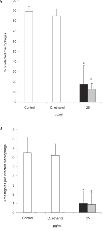
Fig. 1: effect of ethanolic extracts of Brazilian propolis (25 µg/ml) on
Leishmania amazonensis- infected macrophages after 72 h of treatment: control, 0.1% ethanol, BRG ( ) or BRPG ( ). The percentage of infected macrophages (A) and the number of parasites per infected cell (B) were determined as described in Materials and Methods. The results sent the mean ± SD of three independent experiments. Asterisks repre-sent statistically differences (p < 0.01) between control and propolis-treated cultures.
123 123 123
A
Statistical analyses - All experiments were repeated at least three times in triplicate wells. The results were expressed as mean ± SD. Data obtained with different propolis extracts were analyzed by 1-way ANOVA and Student’s t-test (P < 0.01).
RESULTS
Experiments were undertaken to study a possible ef-fect of four typified Brazilian propolis samples on L. amazonensis-infected macrophage cultures. As shown in Fig. 1 murine macrophages were efficiently infected with L. amazonensis amastigotes (around 90% of in-fected cells and 6 intracellular parasites per inin-fected cell). Macrophages infected with the parasite and treated with 25 µg/ml of BRG and BRPG extracts for 72 h have showed significant reduction of both the percentage of infection (Fig. 1A) and of the number of intracellular parasites (Fig. 1B). At concentrations higher than 25 µg/ ml both extracts were toxic to the cells, because light microscopy showed cellular debris and few intact mac-rophages present on the surface of glass cover slips and in the culture supernatants. It must notice that at the per-centage of 0.1% ethanol, amount present in experiments performed, this solvent had no effect on the cultures. The same protocol was employed to test BRP-1 and BRV samples (Fig. 2). The extract from BRP-1 sample was
Fig. 2: effect of ethanolic extracts of Brazilian propolis (3-100 µg/ml) on
Leishmania amazonensis-infected macrophages after 72 h of treatment: control, 0.1% ethanol, BRV ( ) or BRP-1 ( ). The percentage of infected macrophages (A) and the number of parasites per infected cell (B) were determined as described in Materials and Methods. The results repre-sent the mean ± SD of three independent experiments; n.d.: not deter-mined. Asterisks represent statistically differences (p < 0.01) between control and propolis-treated cultures.
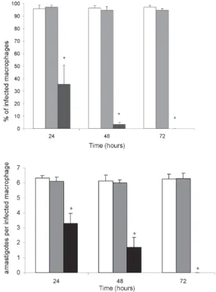
capable to reduce significantly the macrophage infec-tion at concentrainfec-tions ranging from 6 to 100 µg/ml. The treatment with 6 µg/ml of BRV for 72 h led to a reduc-tion of 84.5% of the infecreduc-tion level and at higher con-centrations of the extract no infected macrophage was observed. Since BRV was the most active, further ex-periments were performed, treating the infected cultures for 1, 2, and 3 days with 25 µg/ml of this extract, and being observed a time-dependent decrease of both the percent of infection and of the intracellular prolifera-tion of the parasites (Fig. 3). The viability of macroph-ages treated for 72 h with 0.1% ethanol, 25, 50 or 100 µg/ml BRV was further analyzed by MTT assay. Formazan production was similar between control, macrophages treated with 0.1% ethanol or with 100 µg/ml BRV (Fig. 4). Interestingly, treatment with 25 or 50 µg/ml of the extract induced an increase in the MTT-reducing activity (Fig. 4). Since peritoneal macrophages are non-dividing differentiated cells (Handel-Fernandez & Lopez 2000), we can exclude the possibility that BRV stimulates the proliferation of macrophages.
We also addressed the question concerning whether BRV extracts presented a direct effect on promastigotes and extracellular amastigotes of L. amazonensis. Up to 96 h, at 25 µg/ml, the extract did not affect promastigotes pro-liferation (Fig. 5A). In experiments with lesion-derived
Fig. 3: effect of BRV propolis extract (25 µg/ml) on Leishmania amazonensis-infected macrophages after 24, 48, and 72 h of treatment: control ( ), 0.1% ethanol ( ), and BRV ( ). The percentage of infected macrophages (A) and the number of parasites per infected cell (B) were determined as described in Materials and Methods. The results sent the mean ± SD of three independent experiments. Asterisks repre-sent statistically differences (p < 0.01) between control and propolis-treated cultures.
12 12 12
A
amastigotes, 25 µg/ml BRV did not interfere with their vi-ability (around 98% of control vivi-ability after 24 h of treat-ment) or with their morphology (data not shown). Previous studies indicated that even if amastigotes remain viable, only molecularly undamaged amastigotes are expected to be able to transform into promastigotes (Lemesre et al. 1997, Arrais-Silva et al. 2005). Addition of 25 µg/ml BRV in medium did not affect amastigotes differentiation to promastigotes (48 h) (Fig. 5B).
DISCUSSION
This report provided evidences that ethanolic extracts of Brazilian propolis reduced L. amazonensis infection in macrophage cultures. Based on high performance liq-uid chromatography and nuclear magnetic resonance analysis ethanolic extract of Brazilian propolis samples have been typified in four groups (Marcucci 2000). BRG contains high concentration of coniferaldehyde com-pounds, BRPG contains high concentration of prenylated and coniferaldehyde compounds, BRP-1 contains high concentration of prenylated compounds and BRV con-tains high concentration of prenylated and benzophe-nones compounds (Marcucci & Bankova 1999, Marcucci et al. 2001, Miorin et al. 2003, Sawaya et al. 2004, Trusheva et al. 2006). The four propolis samples (BRG, BRPG, BRP-1, and BRV) evaluated in this study were able to reduce parasite load, as monitored by the per-centage of infected cells and the number of intracellular parasites. Additional experiments with BRG and BRPG samples were abandoned because at concentrations higher than 25 µg/ml both extracts were toxic to mac-rophages. In fact some studies have demonstrated pro-polis toxicity for different cell types (Higashi & de Castro 1994, Chen et al. 2001, Ferguson 2001, Dantas et al. 2006, Tavares et al. 2006). For example, damage to mu-rine macrophages was observed after treatment with ethanolic extract of propolis at concentrations above 30 µg/ml (Higashi & de Castro 1994), and genotoxic effect of ethanolic extract of Brazilian propolis (100 µg/ml) was detected in Chinese hamster ovary cells (Tavares et al. 2006). The mechanism of propolis cytotoxicity is still unknown (Tavares et al. 2006). On the other hand, BRP-1 and BRV extracts were not toxic to macrophages and inhibited the intracellular proliferation of L. ama-zonensis. The BRV extract was the most active and treat-ment of macrophages led to no morphological alterations as judged by light microscopy. Interestingly, 25 or 50 µg/ml BRV extract induced in macrophages an increase of the MTT-reducing activity. These results are appar-ently paradoxical, since macrophages are non-dividing cells (Handel-Fernandez & Lopes 2000), but the present results match those obtained with interferons and plant-derived polyphenols, which induce cell cycle arrest in cancer lineages but increase MTT-reducing activity (Jab-ber et al. 1989, Pagliacci et al. 1993, Bernhard et al. 2003). Such increase may be attributed to an increase in cell volume and mithocondrial number and/or activity of cells treated with the BRV extract (Bernhard et al. 2003). Although BRV is active against intracellular para-sites it presented no direct effect on promastigotes or extracellular amastigotes. Our data corroborate results
Fig. 5: effect of BRV propolis extract on Leishmania amazonensis pro-mastigotes and extracellular apro-mastigotes. A:promastigotes were left untre-ated (), treated at 28oC with 0.1% ethanol ( ), 25 µg/ml BRV extract ( )
up to 96 h. Parasite viability was assessed by microscopic examination; B: extracellular amastigotes were left untreated ( ), treated at 28oC with
0.1% ethanol ( ) or 25 µg/ml BRV extract ( ). The transformation of amastigotes into promastigotes was monitored after 24 h and 48 h. These are the results of a typical experiment, representative of a total of 3. Fig. 4: MTT production by non-infected macrophages: control, 0.1% ethanol, 88 mM H2O2, or BRV extract at 25, 50, 100 µg/ml. The results represent the mean ± SD of three independent experiments. Asterisks represent statistically differences (p < 0.01) between control, H2O2 and BRV-treated cultures.
A
obtained by Higashi and de Castro (1994). The authors observed that concentrations of ethanolic extract of pro-polis that inhibited the levels of T. cruzi infection in macrophages did not affect proliferation of axenic amastigotes. These results and our findings suggest that factors associated with host cell metabolism may con-tribute to intensify the effects of propolis (Higashi & de Castro 1994). Another possibility is that constitu-ents of propolis intensify the mechanism of macroph-age activation, leading to production of cytokines and reactive nitrogen intermediates engaged in the killing of intracellular parasites (Solbach & Laskay 2000). It was demonstrated that Korean propolis induces macrophages by producing interleukin-1, tumor necrosis factor-α, and nitric oxide (Han et al. 2002); these results suggest that propolis may function through macrophage activation. The precise mechanism by which BRV propolis treated macrophages are able to control L. amazonensis infec-tion needs further investigainfec-tions. This sample was col-lected in Alagoas, Brazil and it is a new propolis type called red Brazilian propolis containing high concentra-tion of prenylated and benzophenones compounds (Marcucci 2000, Trusheva et al. 2006). The recent study of Trusheva et al. (2006) identified 14 chemical constitu-ents of red Brazilian propolis, three of them with antibac-terial and antimycotic activities, and encourages further in-vestigations of the chemical constituents which are respon-sible for the leishmanicidal activities of red Brazilian pro-polis showed in this report. The investigation in animal mod-els of Leishmania infection is currently under investiga-tion in our laboratory.
REFERENCES
Abreu PM, Martins ES, Kayser O, Binseil KU, Siems K, Seemann A, Brevet J 1999. Antimicrobial, anitumor and antileisma-nial screening of medicinal plants from Guinea-Bissau.
Phytomedicine 6: 187-195.
Arrais-Silva WW, Colhone MC, Ayres DC, Souto PS, Giorgio S 2005. Effects of hyperbaric oxygen on Leishmania amazo-nensis promastigotes and amastigotes. Parasitol Int 54: 1-7.
Ayres DC, Marcucci MC, Giorgio S 2006. Treatment methods of leishmaniasis with Brazilian propolis. Requested patent. Bra-zilian National Institute for Intellectual Property, INPI no. PI 018060007317, 01/27/2006.
Barbieri CL, Giorgio S, Merjan AJ, Figueiredo EM 1993. Glycosphingolipid antigens from Leishmania (Leishmania) amazonensis amastigotes identified by use of a monoclonal antibody. Infec Immun61: 2132-2137.
Berman JD 2003. Current treatment approaches to leishmania-sis. Curr Opin Infect Dis 16: 397-401.
Bernhard D, Schwaiger W, Crazzolara R, Tinhofer I, Kofler R, Csordas A 2003. Enhanced MTT-reducing activity under growth inhibition by resveratrol in CEM-C7H2 lymphocytic leukemia cells. Cancer Lett 195: 193-199.
Burdock GA 1998. Review of the biological properties and toxicity of bee propolis (propolis). Food Chem Toxicol 36: 347-363.
Carvalho PB, Ferreira EI 2001. Leishmaniasis phytotherapy. Nature's leadership against an ancient disease - Review.
Fitoterapia 72: 599-618.
Chen YJ, Shiao MS, Hsu ML, Tsai TH, Wang SY 2001. Effect of caffeic acid phenethyl ester, an antioxidant from propolis on inducing apoptosis in human leukemic HL-60 cells. J Agric Food Chem 49: 5615-5619.
Colhone MC, Arrais-Silva, WW, Picoli C, Giorgio S 2004. Effect of hypoxia on macrophage infection by Leishmania ama-zonensis. J Parasitol 90: 510-515.
Desjeux P 2004. Leishmaniasis: current situation and new perspectives. Comp Immunol Microbiol Infect Dis 27: 305-318.
Dantas AP, Salomão K, Barbosa HS, de Castro SL 2006. The effect of Bulgarian propolis against Trypanosoma cruzi and during its interaction with host cells. Men Inst Oswaldo Cruz 101: 207-211.
Ferguson LR 2001. Role of plant polyphenols in genomic stabil-ity. Mutation Res475: 89-111.
Freitas SF, Shinohara L, Sforcin JM, Guimarães S 2006. In vitro
effects of propolis on Giardia duodenalis trophozoites.
Phytomedicine 13:170-175.
Grimaldi GJ, Tesh, RB 1993. Leishmaniasis of the new world: current concepts and implications for future research. Clin Microbiol Rev 6: 230-250.
Han S, Sung KH, Yim D, Lee S, Cho K, Lee, CK, Ha NJ, Kim K 2002. Activation of murine macrophage cell line RAW 264.7 by Korean propolis. Arch Pharm Res 25: 895-902.
Handel-Fernandez ME, Lopez DM 2000. Isolation of macroph-ages from tissue, fluids, and immune response sites. In DM Paulnock, Macrophages - A Practical Approach, Oxford University Press, Oxford, p. 1-30.
Higashi KO, de Castro SL 1994. Propolis extracts are effective against Trypanosoma cruzi and have an impact on its inter-action with host cells. J Ethnopharmacol 43: 149-155.
Jabber SAB, Twentyman PR, Watson JV 1989. The MTT assay underestimates the growth inhibitory effects of interferons.
Br J Cancer 60: 523-528.
Lemesre J-P, Sereno D, Daulouede S, Veyret B, Brajon N, Vincendeau P 1997. Leishmania spp.: nitric oxide-mediated metabolic inhibition of promastigote and axenically grown amastigote forms. Exp Parasitol 86: 56-68.
Marcucci MC 1995. Propolis: chemical composition, biological properties and therapeutic activity. Apidologie 26: 517-518.
Marcucci MC 2000. Process to typing natural products. Requested patent. Brazilian National Institute for Intellectual Property, INPI no. PI 0105471-6, 12/22/2000.
Marcucci MC, Bankova V 1999. Chemical composition, plant origin and biological activity of Brazilian propolis. Curr Top Phytochemistry 2: 115-123.
Marcucci MC, Ferreres F, Garcia-Vigueira C, Bankova VS, de Castro SL, Dantas AP, Valente PHM, Paulino N 2001. Phe-nolic compounds from Brazilian propolis with pharmacologi-cal activities. J Ethnopharmacol 74: 105-112.
Miorin PL, Levy Junior NC, Custodio AR, Bretz WA, Marcucci MC 2003. Antibacterial activity of honey and propolis from
Apis mellifera and Tetragonisca angustula against Staphy-lococcus aureus. J Appl Microbiol 95: 913-920.
Pagliacci MC, Spinozzi F, Migliorati G, Fumi G, Smacchia M, Grignani F, Riccardi C, Nicoletti I 1993. Genistein inhibits tumour cell growth in vitro but enhances mitochondrial re-duction of tetrazolium salts: a further pitfall in the use of the MTT assay for evaluating cell growth and survival. Eur J Cancer 29A: 1573-1577.
Rocha LG, Almeida JRGS, Macedo RO, Barbosa-Filho JM 2005. A review of natural products with antileishmanial activity.
Phytomedicine 12: 314-535.
Sawaya AC, Palma AM, Caetano FM, Marcucci MC, da Silva Cunha, IB, Araujo CE, Shimizu, MT 2004. Electrospray ionization mass spectrometry fingerprinting of propolis.
Analyst 129: 739-744.
Solbach W, Laskay T 2000. The host response to Leishmania
infection. Adv Immunol 74: 275-317.
Tavares DC, Barcelos GRM, Silva LF, Tonin CCC, Bastos JK 2006. Propolis-induced genotoxicity and antigenotoxicity in Chinese hamster ovary cells. Toxicol In Vitro 20: 1154-1158.
Trusheva B, Popova M, Bankova V, Simova S, Marcucci MC, Miorin PL, Pasin FR, Tsvetkova I 2006. Bioactive constitu-ents of brazilian red propolis. Evid Based Complement Alternat Med 3: 249-254.