Superoxide Dismutase 2
Mariana Dias Castela de Carvalho, Joelma Freire De Mesquita*
Bioinformatics and Computational Biology Group, Department of Genetics and Molecular Biology, Federal University of Rio de Janeiro State, Rio de Janeiro, Brazil
Abstract
Aging in the world population has increased every year. Superoxide dismutase 2 (Mn-SOD or SOD2) protects against oxidative stress, a main factor influencing cellular longevity. Polymorphisms in SOD2 have been associated with the development of neurodegenerative diseases, such as Alzheimer’s and Parkinson’s disease, as well as psychiatric disorders, such as schizophrenia, depression and bipolar disorder. In this study, all of the described natural variants (S10I, A16V, E66V,
G76R, I82T and R156W) of SOD2 were subjected toin silicoanalysis using eight different algorithms: SNPeffect, PolyPhen-2,
PhD-SNP, PMUT, SIFT, SNAP, SNPs&GO and nsSNPAnalyzer. This analysis revealed disparate results for a few of the algorithms. The results showed that, from at least one algorithm, each amino acid substitution appears to harmfully affect the protein. Structural theoretical models were created for variants through comparative modelling performed using the
MHOLline server (which includes MODELLER and PROCHECK) and ab initio modelling, using the I-Tasser server. The
predicted models were evaluated using TM-align, and the results show that the models were constructed with high accuracy. The RMSD values of the modelled mutants indicated likely pathogenicity for all missense mutations. Structural phylogenetic analysis using ConSurf revealed that human SOD2 is highly conserved. As a result, a human-curated database was generated that enables biologists and clinicians to explore SOD2 nsSNPs, including predictions of their effects and visualisation of the alignment of both the wild-type and mutant structures. The database is freely available at http:// bioinfogroup.com/database/ and will be regularly updated.
Citation:de Carvalho MDC, De Mesquita JF (2013) Structural Modeling andIn SilicoAnalysis of Human Superoxide Dismutase 2. PLoS ONE 8(6): e65558. doi:10.1371/journal.pone.0065558
Editor:Freddie Salsbury, Wake Forest University, United States of America
ReceivedFebruary 28, 2013;AcceptedApril 25, 2013;PublishedJune 13, 2013
Copyright:ß2013 de Carvalho, De Mesquita. This is an open-access article distributed under the terms of the Creative Commons Attribution License, which permits unrestricted use, distribution, and reproduction in any medium, provided the original author and source are credited.
Funding:MDCC received a fellowship from the Federal University of Rio de Janeiro State. The funders had no role in study design, data collection and analysis, decision to publish, or preparation of the manuscript.
Competing Interests:The authors have declared that no competing interests exist. * E-mail: jomesquita@gmail.com
Introduction
Although aging is a multifactorial process, there is significant evidence that shows that oxidative stress is one of the main factors that influences cellular longevity. Interest in the factors that determine longevity has grown recently because the life expec-tancy of the world population is increasing. Additionally, in many countries, the main causes of death are currently comorbidities connected to age and oxidative stress.
Superoxide dismutases (SODs) protect against oxidative stress and have three forms: Cu-Zn SOD (SOD1), located in the cytosol; Mn-SOD (SOD2), located in the mitochondrial matrix; and extracellular SOD (SOD3) [1]. The disproportionate rate of intrauterine death and early fatality in Mn-SOD knock-out animals demonstrated the importance of Mn-SOD, rather than SOD1 and SOD3, in foetal development. [1].
The first 24 amino acids of Mn-SOD are the mitochondrial targeting sequence (MTS), which guides and docks the Mn-SOD protein to mitochondria. [1].
Polymorphisms in SOD2 have been associated with the development of neurodegenerative diseases, such as Alzheimer’s [2] (A16V) and Parkinson’s disease [3] [4] [1] (A16V and I82T), as well as psychiatric disorders, such as schizophrenia [5], depression [6] and bipolar disorder [7]. Similarly, clinical trials showed improvement in symptoms in response to treatment with the glutathione precursor NAC in patients with schizophrenia and
bipolar disorder [8] [9], suggesting that defects in the oxidative stress pathway may contribute to the pathogenesis of various diseases and symptoms. Studies suggest that the effects of all natural variants may primarily reflect functional polymorphism of mitochondrial transport of human MnSOD. As oxidative damage is believed to be an important factor in the pathogenesis of all of these diseases, all of the known variants could possibly contribute to the associated risks. The knowledge of their molecular basis facilitates the diagnosis and design of new drugs.
In this study, we collected the natural variants of SOD2 forin silico analysis, which can determine whether these variants
influence the protein’s three-dimensional structure or stability. Structural theoretical models were created for the variants using comparative modelling performed in MHOLline [10]. MHOLline includes a set of programmes for protein structure analysis, including MODELLER [11] and PROCHECK [12]. I-Tasser [13] was used for ab initio modelling. Afterwards, the predicted
and nsSNPAnalyzer [23] were utilised to predict whether a given single-point protein mutation affected the protein function.
As a result, a database was generated for biologists and clinicians to explore SOD2 nsSNPs and the resulting changes in structure and function. This database is freely available at http:// bioinfogroup.com/database/and will be regularly updated.
Materials and Methods
Sequence Retrieval
The sequence and natural variants of Mn-SOD were retrieved from the UniProt database.
Non-synonymous SNP Analysis
The functional effects of non-synonymous single-nucleotide substitutions (nsSNPs) were predicted using the following pro-grammes: PhD-SNP [17], PMUT [18], PolyPhen-2 [15], SIFT (Sorting Intolerant from Tolerant) [19], SNAP [20,21], SNPs&GO [22] and nsSNPAnalyzer [23]. SNPeffect [16] was used to evaluate aggregation tendency (TANGO), amyloid propensity (WALTZ), chaperone binding tendency (LIMBO) and protein stability (FoldX).
Comparative andab initioModelling
The mutant (E66V, G76R, I82T and R156W) models were built using the MHOLline workflow [10] with the crystallographic structure of human SOD2 (PDB ID: 1LUV) as the template. I-Tasser was utilised for theab initiomodelling of the S10I and A16V
mutants [13]. The TM-scores and root mean square deviations (RMSDs) of the mutant structures with respect to the wild-type structure were calculated using TM-Align [14].
Structural Phylogenetic Analysis
ConSurf was used for high-throughput characterisation of the functional regions in the protein [24]. The degree of conservation of the amino-acid sites among 50 homologues with similar sequences was estimated. The conservation grades were projected onto the molecular surface of the human SOD2 to reveal the patches with highly conserved residues that are often important for biological function.
SOD2 Database Construction
The natural variants listed in the database come from UniProt. For each SNP, we provide predictions of the function effects using SNPeffect, PolyPhen-2, PhD-SNP, PMUT, SIFT, SNAP, SNPs&GO and nsSNPAnalyzer.
The database is web-accessible and can show the following in a comparative table: mutant name; a visualisation of the aligned structures and the predicted functional effects.
Table 1.Summary of identified SOD2 variants.
Position Mutation Feature identifier
10 S10I (S-15I) VAR_019363
16 A16V (A-9V) VAR_016183
66 E66V VAR_019364
76 G76R VAR_025898
82 I82T VAR_007165
156 R156W VAR_019365
doi:10.1371/journal.pone.0065558.t001 Table 2. Predictions of the effect of the missense variations on SOD2 protein function. Non synonymous SNP analysis programs Natural Variant nsSNP Analyzer PhD-SNP PMUT Polyphen-2 SIFT SNAP SNPs&GO
TANGO Aggregation Tendency WALTZ Amyloid Propensity LIMBO Chaperone Binding Tendency
FoldX Protein Stability S10I Unknown Neutral Neutral Benign Tolerated Non-neutral Disease Not Affected Not Affected Not Affected Unknown A16V Unknown Neutral Pathological Benign Tolerated Neutral Neutral N ot Affected Not Affected Not Affected Unknown E66V Disease Disease Neutral Possibly damaging Tolerated Neutral Neutral N ot Affected Not Affected Not Affected Slightly Enhanced G76R Disease Neutral Pathological Benign Tolerated Non-neutral Disease Not Affected Not Affected Not Affected Reduced I82T Neutral Neutral Neutral Benign Affect Protein Function Non-neutral Disease Not Affected Not Affected Decreased Not Affected R156W Neutral Disease Pathological Benign Affect Protein Function Neutral Disease Not Affected Not Affected Not Affected Slyghtly Reduced doi:10.1371/journal.pone. 0065558.t002
Results and Discussion
Sequence Retrieval
The protein sequence and the natural variants of Mn-SOD were retrieved from the UniProt database [25]. The UniProt ID is P04179, and currently, there are natural variants described at six positions. The positions, the substitutions and their references in UniProt are shown in Table 1.
Non-synonymous SNP Analysis
The Mn-SOD variants were subjected to a variety ofin silico
SNP analyses. The results of the non-synonymous SNP analyses are shown in Table 2.
The SNPeffect workflow evaluates aggregation tendency (TANGO), amyloid propensity (WALTZ), chaperone binding tendency (LIMBO) and protein stability (FoldX). The natural variant E66V slightly enhances the protein stability, in contrast with the G76R variant, which reduces the protein stability. The I82T variant decreases the chaperone binding tendency, and the R156W variant slightly reduces the protein stability.
According to PhD-SNP, variants S10I, A16V, G76R and I82T are neutral, whereas variants E66V and R156W cause disease.
The PMUT analysis indicates that the natural variants S10I, E66V and I82T are neutral and that A16V, G76R and R156W are pathological.
The PolyPhen-2 results show that, of the six variants, only E66V may cause damage and that all of the others are benign.
According to SIFT (Sorting Intolerant from Tolerant), tolerance was predicted for the natural variants S10I, A16V, E66V and G76R. I82T and R156W were predicted to affect protein function. The SNAP analysis indicates that variants S10I, G76R and I82T are non-neutral and that A16V, E66V and R156W are neutral.
According to SNPs&GO, variants S10I, G76R, I82T and R156W cause disease, and A16V and E66V are neutral.
The nsSNPAnalyzer results demonstrate that variants S10I and A16V are unknown and variants E66V and G76R cause disease. In contrast, I82T and R156W are neutral.
The SNP analysis, shown in Table 2, indicates that none of the natural variants have only positive results. For each single Figure 1. Superimposed native structures (green) and mutant structures (blue) of the SOD2 produced using comparative modelling.A) mutation E66V (E42V), RMSD: 0.21; B) mutation G76R (G52R), RMSD: 0.38; C) mutation I82T (I58T), RMSD: 0.45; D) mutation R156W (R132W), RMSD: 0.16.
mutation, at least one algorithm indicates a harmful effect on the protein. This result demonstrates the importance of using different algorithms because each algorithm uses different parameters to evaluate the effects of natural variants.
Comparative andab initioModelling
The natural variants were substituted into the wild-type sequence for comparative modelling. These sequences were submitted to the MHOLline workflow [10]. The theoretical models generated using MHOLline are presented in Figure 1.
Figure 2 shows the two chains of SOD2 (PDB ID: 1LUV), four mutations (the ones that are not in the signal peptide) and the
binding site for manganese. This figure indicates that 3 of the variants localise in the interaction surfaces of chains A and B. This localisation may adversely influence dimer formation, especially the I58T mutation, which affects the stability of the tetrameric (dimer-dimer) interface [26].
An alignment between the native and mutant structures was performed using TM-Align [14]. Parameters such as the TM-score and root mean square deviation (RMSD) were used to analyse the topology and structural similarity of the models. TM-score was used to assess the topological similarity of two protein structures, while RMSD was the measure of the average distance between the backbones of the superimposed proteins [27]. The RMSD values for the modelled mutants were significant for pathogenicity for all missense mutations (Figure 1 and Table 3). RMSD values greater than 0.15 were considered significant structural perturbations that could have functional implications for the protein [28].
To analyse the three-dimensional effects of the S10I and A16V mutations, which are located in the signal peptide, ab initio
modelling was necessary because the signalling sequence cannot be resolved experimentally. The I-Tasser server [13] was utilised for theab initio modelling. As shown in Figure 3 and Table 4, the
structural alignment of theab initiomutant models and theab initio
native models reveals that the S10I and A16V mutations exhibited high RMSD values and disrupted the alpha helix in the signal peptide.
Figure 2. 3D structure of human SOD2 with four missense mutation sites.Two subunits are represented as a backbone in green and blue. Four mutation sites are shown in a sphere representation: E66V, G76R, I82T and R156. The manganese binding site is shown in ball-stick form. doi:10.1371/journal.pone.0065558.g002
Table 3.Structure alignment comparing mutant models and wild-type SOD2 models.
Pos. Variant TM-Align
Align RMSD TM-Score
66 E66V (E42V) 1LUV 0.21 0.99834
76 G76R (G52R) 1LUV 0.38 0.995
82 I82T (I58T) 1LUV 0.45 0.995
156 R156W (R132W) 1LUV 0.16 0.995
doi:10.1371/journal.pone.0065558.t003
Structural Phylogenetic Analysis
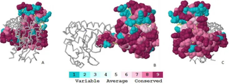
The ConSurf [24] results are based on the concept of identify functional regions in proteins, taking into account by considering the evolutionary relationships among their sequence homologues. An advantage of ConSurf over other methods is the accurate computation of the evolutionary rate using either an empirical
Bayesian method or a maximum likelihood method. Thus, ConSurf can correctly discriminate between the conservation caused by a short evolutionary time and genuine sequence conservation. The surface residues with the most variation are depicted in blue, and the conserved residues are depicted in purple in the protein structures (Figure 4). Our findings revealed that human SOD2 is highly conserved (Figure 4). The sequence alignment of the SOD2 from various species (Figure 5) reveals that residues E66 and G76 are conserved, whereas I82 and R156 are variable.
The conservation analysis of ConSurf used the evolutionary conservation scores of the residues to identify functional regions from proteins with known three-dimensional structures. The degree of conservation of the amino acid sites among the nine homologues with similar sequences (Figure 5) was estimated. The conservation grades were projected onto the molecular surface of the proteins to reveal the patches of highly conserved residues that are often important for biological function. Mutations E66 and G76 are conserved, whereas mutations I82 and R156 are variable. Figure 3. Superimposed native structures (green) and mutant structures (blue) of the SOD2 produced usingab initiomodelling.A) S10I (S-15I) mutation highlighted in red. B) This mutation disrupts the alpha helix, RMSD: 2.02. C) A16V (A-9V) mutation highlighted in red. D) This mutation disrupts the alpha helix, RMSD: 1.94.
doi:10.1371/journal.pone.0065558.g003
Table 4.Structure alignment ofab initioSOD2 mutant
models with theab initiowild-type model.
Pos. Variant I-Tasser TM-Align
C-score TM-score RMSD RMSD TM-Score
Figure 4. Conservation profile of the Mn-SOD (PDB ID: 1LUV) using ConSurf conservational analysis.Mn-SOD is represented as a spacefill model, where the residue conservation scored is colour-coded onto the surface. The backbone model represents the other chain of a Mn-SOD dimer, chain B. The colour-coding bar shows the colouring scheme: conserved amino acids are coloured bordeaux, residues with average conservation are white, and variable amino acids are turquoise.
doi:10.1371/journal.pone.0065558.g004
Figure 5. Multiple protein sequence alignment using ConSurf shows evolutionary conservation of amino acid residues.The colour-coding bar shows the colouring scheme: conserved amino acids are coloured bordeaux, residues of average conservation are white, and variable amino acids are turquoise. SNP positions are marked by an asterisk.
doi:10.1371/journal.pone.0065558.g005
Generally, residues that are implicated in biological processes, such as those located in active sites, involved in protein-protein or protein-ligand interactions, or implicated in protein structure and folding stability, are subject to greater selective pressure and are usually more conserved than other residues.
SOD2 Database
The SOD2 database currently contains all of the natural variants listed in UniProt. For each SNP, we provide the predictions of functional effects, indicated as Disease/Pathological or Neutral/Tolerated, from SNPeffect, PolyPhen-2, PhD-SNP, PMUT, SIFT, SNAP, SNPs&GO and nsSNPAnalyzer.
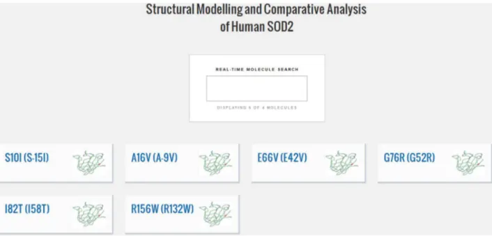
The database interface (Figure 6) allows users to search for a mutation by its non-synonymous SNP.
The database is curated by humans and will be updated as new natural variants are discovered.
The SOD2 database allows a user to quickly retrieve and rapidly analyse the predicted effects of protein variants. In addition
to predicting the effects of variants, an alignment of the wild-type and mutant structures can be visualised using the database.
The major feature that distinguishes the SOD2 database from other databases is that this database can use predictions from several algorithms for all of the known natural variants of Mn-SOD. Furthermore, the user has access to an alignment of the wild type and mutant structures and can thus visualise the damage that a SNP can cause. Our ultimate goal is to turn the database into a toolbox for researchers studying this protein. Thein silicoanalysis
of Mn-SOD in this database will help in the design and prioritisation of further experimental research.
Author Contributions
Conceived and designed the experiments: MDCC JFM. Performed the experiments: MDCC. Analyzed the data: MDCC JFM. Contributed reagents/materials/analysis tools: JFM. Wrote the paper: MDCC JFM.
References
1. Wang V, Chen SY, Chuang TC, Shan DE, Soong BW, et al. (2010) Val-9Ala and Ile+58Thr polymorphism of MnSOD in Parkinson’s disease. Clin Biochem 43: 979–982.
2. Wiener HW, Perry RT, Chen Z, Harrell LE, Go RC (2007) A polymorphism in SOD2 is associated with development of Alzheimer’s disease. Genes Brain Behav 6: 770–775.
3. Shimoda-Matsubayashi S, Matsumine H, Kobayashi T, Nakagawa-Hattori Y, Shimizu Y, et al. (1996) Structural dimorphism in the mitochondrial targeting sequence in the human manganese superoxide dismutase gene. A predictive evidence for conformational change to influence mitochondrial transport and a study of allelic association in Parkinson’s disease. Biochem Biophys Res Commun 226: 561–565.
4. Singh M, Khan AJ, Shah PP, Shukla R, Khanna VK, et al. (2008) Polymorphism in environment responsive genes and association with Parkinson disease. Mol Cell Biochem 312: 131–138.
5. Akyol O, Yanik M, Elyas H, Namli M, Canatan H, et al. (2005) Association between Ala-9Val polymorphism of Mn-SOD gene and schizophrenia. Prog Neuropsychopharmacol Biol Psychiatry 29: 123–131.
6. Galecki P, Smigielski J, Florkowski A, Bobinska K, Pietras T, et al. (2010) Analysis of two polymorphisms of the manganese superoxide dismutase gene (Ile-58Thr and Ala-9Val) in patients with recurrent depressive disorder. Psychiatry Res 179: 43–46.
7. Fullerton JM, Tiwari Y, Agahi G, Heath A, Berk M, et al. (2010) Assessing oxidative pathway genes as risk factors for bipolar disorder. Bipolar Disord 12: 550–556.
8. Berk M, Copolov D, Dean O, Lu K, Jeavons S, et al. (2008) N-acetyl cysteine as a glutathione precursor for schizophrenia–a double-blind, randomized, placebo-controlled trial. Biol Psychiatry 64: 361–368.
9. Berk M, Copolov DL, Dean O, Lu K, Jeavons S, et al. (2008) N-acetyl cysteine for depressive symptoms in bipolar disorder–a double-blind randomized placebo-controlled trial. Biol Psychiatry 64: 468–475.
10. Capriles PV, Guimaraes AC, Otto TD, Miranda AB, Dardenne LE, et al. (2010) Structural modelling and comparative analysis of homologous, analogous and specific proteins from Trypanosoma cruzi versus Homo sapiens: putative drug targets for chagas’ disease treatment. BMC Genomics 11: 610.
11. Sanchez R, Sali A (1997) Evaluation of comparative protein structure modeling by MODELLER-3. Proteins Suppl 1: 50–58.
12. Laskowski RA, MacArthur MW, Moss DS, Thornton JM (1993) PROCHECK: a program to check the stereochemical quality of protein structures. Journal of Applied Crystallography 26: 283–291.
13. Roy A, Kucukural A, Zhang Y (2010) I-TASSER: a unified platform for automated protein structure and function prediction. Nat Protoc 5: 725–738. 14. Zhang Y, Skolnick J (2005) TM-align: a protein structure alignment algorithm
15. Adzhubei IA, Schmidt S, Peshkin L, Ramensky VE, Gerasimova A, et al. (2010) A method and server for predicting damaging missense mutations. Nat Methods 7: 248–249.
16. De Baets G, Van Durme J, Reumers J, Maurer-Stroh S, Vanhee P, et al. (2012) SNPeffect 4.0: on-line prediction of molecular and structural effects of protein-coding variants. Nucleic Acids Res 40: D935–939.
17. Capriotti E, Calabrese R, Casadio R (2006) Predicting the insurgence of human genetic diseases associated to single point protein mutations with support vector machines and evolutionary information. Bioinformatics 22: 2729–2734. 18. Ferrer-Costa C, Orozco M, de la Cruz X (2002) Characterization of
disease-associated single amino acid polymorphisms in terms of sequence and structure properties. J Mol Biol 315: 771–786.
19. Ng PC, Henikoff S (2001) Predicting deleterious amino acid substitutions. Genome Res 11: 863–874.
20. Bromberg Y, Rost B (2007) SNAP: predict effect of non-synonymous polymorphisms on function. Nucleic Acids Res 35: 3823–3835.
21. Bromberg Y, Yachdav G, Rost B (2008) SNAP predicts effect of mutations on protein function. Bioinformatics 24: 2397–2398.
22. Calabrese R, Capriotti E, Fariselli P, Martelli PL, Casadio R (2009) Functional annotations improve the predictive score of human disease-related mutations in proteins. Hum Mutat 30: 1237–1244.
23. Bao L, Zhou M, Cui Y (2005) nsSNPAnalyzer: identifying disease-associated nonsynonymous single nucleotide polymorphisms. Nucleic Acids Res 33: W480– 482.
24. Ashkenazy H, Erez E, Martz E, Pupko T, Ben-Tal N (2010) ConSurf 2010: calculating evolutionary conservation in sequence and structure of proteins and nucleic acids. Nucleic Acids Res 38: W529–533.
25. UNIPROT (2012) Reorganizing the protein space at the Universal Protein Resource (UniProt). Nucleic Acids Res 40: D71–75.
26. Borgstahl GE, Parge HE, Hickey MJ, Johnson MJ, Boissinot M, et al. (1996) Human mitochondrial manganese superoxide dismutase polymorphic variant Ile58Thr reduces activity by destabilizing the tetrameric interface. Biochemistry 35: 4287–4297.
27. Jimenez-Lopez JC, Gachomo EW, Seufferheld MJ, Kotchoni SO (2010) The maize ALDH protein superfamily: linking structural features to functional specificities. BMC Struct Biol 10: 43.
28. Mistri M, Tamhankar PM, Sheth F, Sanghavi D, Kondurkar P, et al. (2012) Identification of novel mutations in HEXA gene in children affected with Tay Sachs disease from India. PLoS One 7: e39122.