Submitted26 August 2014 Accepted 26 January 2015 Published17 February 2015
Corresponding author Steven P. Vensko II, [email protected]
Academic editor Claus Wilke
Additional Information and Declarations can be found on page 9
DOI10.7717/peerj.771
Copyright
2015 Vensko II and Stone
Distributed under
Creative Commons CC-BY 4.0
OPEN ACCESS
X
-to-autosome expression and
msl-2
transcript abundance correlate among
Drosophila melanogaster
somatic tissues
Steven P. Vensko II1and Eric A. Stone2
1Program in Genetics, North Carolina State University, Raleigh, NC, USA
2Department of Biological Sciences, North Carolina State University, Raleigh, NC, USA
ABSTRACT
InDrosophila melanogaster, the male-specific lethal (MSL) complex has been studied extensively for its role in upregulating maleX-linked genes. Recent advances in high-throughput technologies have improved our understanding of how the MSL complex mediates dosage compensation through chromosome-wide chromatin modifications. Most studies, however, have focused on cell line models that cannot reflect any potential heterogeneity ofin vivodosage compensation. Comparisons between cell line and organismal gene-level dosage compensation upregulation sug-gest the possibility of variation in MSL complex activity among somatic tissues. We hypothesize the degree, up to but not exceeding 2-fold, to which the MSL complex upregulates maleX-linked genes varies quantitatively by tissue type. In this model, MSL complex abundance acts as a rheostat to control the extent of upregulation. Using publicly available expression data, we provide evidence for our model in
Drosophilasomatic tissues. Specifically, we findX-to-autosome expression correlates with the tissue-specific expression ofmsl-2which encodes an essential male-specific component of the MSL complex. This result suggests MSL complex mediated dosage compensation varies quantitatively by tissue type. Furthermore, this result has con-sequences for models explaining the organismal-scale molecular and evolutionary consequences of MSL-mediated dosage compensation.
Subjects Computational Biology, Genetics, Genomics
Keywords H4K16Ac, Male-Specific Lethal complex,Drosophila melanogaster, Dosage compensation complex, Dosage compensation
INTRODUCTION
Xchromosome, resulting in acetyaltion of lysine 16 of the fourth histone core (H4K16Ac), and consequently preventing formation of 30 nm chromatin fibers (Shogren-Knaak et al., 2006;Robinson et al., 2008). This induces “looser” chromatin and is believed to increase accessibility for DNA-binding proteins (Bell et al., 2010). Although the mechanism underlying MSL complex-dependent upregulation remains contested, both increased transcriptional elongation and increased RNA polymerase II (PolII) promoter density have been proposed (Larschan et al., 2011;Conrad et al., 2012;Ferrari et al., 2013;Straub & Becker, 2013).
Our understanding ofDrosophilaMSL complex-dependent dosage compensation (hereafter referred to as dosage compensation or DC) has advanced with the development of high-throughput assays over the past decade. These studies contribute insight into many aspects of dosage compensation, including the signals identifying the maleXchromosome as the primary target of the MSL complex (Alekseyenko et al., 2006;Alekseyenko et al., 2008; Straub et al., 2008;Alekseyenko et al., 2012) and the extent to which the MSL complex upregulates maleX-linked genes (Straub et al., 2005;Deng & Meller, 2006;Hamada et al., 2005). S2 cells have been commonly utilized for these studies due to their ease of genetic manipulation and MSL complex activity (Straub et al., 2008;Hamada et al., 2005;Alekseyenko et al., 2006;Alekseyenko et al., 2012;Straub et al., 2005). While S2 cells have been fundamental toward understanding the molecular mechanism of DC, they do not reflect heterogeneity potentially present at an organismal level. Most genes show variable expression patterns across tissues; thus, if dosage compensation shows differential activity across tissues then a cell-line model of dosage compensation cannot faithfully replicate the complex patterns of DC within whole organisms. For this reason, an appreciation of organismal-level DC complexity is essential for models attempting to clarify the global molecular and evolutionary consequences of dosage compensation onX-linked genes (Bachtrog, Toda & Lockton, 2010;Mikhaylova & Nurminsky, 2011). For example, work byMikhaylova & Nurminsky (2011)suggests that tissue specificity may play a role in the migration of genes offof theDrosophila X chromosome to avoid deleterious overexpression by dosage compensation. If the degree of dosage compensation varies by tissue, quantifying tissue-level heterogeneity would help clarify the relationship between DC and expression tissue specificity. This may, in turn, lead to a better understanding of the selective pressures that may have influencedDrosophila X-to-autosome retrotranspositions.
X-linked genes tend to be upregulated in both S2 cells and third instar larval males but at drastically different levels. These differences in upregulation suggest dosage compensation patterns observed within cell lines do not translate to an organismal context, possibly due to variation in dosage compensation activity among third instar larval male somatic tissues. Furthermore, recent work byNozawa et al. (2014)provides support for this hypothesis using interspecific chromosome-level expression comparisons to show dosage compensation on theD. psuedoobscuraneo-Xchromosome varies between male heads, gonads, and male carcasses. Here we present an intraspecific approach providing evidence for extensive variation in dosage compensation among a variety ofDrosophila melanogaster
somatic tissues.
METHODS
FlyAtlas data were collected from Gene Expression Omnibus (GEO) series entry GSE7763 (last updated May 29, 2013) (Chintapalli, Wang & Dow, 2007). Tissues were filtered to include only non-female-specific tissues. Whole organism “tissue” entries were also removed. A complete listing of tissues can be found inTable S1. Probe sets were mapped to FBgn IDs using the FB2014 03 ReleaseDrosophila melanogaster exon summary file (Marygold et al., 2013). In cases where FBgn IDs had multiple representative probe sets, the mean expression intensity was calculated. Genes were filtered for expression on a tissue-by-tissue basis. For each tissue, genes were classified as expressed if they had “present” calls for all four FlyAtlas replicates and hadlog2intensity values≥6 for all
four FlyAtlas replicates (Stenberg et al., 2009;Philip, Pettersson & Stenberg, 2012). Only expressed genes were utilized to estimate X and autosomal expression for each tissue.
Genes were classified asX-linked or autosomal based on the FB2014 03 Release gene map table from FlyBase (Marygold et al., 2013). Sex-bias classifications were acquired from the Sex Bias Database (SEBIDA v3.1) (Gnad & Parsch, 2006). In particular, we used the “meta” sex-bias classifications which utilize expression data from a variety of sources (Ranz et al., 2003;Parisi et al., 2004;Ayroles et al., 2009). Genes were classified as non-sex-biased if classified as “unbiased” by SEBIDA and were classified as sex-biased otherwise.
RESULTS AND DISCUSSION
Variation in maleDrosophila X chromosome upregulation across
somatic tissues
There is ample evidence that the expression patterns of X-linked genes vary in their tissue distribution (Chintapalli, Wang & Dow, 2007). For these genes to exhibit varying levels of dosage compensation on an organismal level, dosage compensation must also be differentially active across the male somatic tissues in which these genes are expressed. Noting that the primary role of the MSL complex in dosage compensation is to offset male monosomy by upregulating a majority ofX-linked genes, it stands to reason that decreased complex activity should associate with a larger difference in expression levels between theXand the autosomes. If the degree of difference varies quantitatively across tissues, this would provide evidence consistent with an analog model of tissue-specific dosage compensation. While this approach appears straight-forward, one must be cognizant of the differing evolutionary histories of theXchromosome and autosomes that may dilute any signal ofXchromosome dosage compensation upregulation. Taking this into consid-eration, we partitioned genes into a non-sex-biased set and a sex-biased set. We expect the non-sex-biased gene set to be dosage compensated, show similar enrichment on theX
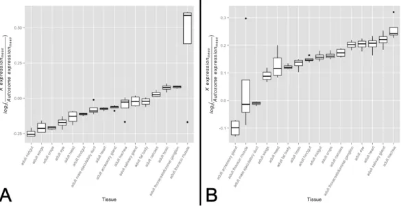
chromosome and autosomes, and be less likely to be regulated sex-specific mechanisms. We expect the sex-biased gene set, on the other hand, to be likely experiencing some level of dosage compensation but for this signal to be diluted by sex-specific regulatory mechanisms and an unequal distribution between theXchromosome and autosomes. These two gene sets, therefore, provide us the ability to detect any signal of dosage compensation in the non-sex-biased gene set while ensuring sex-specific mechanisms and differing gene content between theXchromosome and autosomes are not driving the signal. We tested for variation inX-to-autosome expression among tissues for both gene sets by calculating the difference between meanXchromosome gene expression and mean autosome gene expression. We found significant variation inX-to-autosome expression among somatic adult tissues for both the non-biased gene set (Fig. 1A,
F15,48≈9.40,p<0.005) and sex-biased gene set (Fig. 1B,F15,48≈13.64,p<0.005). These trends were also observed among non-sex-biased genes within larval tissues (Fig. S1,
F7,24≈279.52,p<0.005). In line with our expectations, the adult testis shows the lowest
X-to-autosome expression among adult tissues (seeFig. S2). Interestingly, there is no significant correlation between the non-sex-biased gene set and sex-biased gene set which suggests they are under differing transcriptional regulatory regimes. While these results hint toward the possibility of variation in dosage compensation activity among somatic tissues, other mechanisms may instead be responsible. We sought further evidence of our hypothesis by interrogating tissue-specific variation of the MSL complex itself.
Variation in MSL complex abundance across somatic tissues
Figure 1 X-to-autosome expression variation among somatic tissues.X-to-autosome expression esti-mates were calculated for all four FlyAtlas replicates for each tissue using the log2transformed ratios of the mean expression ofX-linked expressed genes to mean expression of autosomal expressed genes for both the (A) non-sex-biased gene set and (B) sex-biased gene set. Tissues are sorted by their median log2 ratio among the FlyAtlas replicates.
increased binding to the femaleXchromosome. A common strategy in these studies is to modulate transcript levels, specifically those ofmsl-2, which encodes a subunit essential for proper MSL complex formation (Demakova et al., 2003;Dahlsveen et al., 2006;Kelley et al., 1995;Oh, Park & Kuroda, 2003;Park et al., 2002;Hamada et al., 2005). These studies commonly utilize females to study the relationship between MSL complex abundance and dosage compensation through upregulation of the naturally absent MSL-2 protein; however, they establish a relationship that is expected to be relevant in males. While bothmsl-1andmsl-2serve fundamental roles in formation of the MSL complex,
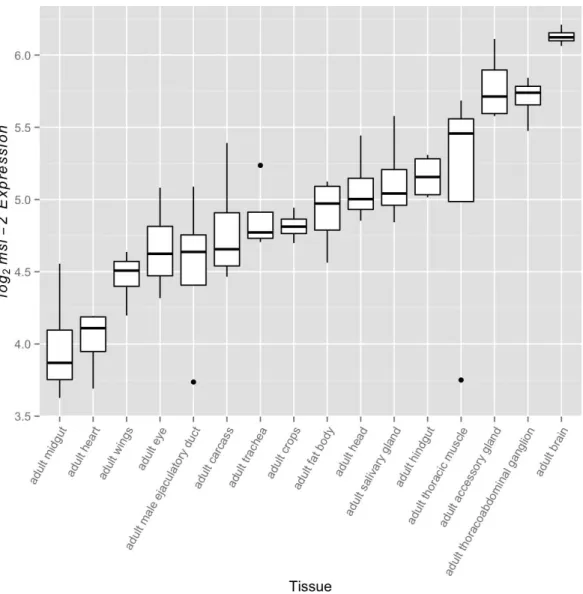
msl-1transcript abundance exceedsmsl-2transcript abundance in every FlyAtlas adult tissue suggesting MSL-2 may be the limiting factor. This led us to examine variation in
Figure 2 msl-2expression variation among somatic tissues.msl-2expression estimates were retrieved for all four FlyAtlas replicates for each tissue. Tissues are sorted by their median log2 msl-2intensity among the FlyAtlas replicates.
Covariation betweenmsl-2 transcript abundance andX
-to-autosome expression across somatic tissues
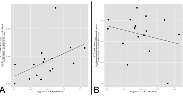
Figure 3 Correlation betweenX-to-autosome expression andmsl-2expression among somatic tis-sues.X-to-autosome mean expression plotted against its correspondingmsl-2mean expression for each somatic adult tissue for the (A) non-sex-biased gene set and (B) sex-biased gene set.
adult somatic tissues. Any other outcome would not provide evidence for our model. In support of our hypothesis, we found a significant positive correlation betweenmsl-2
expression andX-to-autosome expression among somatic adult tissues for non-sex-biased gene set (Fig. 3A,ρ≈0.68,p<0.005). The adult thoracic muscle is an extreme outlier
relative to the other tested tissues suggesting further investigation may yield interesting results. Larval tissues showed a positive but non-significant correlation likely due to its small sample size (Fig. S5,ρ≈0.21,p>0.05). The sex-biased gene set shows no significant
correlation betweenmsl-2expression andX-to-autosome expression among somatic adult tissues (Fig. 3B,ρ≈ −0.19,p>0.05). It is worth noting themsl-2locus is notX-linked
and thusmsl-2expression is not confounded withXchromosome expression. Other MSL complex components, when tested individually, do not show any significant correlation between their expression andX-to-autosome expression among tissues. While only finding significant effects only formsl-2may not seem intuitive, consideringmsl-2’s likely MSL complex-specific role and its required presence for complex formation, it may be the only component with detectable effects.
Implications and considerations
High-throughput studies have improved our understanding ofDrosophiladosage compen-sation; however, substantial questions remain. Here we have contributed evidence for an analog model of dosage compensation in which the degree of transcriptional upregulation activity is tissue specific. This suggests dosage compensation, while bound between 1-fold and 2-fold upregulation, is the product of a gene’s tissue distribution, transcriptional activityandabundance of the MSL complex in tissues in which that gene is expressed.
interpretation of our results. Due to the mixed-sex experimental design of the FlyAtlas data, both tissue-specificmsl-2expression measurements and chromosome-level expres-sion measurements were sampled from a set of equal count of male and female individuals. This is likely diluting the signal of dosage compensation, in which case we may be underestimating bothmsl-2expression variation andX-to-autosome expression variation among somatic adult tissues, as well as the strength of the correlation between them. Specifically, althoughmsl-2is transcribed in both males and females, its lower abundance in females may be biasing tissue-level expression measurements (Zhou et al., 1995). Likewise, tissue-levelX-to-autosome expression measurements may be affected by the contribution of female diploid, non-dosage compensatedXchromosomes to the transcript pool. Nevertheless, we do not believe that signal dilution from the use of mixed-sex data would generate positively misleading results. For a given tissue, one would expect
X-to-autosome expression to increase with an increasing contribution of female mRNA, due to their diploidXchromosomes. Conversely, becausemsl-2is more lowly expressed in females than in males, relativemsl-2abundance should decrease as the proportion of female mRNA to the mixed-sex expression pool grows. Thus, the inclusion of female mRNA would drive a negative correlation betweenmsl-2expression andX-to-autosome expression, as opposed to the significantly positive correlation that we report.
Another consideration is the sole reliance ofmsl-2expression as a marker of MSL complex abundance while using FlyAtlas data. The MSL complex consists of at least seven components that are essential for proper function (Conrad & Akhtar, 2012). That said, modulation ofmsl-2expression has been used repeatedly in the literature as a means to vary MSL complex abundance due its fundamental role in complex formation (Demakova et al., 2003;Dahlsveen et al., 2006;Kelley et al., 1995;Oh, Park & Kuroda, 2003;Park et al., 2002;Hamada et al., 2005). In addition, in every FlyAtlas adult tissue,msl-2transcript abundance is lower than that of another potentially limiting component,msl-1. Taking these relationships into account, we arguemsl-2expression is a faithful surrogate for MSL complex abundance. It is worth noting that other subunits (msl-1, msl-3, mle and mof) do not show any significant correlation between their transcript abundance and
X-to-autosome expression among somatic adult tissues. This result is not unexpected for some subunits, such asmof, that have known roles beyond the MSL complex (Kind et al., 2008). The lack of significant correlations for the remaining subunits support speculation byConrad & Akhtar (2012)for many of them having roles beyond dosage compensation.
Drosophiladosage compensation, while well-studied, remains a perplexing mechanism in several regards. Here we present evidence of varying levels of dosage compensation activity among somatic adult tissues. This hypothesis, motivated by differences inX-linked upregulation between S2 cells and third instar larval males (Deng & Meller, 2006), suggests care must be taken when quantifying the degree to which anX-linked gene is upregulated by dosage compensation inDrosophilamales. Additional data coupled with an improved understanding of the mechanism underlying dosage compensation will be required to conclusively link variation in MSL complex activity among somatic tissues to variation in expression upregulation by the MSL complex in whole organisms. We hope our results motivate further research into better understanding the behavior of dosage compensation within an organismal context.
ADDITIONAL INFORMATION AND DECLARATIONS
Funding
Steven Vensko II was supported in part by RO1-GM70806 from the US National Institutes of Health. The funders had no role in study design, data collection and analysis, decision to publish, or preparation of the manuscript.
Grant Disclosures
The following grant information was disclosed by the authors: US National Institutes of Health: RO1-GM70806.
Competing Interests
The authors declare there are no competing interests.
Author Contributions
• Steven P. Vensko II conceived and designed the experiments, performed the
experi-ments, analyzed the data, contributed reagents/materials/analysis tools, wrote the paper, prepared figures and/or tables, reviewed drafts of the paper.
• Eric A. Stone wrote the paper, reviewed drafts of the paper.
Supplemental Information
Supplemental information for this article can be found online athttp://dx.doi.org/ 10.7717/peerj.771#supplemental-information.
REFERENCES
Alekseyenko AA, Ho JWK, Peng S, Gelbart M, Tolstorukov MY, Plachetka A, Kharchenko PV, Jung YL, Gorchakov AA, Larschan E, Gu T, Minoda A, Riddle NC, Schwartz YB, Elgin SCR, Karpen GH, Pirrotta V, Kuroda MI, Park PJ. 2012.Sequence-specific targeting of dosage compensation in drosophila favors an active chromatin context.PLoS Genetics8(4):e1002646
Alekseyenko AA, Larschan E, Lai WR, Park PJ, Kuroda MI. 2006.High-resolution chip-chip analysis reveals that the drosophilamslcomplex selectively identifies active genes on the male x chromosome.Genes & Development20(7):848–857DOI 10.1101/gad.1400206.
Alekseyenko AA, Peng S, Larschan E, Gorchakov AA, Lee O-K, Kharchenko P, McGrath SD, Wang CI, Mardis ER, Park PJ, Kuroda MI. 2008.A sequence motif within chromatin entry sites directsmslestablishment on theDrosophilax chromosome.Cell134(4):599–609
DOI 10.1016/j.cell.2008.06.033.
Ayroles JF, Carbone MA, Stone EA, Jordan KW, Lyman RF, Magwire MM, Rollmann SM, Duncan LH, Lawrence F, Anholt RRH, Mackay TFC. 2009.Systems genetics of complex traits inDrosophila melanogaster.Nature Genetics41(3):299–307DOI 10.1038/ng.332.
Bachiller D, S´anchez L. 1986.Mutations affecting dosage compensation in Drosophila melanogaster: effects in the germline. Developmental Biology 118(2):379–384
DOI 10.1016/0012-1606(86)90007-2.
Bachtrog D, Toda NR, Lockton S. 2010.Dosage compensation and demasculinization of x chromosomes inDrosophila.Current Biology20(16):1476–1481DOI 10.1016/j.cub.2010.06.076. Bell O, Schwaiger M, Oakeley EJ, Lienert F, Beisel C, Stadler MB, Sch¨ubeler D. 2010.
Accessibil-ity of the drosophila genome discriminates pcg repression, h4k16 acetylation and replication timing.Nature Structural & Molecular Biology17(7):894–900DOI 10.1038/nsmb.1825. Chintapalli VR, Wang J, Dow JA. 2007.Using flyatlas to identify betterDrosophila melanogaster
models of human disease.Nature Genetics39(6):715–720DOI 10.1038/ng2049. Conrad T, Akhtar A. 2012.Dosage compensation in Drosophila melanogaster: epigenetic
fine-tuning of chromosome-wide transcription.Nature Reviews Genetics13(2):123–134
DOI 10.1038/nrg3124.
Conrad T, Cavalli FM, Vaquerizas JM, Luscombe NM, Akhtar A. 2012.Drosophila dosage compensation involves enhanced pol ii recruitment to male x-linked promoters.Science
337(6095):742–746DOI 10.1126/science.1221428.
Dahlsveen IK, Gilfillan GD, Shelest VI, Lamm R, Becker PB. 2006.Targeting determinants of dosage compensation in drosophila.PLoS Genetics2(2):e5DOI 10.1371/journal.pgen.0020005. Demakova OV, Kotlikova IV, Gordadze PR, Alekseyenko AA, Kuroda MI, Zhimulev IF. 2003.
Themslcomplex levels are critical for its correct targeting to the chromosomes inDrosophila melanogaster.Chromosoma112(3):103–115DOI 10.1007/s00412-003-0249-1.
Deng X, Meller VH. 2006.rox rnas are required for increased expression of x-linked genes in
Drosophila melanogastermales.Genetics174(4):1859–1866DOI 10.1534/genetics.106.064568. Ferrari F, Jung YL, Kharchenko PV, Plachetka A, Alekseyenko AA, Kuroda MI, Park PJ. 2013.
Comment on “drosophila dosage compensation involves enhanced pol ii recruitment to male x-linked promoters”.Science340(6130):273DOI 10.1126/science.1231815.
Gnad F, Parsch J. 2006.Sebida: a database for the functional and evolutionary analysis of genes with sex-biased expression.Bioinformatics22(20):2577–2579
DOI 10.1093/bioinformatics/btl422.
Hamada FN, Park PJ, Gordadze PR, Kuroda MI. 2005.Global regulation of x chromosomal genes by themslcomplex inDrosophila melanogaster.Genes & Development19(19):2289–2294
DOI 10.1101/gad.1343705.
Kelley RL, Solovyeva I, Lyman LM, Richman R, Solovyev V, Kuroda MI. 1995.Expression of
Kind J, Vaquerizas JM, Gebhardt P, Gentzel M, Luscombe NM, Bertone P, Akhtar A. 2008. Genome-wide analysis reveals mof as a key regulator of dosage compensation and gene expression inDrosophila.Cell133(5):813–828DOI 10.1016/j.cell.2008.04.036.
Larschan E, Bishop EP, Kharchenko PV, Core LJ, Lis JT, Park PJ, Kuroda MI. 2011. X chromosome dosage compensation via enhanced transcriptional elongation in drosophila.
Nature471(7336):115–118DOI 10.1038/nature09757.
Marygold SJ, Leyland PC, Seal RL, Goodman JL, Thurmond J, Strelets VB, Wilson RJ, FlyBase Consortium. 2013.Flybase: improvements to the bibliography.Nucleic Acids Research
41(D1):D751–D757DOI 10.1093/nar/gks1024.
Meiklejohn CD, Landeen EL, Cook JM, Kingan SB, Presgraves DC. 2011.Sex
chromosome-specific regulation in the drosophila male germline but little evidence for chromosomal dosage compensation or meiotic inactivation.PLoS Biology9(8):e1001126
DOI 10.1371/journal.pbio.1001126.
Mikhaylova L, Nurminsky D. 2011.Lack of global meiotic sex chromosome inactivation, and paucity of tissue-specific gene expression on the drosophila x chromosome.BMC Biology
9(1):29DOI 10.1186/1741-7007-9-29.
Nozawa M, Fukuda N, Ikeo K, Gojobori T. 2014.Tissue- and stage-dependent dosage
compensation on the neo-x chromosome in drosophila pseudoobscura.Molecular Biology
and Evolution31(3):614–624DOI 10.1093/molbev/mst239.
Oh H, Park Y, Kuroda MI. 2003.Local spreading ofmslcomplexes from rox genes on the drosophila x chromosome.Genes & Development17(11):1334–1339DOI 10.1101/gad.1082003. Parisi M, Nuttall R, Edwards P, Minor J, Naiman D, L¨u J, Doctolero M, Vainer M, Chan C,
Malley J, Eastman S, Oliver B. 2004.A survey of ovary-, testis-, and soma-biased gene expression inDrosophila melanogasteradults.Genome Biology5(6):R40
DOI 10.1186/gb-2004-5-6-r40.
Park Y, Kelley RL, Oh H, Kuroda MI, Meller VH. 2002.Extent of chromatin spreading determined by rox rna recruitment ofmslproteins.Science298(5598):1620–1623
DOI 10.1126/science.1076686.
Philip P, Pettersson F, Stenberg P. 2012.Sequence signatures involved in targeting the male-specific lethal complex to x-chromosomal genes inDrosophila melanogaster.BMC Genomics13(1):97DOI 10.1186/1471-2164-13-97.
Philip P, Stenberg P. 2013.Male x-linked genes inDrosophila melanogasterare compensated independently of the male-specific lethal complex.Epigenetics & Chromatin6(1):35
DOI 10.1186/1756-8935-6-35.
Prestel M, Feller C, Becker PB. 2010.Dosage compensation and the global re-balancing of aneuploid genomes.Genome Biology11(8):216DOI 10.1186/gb-2010-11-8-216.
R Core Team. 2013.R: a language and environment for statistical computing. Vienna: R Foundation for Statistical Computing.
Ranz JM, Castillo-Davis CI, Meiklejohn CD, Hartl DL. 2003.Sex-dependent gene expression and evolution of the drosophila transcriptome.Science300(5626):1742–1745
DOI 10.1126/science.1085881.
Rastelli L, Kuroda MI. 1998.An analysis ofmalelessand histone h4 acetylation inDrosophila melanogasterspermatogenesis.Mechanisms of Development71(1):107–117
Robinson PJ, An W, Routh A, Martino F, Chapman L, Roeder RG, Rhodes D. 2008.30 nm chromatin fibre decompaction requires both h4-k16 acetylation and linker histone eviction.
Journal of Molecular Biology381(4):816–825DOI 10.1016/j.jmb.2008.04.050.
Shogren-Knaak M, Ishii H, Sun J-M, Pazin MJ, Davie JR, Peterson CL. 2006.Histone h4-k16 acetylation controls chromatin structure and protein interactions.Science311(5762):844–847
DOI 10.1126/science.1124000.
Stenberg P, Lundberg LE, Johansson A-M, Ryd´en P, Svensson MJ, Larsson J. 2009.Buffering of segmental and chromosomal aneuploidies inDrosophila melanogaster.PLoS Genetics
5(5):e1000465DOI 10.1371/journal.pgen.1000465.
Straub T, Becker PB. 2013.Comment on “drosophila dosage compensation involves enhanced pol ii recruitment to male x-linked promoters”.Science340(6130):273
DOI 10.1126/science.1231895.
Straub T, Gilfillan GD, Maier VK, Becker PB. 2005.The drosophilamslcomplex activates the transcription of target genes.Genes & Development19(19):2284–2288
DOI 10.1101/gad.1343105.
Straub T, Grimaud C, Gilfillan GD, Mitterweger A, Becker PB. 2008. The chromosomal high-affinity binding sites for the drosophila dosage compensation complex.PLoS Genetics
4(12):e1000302DOI 10.1371/journal.pgen.1000302.
Wickham H. 2009.ggplot2: elegant graphics for data analysis. New York: Springer.