Generation of a Novel Bacteriophage Library
Displaying scFv Antibody Fragments from the
Natural Buffalo Host to Identify Antigens
from Adult
Schistosoma japonicum
for
Diagnostic Development
Christopher G. Hosking1*, Hamish E. G. McWilliam2, Patrick Driguez3, David Piedrafita4, Yuesheng Li3, Donald P. McManus3, Leodevico L. Ilag5, Els N. T. Meeusen6, Michael J. de Veer1¤
1Department of Physiology, Monash University, Clayton, Victoria, Australia,2Department of Microbiology and Immunology, The University of Melbourne, the Peter Doherty Institute for Infection and Immunity, Parkville, Victoria, Australia,3QIMR Berghofer Medical Research Institute, Brisbane, Queensland, Australia,4School of Applied Sciences and Engineering, Federation University, Churchill, Victoria, Australia,5Bio21 Molecular Sciences and Biotechnology Institute, University of Melbourne, Parkville, Victoria, Australia,6Department of Microbiology, Monash University, Clayton, Victoria, Australia
¤ Current address: Department of Physiology, Monash University, Clayton, Victoria, Australia *[email protected]
Abstract
The development of effective diagnostic tools will be essential in the continuing fight to reduce schistosome infection; however, the diagnostic tests available to date are generally laborious and difficult to implement in current parasite control strategies. We generated a series of single-chain antibody Fv domain (scFv) phage display libraries from the portal lymph node of field exposed water buffaloes,Bubalus bubalis, 11–12 days post challenge withSchistosoma japonicumcercariae. The selected scFv-phages showed clear enrichment towards adult schistosomes and excretory-secretory (ES) proteins by immunofluorescence, ELISA and western blot analysis. The enriched libraries were used to probe a schistosome specific protein microarray resulting in the recognition of a number of proteins, five of which were specific to schistosomes, with RNA expression predominantly in the adult life-stage based on interrogation of schistosome expressed sequence tags (EST). As the libraries were enriched by panning against ES products, these antigens may be excreted or secreted into the host vasculature and hence may make good targets for a diagnostic assay. Further selection of the scFv library against infected mouse sera identified five soluble scFv clones that could selectively recognise soluble whole adult preparations (SWAP) relative to an irrel-evant protein control (ovalbumin). Furthermore, two of the identified scFv clones also selec-tively recognised SWAP proteins when spiked into naïve mouse sera. These host B-cell derived scFvs that specifically bind to schistosome protein preparations will be valuable reagents for further development of a cost effective point-of-care diagnostic test.
a11111
OPEN ACCESS
Citation:Hosking CG, McWilliam HEG, Driguez P, Piedrafita D, Li Y, McManus DP, et al. (2015) Generation of a Novel Bacteriophage Library Displaying scFv Antibody Fragments from the Natural Buffalo Host to Identify Antigens from Adult
Schistosoma japonicumfor Diagnostic Development. PLoS Negl Trop Dis 9(12): e0004280. doi:10.1371/ journal.pntd.0004280
Editor:John Pius Dalton, McGill University, CANADA
Received:June 24, 2015
Accepted:November 13, 2015
Published:December 18, 2015
Copyright:© 2015 Hosking et al. This is an open access article distributed under the terms of the
Creative Commons Attribution License, which permits unrestricted use, distribution, and reproduction in any medium, provided the original author and source are credited.
Data Availability Statement:All relevant data are within the paper and its Supporting Information files.
Author Summary
Mass drug administration using the highly effective drug praziquantel (PZQ) is currently the method of choice to combat schistosomiasis. However, this treatment regime has limi-tations; in particular, it does not prevent re-infection and sporadic parasite resistance against PZQ is a continuing threat. The path to the successful control of schistosomiasis is highly challenging and must consider, not only the complex nature of the host-parasite interaction, but also the capacity to assess disease burden and parasite re-emergence in communities where successful control has been achieved. Furthermore, control programs must be economically sustainable in endemic countries and despite significant recent advancements the elimination of schistosomiasis may still be some time away. Accord-ingly, there is a definitive need to formulate innovative approaches for the development of improved diagnostic tools to accurately assess the disease burden associated with active schistosome infections. Here we describe the usefulness of a phage display library to mature antibody fragments derived from lymph node RNA of the natural buffalo host of the Asian schistosome,Schistosoma japonicum, following an experimental infection. These mature antibody fragments were able to bind native parasite proteins and could thus be used to develop a low cost and accurate point-of-care diagnostic test.
Introduction
Schistosomiasis is one of the most insidious of all the tropical parasitic infections and threatens the health of hundreds of millions of people worldwide [1]. The last 20 years has seen remark-able progress in disease control through the use of praziquantel (PZQ), but this drug does not protect against re-infection and mass drug administration programmes based around its use are probably untenable long term [2–4]. Recently there has been a major focus on the develop-ment of anti-schistosome vaccines, but to date a protective commercial vaccine remains elusive [1]. As mass drug administration decreases worm burdens within endemic areas, the need for improved diagnostic tests should be given research priority [5]. However, in countries where elimination of schistosomiasis has been given precedence, case detection of infected individuals remains problematic as the commonly used methods for diagnosis lack the necessary sensitiv-ity and specificsensitiv-ity to accurately determine parasite burden [6]. Although application of modern research laboratory techniques has seen improvements in the diagnosis of helminth infection, uptake has not been uniform and proof of concept studies that show promise have often not been followed through with much needed product development [7].
Currently the Kato-Katz thick smear stool method, based on detection of eggs in faeces, is the test sanctioned by the World Health Organization (WHO) for qualitative and quantitative diagnosis of intestinal schistosomiasis [8,9]. This test is generally specific, simple and relatively inexpensive, but like many parasitological tests, sensitivity can be insufficient, particularly when worm burdens are low [7]. Consequently, the use of single Kato-Katz measurements can underestimate the prevalence of infection and can confound confirmation of cure assessment following chemotherapy [10]. This is of particular importance in the Peoples Republic (PR) of China as the country moves towards programs aimed at the elimination of schistosomiasis japonica [11,12]. Since the 1950’s the prevalence of schistosomiasis japonica within Chinese provinces has dramatically decreased [13,14] and the requirements for a diagnostic tool has moved from the detection of parasitic infection to the ability to effectively assess disease preva-lence [14].
Evaluation of the Gates-funded SCORE project in African countries demonstrated that a rapid, accurate point-of-care (POC) diagnostic test that detects a circulating cathodic antigen (CCA) could identifyS.mansoniantigens [15,16]. The CCA and circulating anodic antigen (CAA) have been investigated as potential diagnostic candidates and can be detected in the serum and urine of infected individuals [17,18]. These antigens are cleared from the serum and urine of schistosomiasis patients within weeks following curative treatment [19]. However, success of these tests has only been validated for areas of high and moderate endemicity [18,
20]. Whilst CCA and CAA appear to be excellent antigen based tests, we have taken a different approach that may offer advantages for the development of reagents aimed at detecting very low infection levels.
Recently McWilliam et al., demonstrated, in a rat model of schistosomiasis, that the devel-oping schistosome worm can elicit a distinct immune response in discrete tissue sites [21]. Building on this concept we previously published the construction of an scFv-phage library for the detection of larval stage antigens as potential vaccine candidates [22]. However, theS. japo-nicumlarval stages are small, transient and rapidly migrate between tissues. The adult parasites are much larger, more persistent and shed antigen directly into the blood which makes them much more attractive targets for an antigen based diagnostic. Here we describe the construc-tion and characterisaconstruc-tion of scFv libraries derived from the portal lymph nodes ofS.japonicum
infectedB.bubalis, and demonstrate their ability to bind to the surface of adultS.japonicum
worms and excretory-secretory (ES) products. These reagents offer many advantages for diag-nostic development, including the ability to affinity mature the reagents, easy selection in a number of modalities, existing detection reagents and strong binding. It is hoped these reagents can be developed into a rapid POC diagnostic to aid in the surveillance and eventual elimina-tion ofS.japonicum.
Materials and Methods
Ethics statement
Written approval for animal experiments was provided by the Ethical Review Board of the Hunan Institute of Parasitic Diseases (approval # 110818) and from Monash University Ani-mal Ethics Committee (approval # 2011-124-FW). AniAni-mals were maintained and cared for according to the Animal Ethics and Procedures Guidelines of the PR China.
Parasite collection and crude protein extracts
Cercariae fromS.japonicumwere shed from infectedOncomelania hupensissnails collected from an endemic region in the People’s Republic (PR) of China using described methods [23,
24]. AdultS.japonicumworm pairs were collected from infected mice at QIMR Berghofer Institute for Medical Research as described [25]. Extracts of soluble whole adult preparations (SWAP) or excretory-secretory (ES) products from live adult worms were prepared as described [26,27]. Soluble egg antigen (SEA) was generated from eggs extracted from infected livers digested in 400μg/ml collagenase B overnight at 37°C. The digested liver solution was
centrifuged (400g, 5 min) and washed extensively in PBS. The washed solution was then passed though sieves (250μm and 150μm, respectively) and eggs were separated out of the
Experimental animal infections and sample collection
Animal experiments and sample collection were conducted in the PR China as described [28]. Briefly, six mixed sexS.japonicuminfectedB.bubaliswere obtained from anS.japonicum -endemic region in Hunan Province, PR China. Pre-existing infection status was confirmed by faecal egg count to provide animals where natural immunity could be“boosted”by subsequent experimental schistosome infection. All animals were drenched upon arrival with PZQ (25 mg/ kg) and randomly assigned into two experimental groups (n= 3 per group). Group 1 was infected with 400 liveS.japonicumcercariae percutaneously on the inner thigh. Group 2 was the uninfected control group. Animals were sacrificed 11–12 days post infection (days p.i.).
Collection of liver-draining lymph node samples
Lymph nodes draining the liver ofB.bubaliswere collected and cells isolated as described pre-viously [29]. For RNA preparation, 1 x 10⁹cells were centrifuged and the cell pellet resus-pended in 2 ml of QIAzol lysis buffer (QIAGEN, Netherlands) and RNA was prepared according to the manufacturer’s recommendations. RNA was further purified using an RNeasy Mini Kit (QIAGEN, Netherlands) as per the manufacturer’s recommendations. RNA was quantitated via absorbance at 260 nm using a NanoDrop spectrophotometer (Thermo Fisher Scientific, USA) and stored at -80°C until required.
Construction and amplification of B-cell scFv-phage antibody libraries
The preparation and characterisation of a scFv-phage display library similar to that used in this study has been described previously [22]. Briefly, full length variable light (VL) and variable heavy (VH) chain genes were amplified by PCR from portal-lymph node RNA derived from buffalo 11–12 days following an experimentalS.japonicuminfection. The scFv fragment was cloned into a phage display vector (pAK100; Plückthan Laboratory, University of Zurich, Swit-zerland) with the VL and VH genes separated by DNA encoding a flexible linker sequence (VL-(G4S)4-VH). The scFv fragment was directionally cloned into the pAK100 vector usingdifferentialSfiI restriction sites and was fused in-frame to the phage gene III coding DNA. This library was transformed in XL-1 BlueEscherichia colicells and approximately 5 x 10⁷ transfor-mants were recovered following overnight incubation at 37°C. The panning library was ampli-fied by inoculating 50 ml of non-expressing (NE) medium (2 X Yeast Tryptone (2YT), 1% glucose, 25 mg/ml chloramphenicol) with approximately 109XL-1 BlueE.colicells containing scFv phagemids and shaken at 37°C. At OD600= 0.5, 1 x 10¹¹transducing units per ml (TU/
ml) M13K07 helper-phage (New England Biolabs, USA) and 25μl 1 M Isopropylβ
Selection of scFv-phage binders to adult worms or excretory-secretory
products
For selection of high affinity binders, 1 x 10¹¹scFv-phage particles in 1ml PBS were initially pre-absorbed in 1.7 ml microfuge tubes (Axygen, Corning Life Sciences, USA) or 96 well microplates (Corning Life Sciences, USA) for 1 hr at RT with agitation. This was repeated a total of four times to eliminate scFv-phages that preferentially bind plastic. Following pre-absorption, 1 x 10¹¹TU/ml scFv-phages were added to 10 ± 2 formaldehyde-fixed adult schis-tosome pairs (10% formaldehyde for 30 mins followed by 3 washes with PBS) or 1μg of ES
worm products (0.1μg coated per well on a on 96 well plate) and allowed to bind for 2 hr at RT
with gentle agitation. Control reactions of scFv-phages without the addition of parasite mate-rial were also prepared. Tubes and microplates were then washed 10 times with PBS with 0.05% (v/v) Tween-20 (PBS-T), followed by an additional two washes with PBS. Bound scFv-phages were eluted with 0.2 M glycine/HCl, pH 3.0 for 15 min at RT. Supernatant from tubes and microplates was then collected and immediately neutralised with appropriate volume of 1 M Tris-HCL. Eluted scFv-phages (typically 1 x 10⁴–1 x10⁶TU/ml) were amplified as previously outlined and resuspended in PBS. Amplified scFv-phages were used for further panning rounds or parasite binding analysis. Libraries panned against adults or ES are termed R3-A or Bp-R3-ES respectively and an equal mix of each scFv-pool is termed Bp-R3-AES. Combined BP-R3-AES phage pools were used to minimise amount ofS.japonicummaterial required.
Selection against infected mouse sera
Naïve andS.japonicuminfected mouse sera (21 days p.i.; kindly supplied by Dr. Patrick Dri-guez, QIMR Berghofer Medical Research Institute, Australia) were treated with Affi-Gel Blue to remove albumin as per the manufacturer’s instructions (Bio-Rad, USA). The Bp-R3-AES phage pool was adjusted to 1 x 10¹¹TU/ml and absorbed against depleted naïve mouse sera (diluted 1:10 in PBS) coated onto 96 well microtitre plates (100μl/well; Maxisorb; NUNC,
Denmark). Pre-absorbed Bp-R3-AES were then panned against depleted infected mouse sera (diluted 1:10 in PBS) coated onto 96 well microtitre plates (100μl/well; Maxisorb; NUNC,
Denmark). Following each panning round phages were eluted and amplified as previously out-lined and again absorbed against depleted naïve mouse sera, before being further panned against depleted infected mouse sera. This process was repeated for a total of three rounds. This post infected mouse sera panned library will be termed Bp-R3-post infected mouse sera (Bp-R3-PIMS).
Generation of soluble scFv clones
Selected scFv coding regions from Bp-R3 phages following infected mouse sera panning (Bp-R3-PIMS) were sub-cloned into theE.coliexpression vector pAK600 (Plückthun Laboratory, University of Zurich, Switzerland) and transformed using electroporation into TOP 10 F’E.
Binding of Bp-R3 scFv-phages and soluble scFvs clones to schistosome
extracts by ELISA
Microplates (Corning Life Sciences, USA) were coated with 100μl of eitherS.japonicum
SWAP, ES, SEA, ovalbumin (0.5μg/well) or SWAP spiked into uninfected mouse sera
(equiva-lent to 0.5μg/well in 1:10 diluted naïve mouse sera) in carbonate coating buffer (0.05 M
car-bonate-bicarbonate, pH 9.6) and incubated overnight at 4°C. The next day microplates were washed three times and blocked in PBS-T for 1 hr at 37°C. The scFv-phage pools (pre, Bp-R3-A, Bp-R3-ES, Bp-R3-AES or Bp-R3-PIMS) diluted 1:5 in PBS-T and soluble scFv-AP clones diluted 1:10 in PBS-T were added to triplicate wells and incubated for 1 hr at 37°C. Specific binding of scFv-phage pools was detected using an anti-M13 pIII monoclonal antibody (New England Biolabs, USA) followed by biotin-conjugated anti-mouse antibody (goat anti-mouse IgG Fc, Jackson Immunoresearch), then streptavidin-HRP (BioRad, USA). Reactivity of scFv-AP clones towards schistosome antigens was detected using rabbit anti-alkaline phosphatase (ABCAM, USA), then swine rabbit-HRP conjugate (Dako, Germany) All detection anti-bodies were diluted 1:1000 in PBS-T and incubated for 1 hr at 37°C. Following incubation, all plates were washed three times with PBS-T and developed with 3,3’,5,5’-tetramethylbenzidine (TMB) solution (Life Technologies, USA) for 15 minutes and the reaction was stopped with 2 M H2SO4. Antibody or scFv-phage binding was detected using O.D. measurements at 450 nm.
Western blotting
Protein preparations (SWAP or SEA) were resolved by reducing SDS-PAGE and transferred to nitrocellulose membranes as per the manufactures recommendations (iBlot Dry Blotting Sys-tem; Life Technologies, USA). Membranes were visualised with Ponceau S (0.1% (w/v) Pon-ceau S in 5% acetic acid; Sigma-Aldrich, USA), cut into individual lanes and blocked for 2 hr at RT in PBS-T. Membranes were incubated overnight at 4°C with Bp-R3-AES phages (diluted 1:5 in PBS-T). Individual lanes were washed three times with PBS-T and Bp-R3-AES phages were detected with anti-M13 pIII monoclonal antibody (New England Biolabs, USA), followed by biotin-conjugated anti-mouse antibody (goat anti-mouse IgG Fc, Jackson Immunoresearch, USA), then streptavidin-HRP (BioRad, USA). All detection antibodies were diluted 1:1000 in PBS-T and incubated for 1 hr at RT. Protein bands were detected using the metal enhanced DAB peroxidase substrate detection system (Thermo Fisher Scientific, USA).
Carboxyfluorescein succinimidyl ester (CFSE) conjugation and
fluorescent microscopy
Discrete amplified scFv-phages (Bp-pre, Bp-R3-A or Bp-R3-ES) were diluted to 1 x 10¹¹TU/ ml and labelled with 120μg of carboxyfluorescein succinimidyl ester (CFSE; 10 mg/ml in
parasites. Two hundred images were captured at 10.8μm steps with fixed exposure times and
an SHR Plan Apo 2x objective. The images shown were reconstructed and rendered using Ima-geJ software [34].
Protein microarray screening and scanning
A microarray consisting of 232 schistosome specific proteins was prepared as described [35]. Briefly, array pads were hydrated with Whatman Blocking Buffer (WBB; Whatman, GE Healthcare, UK). Phages (M13K07 helper-phage control, Bp-R3-A or Bp-R3-ES phages) were pre-absorbed in WBB containing 10% (w/v) reconstitutedE.colilysate (EBB; Mc Lab, USA). Binding of scFv-phage particles was detected by addition of M13 pIII monoclonal anti-body (diluted 1:1000 in WBB; New England Biolabs, USA), biotin-anti-mouse antianti-body (diluted 1:1000 in WBB; Jackson Immunoresearch, USA), then streptavidin-conjugated Cy5 fluorophore (diluted 1:200 in WBB; Surelight P3, Columbia Biosciences, USA). Microarray pads were washed 3 times with TBS-T, 3 times with TBS, then in ultrapure water and dried by centrifugation at RT in a 50 ml Falcon tube (BD Biosciences, USA) for 5 min at 500g.
Microarray slides were scanned using a confocal laser microarray scanner (Genepix 4300A, Molecular Devices, USA). The signals were quantified with image analysis software (Genepix Pro 7, Molecular Devices, USA) and the reported feature intensity was calculated by subtract-ing the median local background signal from the feature signal.Data was analysed ussubtract-ing a varia-tion of the“group average”method [36,37]. Signal intensity (S.I.) greater than the average of the“No DNA”negative controls plus 2.5 standard deviations (S.D.) and no binding by
M13K07 helper-phage was used to determine positive recognition. Binding intensity was desig-nated on a plus minus scale. Sequences of identified antigens were then analysed for develop-mental expression based on previous work [38] and EST Profile Viewer (http://www.ncbi.nlm. nih.gov).
Statistical analysis
Differences between scFv-phages or soluble scFv clones was determined using a one- or two-way analysis of variance test (ANOVA) followed by a Tukey’s post-hoc test. Statistical results were reported when significance was achieved atP<0.05. Statistical analysis was performed using GraphPad Prism, version 6.01 software.
Results
Generation and enrichment of a scFv-phage library from infected buffalo
portal-lymph nodes
The VH and VL antibody regions were amplified from buffalo portal lymph nodes 11–12 days post infection withS.japonicumand cloned into a scFv-phage vector (S1A Fig). Sequencing of assembled scFv fragments revealed framework similarities and distinct variation in the comple-mentarity determining regions (CDR;S1B Fig). Transformation into XL-1 BlueE.colicells produced approximately 1.5 x 10⁷colonies. Panning against whole fixed adult worm pairs and ES products resulted in relative enrichment levels of greater than 800 or 80 fold, respectively, (S2 Fig). Libraries generated from this material are prefixed with Bp-R3.
to adult worm pairs under constant exposure conditions compared with CFSE labelled phage prior to selection (Bp-pre) and control M13K07 helper phage (Fig 1A3–4 compared to 1–2). To determine where on the adult schistosome the Bp-R3 phage bound we performed confocal microscopy using CFSE labelled Bp-R3 phages and adult schistosomes (Fig 1B). It demon-strated that binding to fixed parasites was primarily restricted to the surface, with no apparent binding to internal structures (Fig 1B-2). Interestingly, although initially binding appeared to be uniform (Fig 1A), confocal examination indicated there are regions on the surface of the parasite where phage do not bind in sufficient numbers to obtain a positive fluoresce signal (Fig 1B-1arrows). To quantitate phage binding to schistosome ES products we coated an ELISA plate with ES antigens and added different phage libraries, this showed that the enriched Bp-R3-ES phage library displayed significantly greater binding to ES products than any other phage library (Fig 1C). The Bp-R3-A library selected for adult binding also bound ES antigens more strongly than control phage, indicating the ES and adult surface likely share cross-reac-tive epitopes (Fig 1C).
Bp-R3 scFv-phage libraries recognise stage-specific antigens on protein
microarrays
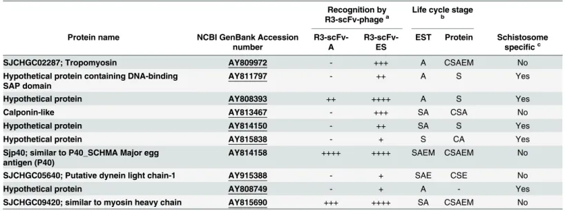
Screening of a schistosome specific protein microarray with Bp-R3 phages (Adult or ES) resulted in significant recognition of ten antigens. All ten of these antigens were recognised by the Bp-R3-ES phages, however only three antigens were consistently recognised by both the Bp-R3-A and Bp-R3-ES phages. These were a hypothetical protein (NCBI GenBank accession numberAY808393), a protein similar to the myosin heavy chain (SJCHGC09420;
AY8153690) and the previously defined Sjp40 protein (AY814158) (Table 1).
Other antigens recognised by the Bp-R3 phages included an additional four hypothetical proteins (AY811797,AY814150,AY815838andAY808749) and a further four known pro-teins, including tropomyosin (AY809972), a calponin-like protein (AY813467), a putative dynein light chain-1 protein (DLC1;AY915388) and a protein similar to myosin heavy chain (AY815690). The tropomyosin antigen (SJCHGC02287) is a 25 kDa protein expressed in a range of tissues at different developmental stages and is associated with muscle contraction and cystoskeletal structure and function [39]. Although tropomysins are not specific to schisto-somes, the SJCHGC02287 tropomyosin variant shares less than 50% homology with any other organism outside the schistosome genus. The calponin-like antigen shares homology with a 38 kDaS.japonicumcalponin, which has been previously investigated [40]. The DLC1 fromS.
japonicumhas previously been investigated as a potential target for vaccine or drug develop-ment [41]. The function of theAY815838hypothetical protein is currently unknown, but the sequence has some homology to theS.mansonisurface antigens Sm13 (AAC25419.1; 30%) and Sm25 (AAA29943; 34%). Interestingly, all of the schistosome specific antigens identified using the array show increased binding with the Bp-R3-ES selected phage indicating that these antigens may be excreted or secreted into the host vasculature. It should be noted that the pro-teins observed inTable 1were often identified in one life-stage by proteomic analysis, but had expressed sequence tags (ESTs) in many life-stages or vice versa. This apparent inconsistency may reflect the incompleteness of the proteomic datasets available for analysis or the fact that mRNA levels do not necessarily correlate with protein expression.
exposure time. Arrows represent areas of minimal binding (B1). Cross sectional view of scFv-phage binding to adult schistosome showing binding is restricted to the surface of the parasite, with no observable binding to internal structures (B2). Bp-R3 scFv-phages bind toS.japonicumexcretory-secretory (ES) protein extracts by ELISA (C). Data represent mean±S.E.M. of the O.D.450nmfor discrete scFv-phage pools binding to ES protein preparations (C).
Statistical significance was determined by one-way ANOVA followed by Tukey’s post-hoc test, where**P<0.01,***P<0.001,****P<0.0001.
Bp-R3-AES scFv-phage pools recognise adult schistosome protein
extracts
We pooled the Bp-R3-A and Bp-R3-ES to widen the pool of potential phage sequences selected for adult and secreted schistosome epitopes and analysed the specificity of the pooled Bp-R3-AES library to bind to different schistosome antigen preparations by ELISA (Fig 2). The Bp-R3-AES phages showed significant levels of binding towards SWAP (p<0.05) (Fig 2A) indicating they recognised adult proteins. There was no observed binding of the Bp-R3-AES pool to the SEA preparation (Fig 2B) and background binding by M13K07 helper-phage to any antigen preparation was negligible (Fig 2A and 2B). Western blotting of Bp-R3-AES phages was also performed againstS.japonicumderived SWAP and SEA preparations (Fig 2C). The Bp-R3-AES phages bound to a broad range of epitopes within the SWAP preparation, but as observed using ELISA, did not recognise any epitopes within the SEA preparation (Fig 2C; Lane 2). Although the lack of SEA reactivity was unexpected, for a diagnostic aimed to detect an active adult infections, lack of SEA reactivity could be advantageous.
Isolated soluble scFv clones preferentially recognise SWAP antigens
The buffalo derived Bp-R3-AES phages were further panned against infected mouse sera to eliminate scFv-phage sequences that bind epitopes within sera, and enrich for antigens that are excreted into the serum during an experimental infection. This process generated a library des-ignated Bp-R3-PIMS, which displayed similar binding to SWAP proteins when compared to the Bp-R3-AES libraries as determined by ELISA (Fig 3A). Five soluble alkaline phosphatase (AP) scFv fusion proteins were generated from the R3-PIMS library (scFv-1 to Bp-scFv-5;S3 Fig).Table 1. Schistosoma japonicumprotein microarray antigen recognition by buffalo portal-round three selected (Bp-R3) single-chain antibody Fv domain (scFv) phages.
Recognition by R3-scFv-phagea
Life cycle stage
b
Protein name NCBI GenBank Accession
number
R3-scFv-A
R3-scFv-ES
EST Protein Schistosome specificc
SJCHGC02287; Tropomyosin AY809972 - +++ A CSAEM No
Hypothetical protein containing DNA-binding SAP domain
AY811797 - ++ A S Yes
Hypothetical protein AY808393 ++ ++++ A S Yes
Calponin-like AY813467 - +++ SA CSA No
Hypothetical protein AY814150 - ++ SA S Yes
Hypothetical protein AY815838 - + S CA Yes
Sjp40; similar to P40_SCHMA Major egg antigen (P40)
AY814158 ++++ ++++ SAEM CSAEM No
SJCHGC05640; Putative dynein light chain-1 AY915388 - + SAE CSE No
Hypothetical protein AY808749 - + A - Yes
SJCHGC09420; similar to myosin heavy chain AY815690 +++ ++++ SA CSAEM No
aRecognition level score: the average signal intensity of binding which was greater than 2.5 standard deviations of controls. Very high (++++)>1000
signal intensity (SI), High (+++) 500–999 SI; Moderate (++) 100–499 SI; low (+) 0–99 SI; (-) not a significant hit.
bLife stage with highest expression is based on previous work [38]: cercariae (C), schistosomula (S), adult (A), egg (E) or miracidium (M). cSchistosome speci
fic proteins are defined as those with no orthologues protein in other species via proteomic analysis [38].
Fig 2. Binding of buffalo portal-round three selected-adult and ES pooled (Bp-R3-AES) single-chain Fv domain (scFv) phage library to
Schistosoma japonicumprotein extracts.Data represent mean±S.E.M. of the O.D.450nmfor Bp-R3-AES phage binding to soluble whole adult
preparations (SWAP) (A) and soluble egg antigens (SEA) (B). Statistical significance was determined by one-way ANOVA followed by Tukey’s post-hoc test, where*P<0.05. Antigens from SWAP (Lane 1), and SEA (Lane 2) ofS.japonicumwere probed with Bp-R3-AES phage (C). Molecular weights in
kilodaltons (kDa) are indicated on the left hand side. Regions of intense binding by Bp-R3 scFv phages to antigen preparations is indicated by arrows.
Fig 3. Binding of buffalo portal-round three selected (Bp-R3) single-chain antibody Fv domain (scFv) phage panned against infected mouse sera and scFv-alkaline phosphatase (scFv-AP) fusion proteins toSchistosoma japonicumsoluble whole adult preparation (SWAP) by ELISA.Data represent mean±S.E.M. of the O.D.450nmfor Bp-R3-A and ES pooled phage libraries (Bp-R3-AES) and Bp-R3-post-infected-mouse-sera (Bp-R3-PIMS)
phage binding toS.japonicumSWAP (A), and scFv-AP proteins (Bp-scFv-1 to Bp-scFv-5) binding to SWAP (B and C) or SWAP spiked naive mouse sera (D). Antibodies derived from anS.japonicuminfected rabbit (infected antibodies) were used as a positive control (D#). Statistical significance was determined by one- or two-way ANOVA followed by Tukey’s post-hoc test, where*P<0.05,***P<0.001,****P<0.0001.
All identified scFv-AP proteins (Bp-scFv-1 to Bp-scFv-5) showed significantly higher bind-ing to SWAP relative to an ovalbumin protein bindbind-ing control as examined by ELISA (Fig 3C). AnE.colilysate control, containing no expressed scFv-AP protein, showed no reactivity against SWAP protein extract (Fig 3B). Two of the scFv-AP clones (Bp-scFv-1 and Bp-scFv-2) were also able to significantly and preferentially recognise SWAP proteins that had been spiked into naïve mouse sera by ELISA (Fig 3D)
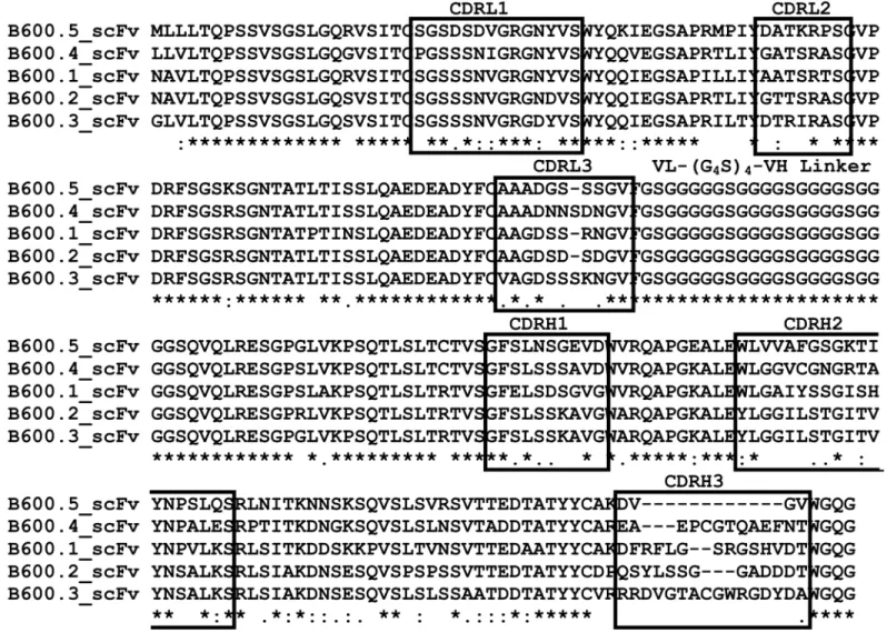
Sequencing of the scFv-AP clones (Fig 4) revealed high conservation of framework
sequences within both the VH and VL regions with significant variations only within the com-plementarity determining (CDR) regions. As expected within antibody sequences, the major region of diversity observed was within the CDR 3 of the heavy chain (CDRH3;Fig 4) with amino acid sequence length ranging from 4–16 amino acids.
Discussion
There has been considerable success at reducing transmission and infection rates ofS. japoni-cumin the PR China [42], however, there is a need to ensure the parasite life cycle is completely broken and that the national elimination program does not falter due to a significant number of low-level infections not being detected [11]. In areas where schistosomiasis japonica remains a problem there is a need to measure and target treatments as well as to educate communities [43]. If a significant number of low-level infections go undetected, there is the risk that the efforts already employed to control transmission will be in vain [11].
The Kato-Katz stool smear technique has been the backbone of intestinal schistosomiasis diagnosis in epidemiological studies, and in the case ofS.japonicumis the only approved mea-sure for diagnosis of current infection [20,44]. However, the technique is becoming less useful in regions where control programs have resulted in light parasite infections [45,46]. The vast majority of diagnostic measures for schistosomiasis are underestimating parasite burdens [47,
48]. This is of particular concern for prevention strategies being employed in regions of PR China where accurate diagnosis is crucial for the effective control and surveillance of the dis-ease [49]. The most recent epidemiological surveys suggest that the prevalence ofS.japonicum
infection in areas where transmission has not yet been controlled is 5.1% [49] and, as the coun-try strives for elimination of schistosomiasis, the need for more reliable diagnostic tests to sur-vey parasite prevalence is essential [50].
A number of diagnostic tests that detect the host antibody response to schistosome infection have been developed and are being integrated into national control programs in endemic areas of PR China. These include the circumoval precipitin test (COPT), the silver-enhanced colloi-dal gold metalloimmunoassay, enzyme-linked immunosorbent assay (ELISA), indirect hemag-glutination assay (IHA), dot immunogold filtration assay (DIGFA) and dipstick dye
immunoassay (DDIA), [44,51–53]. These tests provide a more accurate measure of infection than stool based parasitological tests with greater patient compliance as they are less invasive [44]. The DDIA test is now commercially available as a test forS.japonicumin low endemic regions [53]. However, it should be noted that due to the lack of strict approval guidelines in PR China, many poor performing diagnostic tests are also being used [44]. A study by Cai and colleagues further confirmed three of the antibody based diagnostic tests (IHA, ELISA and DDIA) successfully identify schistosomiasis infected patients [49]; however none are able to accurately distinguish between current or cured infection status and thus assessment of treat-ment outcomes is difficult.
schistosomiasis. A point-of-care (POC) CCA test is now commercially available and has proven successful for detection of intestinalS.mansoni, and has been proposed as an alterna-tive to the replicate Kato-Katz measurements [20,54]. The POC-CCA test is effective in endemic regions, yet its efficacy in areas of low intensity infections requires validation [14,18,
55,56]. Recent work by van Dam and colleagues demonstrated that a new technique to enhance CAA detection in sera and urine was six fold more sensitive at diagnosing an active schistosome infection when compared to Kato-Katz measurements [14]. The significant advantage of a sensitive and reliable antigen based diagnostic for schistosome infection is the ability to discriminate active infection. Yet, immunological tests are currently still expensive and often require trained technicians for the administration and analysis, which increases cost and demand for infrastructure [57]. Moreover, the ability of these tests to assess drug efficacy and provide information regarding the impact of control interventions stills needs to be evalu-ated [20].
Fig 4. Clustal Omega alignment of soluble single-chain antibody Fv domain-alkaline phosphatase (scFv-AP) antibody sequences.Complementarity determining regions (CDR) regions of variable light (CDRL1-3) and variable heavy (CDRH1-3) chains are indicated in boxes. Respective VL and VH regions show similarity within the framework regions and distinct differences within CDR regions, particularly within CDRH3. (*) indicates a fully conserved residue, (:) a strongly similar and (.) a weakly similar residue.
The zoonotic nature ofS.japonicummakes animals, particular water buffaloes, significant infection reservoirs. Water buffalo have been thought to contribute to 75% of human infections [13,58] and prevalence of infection within buffalo populations has been reported to be as high as 10% [59]. It is likely that control within humans living in close proximity to water buffalo will require accurate diagnosis of residual infection within these buffalo populations. This will likely require a low cost easy to administer test that accurately determines infection status.
The novel phage display approach used in this study incorporates the natural reservoir host ofS.japonicumto generate mature antibody fragments that bind to adult schistosomes and ES products. The matured buffalo antibody fragments should show little cross-reactivity with buf-falo proteins and may help form the basis of a reagent to diagnose infection in bufbuf-falo. Further-more, we have recently shown that buffalo CDRH3 regions selected for binding to
schistosomules using the antibody phage display system are longer than those from non-selected libraries, suggesting that long CDRH3 regions may be important in host parasite anti-gen recognition [22]. Recent studies have shown that long CDRH3 regions are pivotal in the successful defence against dangerous viral infections such as HIV [60] and although the func-tional mechanisms for ultra-long CDRH3 regions in cattle has yet to be elucidated [61] they may offer advantages for diagnostic development. Although we did not observe ultra-long CDRH3 regions in the five soluble scFv-AP clones characterised they are only a very small sub-set of the total number of clones generated.
Previous studies have utilised phage display technology to investigate the maturation of dis-played antibody or peptide fragments toward specificS.japonicummolecules [62–64]. These studies primarily involved the identification of peptide binders through the use of commercial peptide-phage display libraries. The present study used the immunological status of infected host animals to select from a B-cell repertoire primed to produce variable regions that recog-nise immune exposed epitopes present in the host at the time of sacrifice, i.e. when adult para-sites reach the hepatoportal circulation. Previous studies by McWilliam et al., have illustrated the ability of selected activated lymph node antibodies to discriminate against particular stages ofS.japonicum[21,28]. We have expanded on this principle by producing scFv-phage libraries from activated lymph nodes to allow molecular characterisation and easier production of the binding moity, which can be difficult to isolate from lymph node antibody preparations.
This present work also identified and characterised five unique soluble scFv-AP clones selected from the Bp-R3-PIMS phage library, which had been panned against serum obtained fromS.japonicuminfected mice. This blood panning, in particular the initial selection against naïve sera, was performed to both eliminate scFvs that preferentially bind to blood proteins and enrich for scFv-phages that bind schistosome antigens present in blood following infec-tion. All of the isolated soluble scFv-AP clones (Bp-scFv-1 to Bp-scFv-5) significantly bound to SWAP proteins. Importantly a lysate control, containing no expressed scFv, did not recognise SWAP proteins and no purified scFv-AP clone specifically recognised ovalbumin, a non-related protein binding control. Only two of the five scFv-AP clones were able to significantly detect SWAP above the level of background mouse blood proteins in an ELISA using naïve mouse sera spiked with SWAP antigens. This suggests that the scFv-phage technique can iso-late antibody fragments that recognise schistosome antigens in blood. However, further valida-tion and isolavalida-tion of these scFv antibody fragments is required to ensure they exclusively recognise schistosome antigens in naturally infected animals.
The development of a range of approaches to deliver accurate point of care, rapid schisto-some diagnosis will be critical in the fight to eradicate schistosomiasis japonica in areas of low but continuing transmission in PR China. Here we describe the use of the natural buffalo host to develop and identify recombinant scFv fragments that bind adult stage and ES antigens fromS.japonicum. The isolated soluble scFv clones require further development, purification and testing within a true schistosomiasis japonica infection setting, but once completed could be valuable reagents in the construction of a cost effective point-of-care diagnostic test.
Supporting Information
S1 Fig. Cloning procedure and amplification of single-chain antibody Fv domain (scFv) inserts.Total RNA from the portal-LN collected 11–12 days postSchistosoma japonicum cer-carial infection was reverse transcribed to cDNA. Variable heavy (VH) and variable light (VL) genes were amplified (A). Full length scFv fragments were assembled by splice overlap PCR and the full length scFv construct was amplified using scFv specific primers (A#). Molecular weights in base-pairs (bp) are indicated. Full length scFv fragments were inserted into pAK100 vector viaSfiI cloning. Selected phagemid were sequenced and aligned to cattle variable regions (B). Shaded regions represent complementarity determining regions (CDR) for VH and VL genes; () indicates a fully conserved residue, (:) a strongly similar and (.) a weakly similar resi-due.
(TIF)
S2 Fig. Relative enrichment of functional single-chain Fv domain (scFv) phage particles.
Binding to adultSchistosoma japonicumworms and excretory secretory (ES) products was determined using the relationship between the output titre from scFv-phage eluted from adult
S.japonicumworms or ES products at each round and control reactions containing no parasite material.
(TIF)
S3 Fig. Expression of soluble single-chain Fv domain (scFv) alkaline phosphatase (AP) fusion proteins.Soluble scFv-AP clones were expressed and probed with anti-AP-HRP and positive expression is indicated. A soluble scFv-His tag control protein was expressed and probed with anti-His-HRP and is also indicated. Molecular weights in kilodaltons (kDa) are indicated on the left hand side.
Acknowledgments
The authors wish to thank Prof. Andreas Plückthun at the University of Zurich, Switzerland for kindly supplying the pAK vectors used in this study.
Author Contributions
Conceived and designed the experiments: CGH HEGM DP ENTM MJdV YL. Performed the experiments: CGH HEGM DP ENTM. Analyzed the data: CGH MJdV. Contributed reagents/ materials/analysis tools: PD DPM YL. Wrote the paper: CGH HEGM PD DPM LLI ENTM MJdV.
References
1. Xu J, Ren Y, Xu X, Chen J, Li Y, Gan W, et al.,Schistosoma japonicumtegumental protein 20.8, role in reproduction through its calcium binding ability. Parasitol Res, 2014 Feb; 113(2): 491–7. doi:10.1007/ s00436-013-3678-7PMID:24276643
2. Bergquist R, Al-Sherbiny M, Barakat R, and Olds R, Blueprint for schistosomiasis vaccine develop-ment. Acta Trop, 2002 May; 82(2): 183–92. PMID:12020891
3. McManus DP and Bartley PB, A vaccine against Asian schistosomiasis. Parasitol Int, 2004 Jun; 53(2): 163–73. doi:10.1016/j.parint.2004.01.006PMID:15081948
4. Bethony JM, Cole RN, Guo X, Kamhawi S, Lightowlers MW, Loukas A, et al., Vaccines to combat the neglected tropical diseases. Immunol Rev, 2011 Jan; 239(1): 237–70. doi:10.1111/j.1600-065X.2010. 00976.xPMID:21198676
5. You H and McManus DP, Vaccines and diagnostics for zoonotic schistosomiasis japonica. Parasitol-ogy, 2014; 14(2): 271–89.
6. Utzinger J, Zhou XN, Chen MG, and Bergquist R, Conquering schistosomiasis in China: the long march. Acta Trop, 2005 Nov-Dec; 96(2–3): 69–96. PMID:16312039
7. McCarthy JS, Lustigman S, Yang GJ, Barakat RM, Garcia HH, Sripa B, et al., A Research Agenda for Helminth Diseases of Humans: Diagnostics for Control and Elimination Programmes. PLoS Negl Trop Dis, 2012; 6(4): e1449.
8. Kongs A, Marks G, Verle P, and Van der Stuyft P, The unreliability of the Kato-Katz technique limits its usefulness for evaluatingS.mansoniinfections. Trop Med Int Health, 2001 Mar; 6(3): 163–9. PMID: 11299032
9. Zhou YB, Zheng HM, and Jiang QW, A diagnostic challenge for Schistosomiasis japonica in China: consequences on praziquantel-based morbidity control. Parasit Vectors, 2011; 4: 194. doi:10.1186/ 1756-3305-4-194PMID:21981948
10. Berhe N, Medhin G, Erko B, Smith T, Gedamu S, Bereded D, et al., Variations in helminth faecal egg counts in Kato-Katz thick smears and their implications in assessing infection status withSchistosoma mansoni. Acta Trop, 2004 Nov-Dec; 92(3): 205–12. doi:10.1016/j.actatropica.2004.06.011PMID: 15533288
11. Lodh N, Mwansa JC, Mutengo MM, and Shiff CJ, Diagnosis ofSchistosoma mansoniwithout the stool: comparison of three diagnostic tests to detectSchistosoma mansoniinfection from filtered urine in Zambia. Am J Trop Med Hyg, 2013 Jul; 89(1): 46–50. doi:10.4269/ajtmh.13-0104PMID:23716406
12. Wang LD, Guo JG, Wu XH, Chen HG, Wang TP, Zhu SP, et al., China's new strategy to block Schisto-soma japonicumtransmission: experiences and impact beyond schistosomiasis. Tropical medicine & international health: TM & IH, 2009 Dec; 14(12): 1475–83. doi:10.1111/j.1365-3156.2009.02403.x
13. McManus DP, Gray DJ, Li Y, Feng Z, Williams GM, Stewart D, et al., Schistosomiasis in the People's Republic of China: the era of the Three Gorges Dam. Clin Microbiol Rev, 2010 Apr; 23(2): 442–66. doi: 10.1128/cmr.00044-09PMID:20375361
14. van Dam GJ, Xu J, Bergquist R, de Dood CJ, Utzinger J, Qin ZQ, et al., An ultra-sensitive assay target-ing the circulattarget-ing anodic antigen for the diagnosis ofSchistosoma japonicumin a low-endemic area, People's Republic of China. Acta Trop, 2015 Jan; 141(Pt B): 190–7. doi:10.1016/j.actatropica.2014. 08.004PMID:25128703
16. Shane HL, Verani JR, Abudho B, Montgomery SP, Blackstock AJ, Mwinzi PN, et al., Evaluation of urine CCA assays for detection of Schistosoma mansoni infection in Western Kenya. PLoS Negl Trop Dis, 2011; 5(1): e951. doi:10.1371/journal.pntd.0000951PMID:21283613
17. van Dam GJ, Bogitsh BJ, van Zeyl RJ, Rotmans JP, and Deelder AM,Schistosoma mansoni: in vitro and in vivo excretion of CAA and CCA by developing schistosomula and adult worms. J Parasitol, 1996 Aug; 82(4): 557–64. PMID:8691363
18. Coulibaly JT, Knopp S, N'Guessan NA, Silue KD, Furst T, Lohourignon LK, et al., Accuracy of urine cir-culating cathodic antigen (CCA) test for Schistosoma mansoni diagnosis in different settings of Cote d'Ivoire. PLoS Negl Trop Dis, 2011 Nov; 5(11): e1384. doi:10.1371/journal.pntd.0001384PMID: 22132246
19. van Lieshout L, De Jonge N, Mansour MM, Bassily S, Krijger FW, and Deelder AM, Circulating cathodic antigen levels in serum and urine of schistosomiasis patients before and after chemotherapy with prazi-quantel. Trans R Soc Trop Med Hyg, 1993 May-Jun; 87(3): 311–2. PMID:8236404
20. Coulibaly JT, N'Gbesso YK, Knopp S, N'Guessan NA, Silue KD, van Dam GJ, et al., Accuracy of urine circulating cathodic antigen test for the diagnosis of Schistosoma mansoni in preschool-aged children before and after treatment. PLoS Negl Trop Dis, 2013; 7(3): e2109. doi:10.1371/journal.pntd.0002109 PMID:23556011
21. McWilliam HE, Driguez P, Piedrafita D, Maupin KA, Haab BB, McManus DP, et al., The developing schistosome worms elicit distinct immune responses in different tissue regions. Immunol Cell Biol, 2013 Aug; 91(7): 477–85. doi:10.1038/icb.2013.33PMID:23856766
22. Hosking CG, Driguez P, McWilliam HEG, Ilag LL, Gladman S, Li Y, et al., Using the local immune response from the natural buffalo host to generate an antibody fragment library that binds the early lar-val stages ofSchistosoma japonicum. Int J Parasitol, 2015; in press:
23. Gobert GN, Moertel L, Brindley PJ, and McManus DP, Developmental gene expression profiles of the human pathogenSchistosoma japonicum. BMC Genomics, 2009; 10: 128. doi: 10.1186/1471-2164-10-128PMID:19320991
24. Moertel L, McManus DP, Piva TJ, Young L, McInnes RL, and Gobert GN, Oligonucleotide microarray analysis of strain- and gender-associated gene expression in the human blood fluke,Schistosoma japonicum. Mol Cell Probes, 2006 Oct; 20(5): 280–9. doi:10.1016/j.mcp.2006.02.002PMID: 16647836
25. Jones MK, McManus DP, Sivadorai P, Glanfield A, Moertel L, Belli SI, et al., Tracking the fate of iron in early development of human blood flukes. Int J Biochem Cell Biol, 2007; 39(9): 1646–1658. doi:10. 1016/j.biocel.2007.04.017PMID:17556009
26. You H, Zhang W, Jones MK, Gobert GN, Mulvenna J, Rees G, et al., Cloning and characterisation of Schistosoma japonicum insulin receptors. PLoS One, 2010; 5(3): e9868. doi:10.1371/journal.pone. 0009868PMID:20352052
27. Perez-Sanchez R, Ramajo-Hernandez A, Ramajo-Martin V, and Oleaga A, Proteomic analysis of the tegument and excretory-secretory products of adultSchistosoma bovisworms. Proteomics, 2006 Apr; 6 Suppl 1: S226–36. doi:10.1002/pmic.200500420PMID:16511809
28. McWilliam HEG, Piedrafita D, Li Y, Zheng M, He Y, Yu X, et al., Local Immune Responses of the Chi-nese Water Buffalo, Bubalus bubalis, against Schistosoma japonicum Larvae: Crucial Insights for Vac-cine Design. PLoS Negl Trop Dis, 2013; 7(9): e2460. doi:10.1371/journal.pntd.0002460PMID: 24086786
29. Meeusen EN and Brandon MR, Antibody secreting cells as specific probes for antigen identification. J Immunol Methods, 1994 Jun 3; 172(1): 71–6. PMID:8207267
30. Sambrook J, Fritsch EF, and Maniatis T, Molecular Cloning: A Laboratory Manual. Cold Spring Harbor Laboratory Press. Plainview, N.Y.:, 1989; 2nd ed:
31. McWilliam H, Li W, Uludag M, Squizzato S, Park YM, Buso N, et al., Analysis Tool Web Services from the EMBL-EBI. Nucleic Acids Res, 2013 Jul; 41(Web Server issue): W597–600. doi:10.1093/nar/ gkt376PMID:23671338
32. Krebber A, Bornhauser S, Burmester J, Honegger A, Willuda J, Bosshard HR, et al., Reliable cloning of functional antibody variable domains from hybridomas and spleen cell repertoires employing a reengi-neered phage display system. J Immunol Methods, 1997 Feb 14; 201(1): 35–55. PMID:9032408
33. Lindner P, Bauer K, Krebber A, Nieba L, Kremmer E, Krebber C, et al., Specific detection of his-tagged proteins with recombinant anti-His tag scFv-phosphatase or scFv-phage fusions. Biotechniques, 1997 Jan; 22(1): 140–9. PMID:8994661
35. Driguez P, Doolan D, Molina D, Loukas A, Trieu A, Felgner P, et al., Protein Microarrays for Parasite Antigen DiscoveryParasite Genomics Protocols, ed. Peacock C.. Vol. 1201. 2015: Springer New York. 221–233.
36. Sundaresh S, Doolan DL, Hirst S, Mu Y, Unal B, Davies DH, et al., Identification of humoral immune responses in protein microarrays using DNA microarray data analysis techniques. Bioinformatics, 2006 Jul 15; 22(14): 1760–6. doi:10.1093/bioinformatics/btl162PMID:16644788
37. Gaze S, Driguez P, Pearson MS, Mendes T, Doolan DL, Trieu A, et al., An immunomics approach to schistosome antigen discovery: antibody signatures of naturally resistant and chronically infected indi-viduals from endemic areas. PLoS Pathog, 2014 Mar; 10(3): e1004033. doi:10.1371/journal.ppat. 1004033PMID:24675823
38. Liu F, Lu J, Hu W, Wang SY, Cui SJ, Chi M, et al., New Perspectives on Host-Parasite Interplay by Comparative Transcriptomic and Proteomic Analyses of Schistosoma japonicum. PLoS Pathog, 2006 Apr; 2(4): doi:10.1371/journal.ppat.0020029
39. Silas S, Fitzsimmons CM, Jones FM, Pinot de Moira A, Wawrzyniak J, Tukahebwa EM, et al., Human IgE responses to different splice variants ofSchistosoma mansonitropomyosin: associations with immunity. Int J Parasitol, 2014 May; 44(6): 381–90. doi:10.1016/j.ijpara.2014.02.004PMID:24657550
40. Yang W, Zheng YZ, Jones MK, and McManus DP, Molecular characterization of a calponin-like protein fromSchistosoma japonicum. Mol Biochem Parasitol, 1999 Jan 25; 98(2): 225–37. PMID:10080391
41. Yang W, Jones MK, Fan J, Hughes-Stamm SR, and McManus DP, Characterisation of a family of
Schistosoma japonicumproteins related to dynein light chains. Biochim Biophys Acta, 1999 Jun 15; 1432(1): 13–26. PMID:10366724
42. Collins C, Xu J, and Tang S, Schistosomiasis control and the health system in P.R. China. Infect Dis Poverty, 2012; 1(1): 8. doi:10.1186/2049-9957-1-8PMID:23849320
43. McManus DP, Bieri FA, Li YS, Williams GM, Yuan LP, Henglin Y, et al., Health education and the con-trol of intestinal worm infections in China: a new vision. Parasit Vectors, 2014; 7: 344. doi:10.1186/ 1756-3305-7-344PMID:25060336
44. Xu J, Peeling RW, Chen JX, Wu XH, Wu ZD, Wang SP, et al., Evaluation of immunoassays for the diag-nosis of Schistosoma japonicum infection using archived sera. PLoS Negl Trop Dis, 2011; 5(1): e949. doi:10.1371/journal.pntd.0000949PMID:21267065
45. Wu G, A historical perspective on the immunodiagnosis of schistosomiasis in China. Acta Trop, 2002 May; 82(2): 193–8. PMID:12020892
46. Zhou YB, Yang MX, Wang QZ, Zhao GM, Wei JG, Peng WX, et al., Field comparison of immunodiag-nostic and parasitological techniques for the detection of Schistosomiasis japonica in the People's Republic of China. Am J Trop Med Hyg, 2007 Jun; 76(6): 1138–43. PMID:17556625
47. Enk MJ, Oliveira e Silva G, and Rodrigues NB, Diagnostic accuracy and applicability of a PCR system for the detection ofSchistosoma mansoniDNA in human urine samples from an endemic area. PLoS One, 2012; 7(6): e38947. doi:10.1371/journal.pone.0038947PMID:22701733
48. Enk MJ, Lima AC, Drummond SC, Schall VT, and Coelho PM, The effect of the number of stool sam-ples on the observed prevalence and the infection intensity withSchistosoma mansoniamong a popu-lation in an area of low transmission. Acta Trop, 2008 Nov-Dec; 108(2–3): 222–8. doi:10.1016/j. actatropica.2008.09.016PMID:18973744
49. Cai YC, Xu JF, Steinmann P, Chen SH, Chu YH, Tian LG, et al., Field comparison of circulating anti-body assays versus circulating antigen assays for the detection of schistosomiasis japonica in endemic areas of China. Parasit Vectors, 2014; 7: 138. doi:10.1186/1756-3305-7-138PMID:24684924
50. Wang LD, Chen HG, Guo JG, Zeng XJ, Hong XL, Xiong JJ, et al., A strategy to control transmission of
Schistosoma japonicumin China. N Engl J Med, 2009 Jan 8; 360(2): 121–8. doi:10.1056/ NEJMoa0800135PMID:19129526
51. Zhou XN, Guo JG, Wu XH, Jiang QW, Zheng J, Dang H, et al., Epidemiology of schistosomiasis in the People's Republic of China, 2004. Emerg Infect Dis, 2007 Oct; 13(10): 1470–6. doi:10.3201/eid1310. 061423PMID:18257989
52. Zhu YC, Immunodiagnosis and its role in schistosomiasis control in China: a review. Acta Trop, 2005 Nov-Dec; 96(2–3): 130–6. doi:10.1016/j.actatropica.2005.07.007PMID:16143288
53. Gomes LI, Enk MJ, and Rabello A, Diagnosing schistosomiasis: where are we? Rev Soc Bras Med Trop, 2014 Jan-Feb; 47(1): 3–11. doi:10.1590/0037-8682-0231-2013PMID:24553804
55. van Dam GJ, Odermatt P, Acosta L, Bergquist R, de Dood CJ, Kornelis D, et al., Evaluation of banked urine samples for the detection of circulating anodic and cathodic antigens inSchistosoma mekongi
andS.japonicuminfections: a proof-of-concept study. Acta Trop, 2015 Jan; 141(Pt B): 198–203. doi: 10.1016/j.actatropica.2014.09.003PMID:25225158
56. Colley DG, Binder S, Campbell C, King CH, Tchuem Tchuente LA, N'Goran EK, et al., A five-country evaluation of a point-of-care circulating cathodic antigen urine assay for the prevalence ofSchistosoma mansoni. Am J Trop Med Hyg, 2013 Mar; 88(3): 426–32. doi:10.4269/ajtmh.12-0639PMID:23339198
57. Nausch N, Dawson EM, Midzi N, Mduluza T, Mutapi F, and Doenhoff MJ, Field evaluation of a new anti-body-based diagnostic for Schistosoma haematobium and S. mansoni at the point-of-care in northeast Zimbabwe. BMC Infect Dis, 2014; 14: 165. doi:10.1186/1471-2334-14-165PMID:24666689
58. Li YS, McManus DP, Lin DD, Williams GM, Harn DA, Ross AG, et al., TheSchistosoma japonicum self-cure phenomenon in water buffaloes: potential impact on the control and elimination of schistosomiasis in China. Int J Parasitol, 2014 Mar; 44(3–4): 167–71. doi:10.1016/j.ijpara.2013.10.007PMID: 24440417
59. Balen J, Zhao ZY, Williams GM, McManus DP, Raso G, Utzinger J, et al., Prevalence, intensity and associated morbidity ofSchistosoma japonicuminfection in the Dongting Lake region, China. Bulletin of the World Health Organization, 2007 Jul; 85(7): 519–26. PMID:17768500
60. Doria-Rose NA, Schramm CA, Gorman J, Moore PL, Bhiman JN, DeKosky BJ, et al., Developmental pathway for potent V1V2-directed HIV-neutralizing antibodies. Nature, 2014 May 1; 509(7498): 55–62. doi:10.1038/nature13036PMID:24590074
61. Wang F, Ekiert Damian C, Ahmad I, Yu W, Zhang Y, Bazirgan O, et al., Reshaping Antibody Diversity. Cell, 2013; 153(6): 1379–1393. doi:10.1016/j.cell.2013.04.049PMID:23746848
62. Liu Y, Brindley PJ, Zeng Q, Li Y, Zhou J, Chen Y, et al., Identification of phage display peptides with affinity for the tegument ofSchistosoma japonicumschistosomula. Mol Biochem Parasitol., 2011; 180 (2): 86–98. doi:10.1016/j.molbiopara.2011.09.001PMID:21930161
63. Arnon R, Tarrab-Hazdai R, and Steward M, A mimotope peptide-based vaccine againstSchistosoma mansoni: synthesis and characterization. Immunology, 2000 Dec; 101(4): 555–62. PMID:11122460