)228(
COPYRIGHT 2016 © BY THE ARCHIVES OF BONE AND JOINT SURGERY
Arch Bone Jt Surg. 2016; 4(2): 228-230. http://abjs.mums.ac.ir
the online version of this article abjs.mums.ac.ir
Dirk P. ter Meulen, MD; Sjoerd P.F.T. Nota, MD; Michiel G.J.S. Hageman, MD; David C. Ring, MD
Research performed at the Hand Surgery Service, Massachusetts General Hospital, Boston, MA, USA
Corresponding Author:David Ring, 1400 Barbara Jordan Blvd 1.114, Austin, TX 78723, USA
Email: [email protected]
RESEARCH ARTICLE
Received: 30 May 2016 Accepted: 26 June 2016
Progression of Heterotopic Ossification around
the Elbow after Trauma
Abstract
Background: This study addresses the null hypothesis that there is no expansion of heterotopic ossiication (HO) in the
elbow beyond what can be seen early on.
Methods: The area of HO was measured on lateral radiographs of 38 consecutive patients that had operative treatment of HO between 2000 and 2013. Measurements from radiographs obtained between 3 to 7 weeks were compared to measurements from radiographs made 3 months or more after injury.
Results: There was no signiicant difference between the average area of HO on the irst (median 2.8 square centimeters, Q1: 1.5, Q3: 5.1) and later radiographs (median of 2.8 square centimeters, Q1: 1.4, Q3: 5.0) (P = 0.99).
Conclusion:According to our results the area of HO does not expand beyond what can be seen early in the disease
process.
Keywords: Disease progression, Elbow, Heterotopic, Injuries, Ossiication
Introduction
P
atients with elbow trauma or burn injuries often develop heterotopic ossification (HO) that limits elbow motion (1, 2). Proposed risk factors for developing HO after injury include the severity of the injury, the presence of dislocation, and the timing of surgery (3, 4). Among surgically treated fractures and fracture-dislocations involving the proximal aspect of the radius or ulna about 20% of patients develop HO that interferes with motion (1, 2).It is our experience that the extent of HO seems apparent within a few weeks. HO tends to ossify and become radiographically visible before fracture callus. It is our impression that HO does not “expand” beyond what can be seen early on. Abrams and colleagues found that--among patients who eventually develop HO--it is already present on radiographs obtained 2 weeks after injury in 86% of patients (4).
This study addressed the null hypothesis that there is no difference in the area of heterotopic ossification (HO) seen on radiographs 3 to 7 weeks after injury and the area of HO seen more than 3 months after injury.
Materials and Methods
With approval from our institutional review board, 98 patients with the diagnosis code for heterotopic ossification and a billing code for elbow surgery between 2000 and 2013 were retrospectively identified.
Inclusion criteria were: (1) Patients age 18 years or greater, (2) fracture of the proximal ulna or radius or fracture of the distal humerus, (3) surgical treatment for elbow trauma, (4) surgery within 10 days after trauma to minimize influence by secondary surgery as additional trauma, (5) a lateral elbow radiograph taken between 21 and 49 days after surgery, and (6) a lateral elbow radiograph taken between 90 and 365 days after surgery. Exclusion criteria were: (1) Patients with prior injury or deformity and (2) surgery in between the study radiographs. Thirty-eight patients met these inclusion and exclusion criteria.
The mean age of the study population was 50 years (SD 13, range 24 – 74). There were 25 male patients (66%) and 19 right-sided fractures (50%).
PROGRESSION OF HO AROUND THE ELBOW THE ARCHIVES OF BONE AND JOINT SURGERY. ABJS.MUMS.AC.IR
VOLUME 4. NUMBER 3. JULY 2016
)229(
9 columnar fractures of the distal humerus, 6 fractures of the capitellum and trochlea, 3 posterior dislocation with radial head fractures, 3 varus posteromedial rotational injuries/ anteromedial facet coronoid fractures, 2 posterior olecranon fracture dislocations, 2 anterior olecranon fracture dislocations, and 1 diaphyseal ulna fracture with a fracture of the radial head.
On average the first radiograph was taken after 35 days (SD 7, range 23 - 49) and the second after 132 days (SD 40, range 91 - 266). The average number of days between the first and second radiograph was 97 (SD 41, range 50-231).
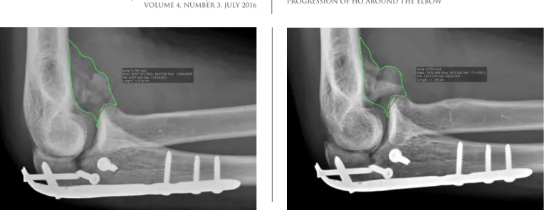
Radiographs were retrieved as DICOM (Digital Imaging and Communications in Medicine) images and were then loaded into Osirix (v5.6, Bernex, Switzerland). In Osirix, using the regions-of-interest tool the area of HO was measured in square centimeters in both subsequent lateral elbow radiographs [Figure 1; 2]. Areas of HO overlying host bone or implants were not
measured.
Statistical Analysis
The Wilcoxon signed rank test was used to test for a significant difference in the measured area of HO area between early and late radiographs.
Results
There was no significant difference between the average area of HO on the first (median 2.8 square centimeters, Q1: 1.5, Q3: 5.1) and second (median of 2.8 square centimeters, Q1: 1.4, Q3: 5.0) radiographs (P = 0.99).
Discussion
We found that there is no significant difference in the area of heterotopic ossification (HO) seen on radiographs 3 to 7 weeks after injury and the area of HO seen more than 3 months after injury. In other words, heterotopic bone does not grow or expand beyond where it is initially seen early on.
When interpreting the results of this study, certain limitations should be taken into account: 1) Due to the retrospective nature of the study data was collected with
Figure 1. Lateral radiographs a few weeks (A) and several months (B) after open reduction and internal fixation of an olecranon fracture-dislocation shows little change in the area of heterotopic ossification.
PROGRESSION OF HO AROUND THE ELBOW THE ARCHIVES OF BONE AND JOINT SURGERY. ABJS.MUMS.AC.IR
VOLUME 4. NUMBER 3. JULY 2016
)230(
Dirk P. ter Meulen MD Sjoerd P.F.T. Nota MD Michiel G.J.S. Hageman MD David C. Ring MD
Dell Medical School, The University of Texas at Austin, Austin, USA
References
a relative wide variation in time points. 2) We measured area of HO two-dimensionally, ideally measurements should be three-dimensionally (3D) to asses the volume of the HO, but that would require routine CT scanning (5). 3) Areas of HO with overlying host bone or implants could not be measured. Potential progression of HO in these areas has not been included in this study 4) Measurements were done by 1 author not blinded to timing. 5) Borders of HO were more distinct on the second set series of radiograph; this made measurements more accurate on the second set.
It is hard to predict which patients will develop HO and a major concern is that currently available prophylaxis can cause impaired wound or bone healing (6, 7). One study noted that in 86% of the patients who eventually develop HO it is already present on radiographs obtained 2 weeks after injury (4). Once HO begins to form it is unclear whether anything can or should be done to prevent further growth (8).
According to our study the area of HO on lateral
radiographs does not expand beyond what can be seen early in the disease process. This suggests that treatment with radiation or non-steroidal anti-inflammatory medication is most helpful before HO begins to form and may not have much of a role in limiting HO once it is noticed. Issues of the timing of HO excision are best decided on the basis of data regarding the safety and ease of earlier vs. a few month delayed surgery (delay provides more mobile skin with less edema and more mature bone that is easier to isolate and remove), and the risk of recurrence which is felt to potentially be higher when operating on immature HO (9).
1. Foruria AM, Augustin S, Morrey BF, Sanchez-Sotelo J. Heterotopic ossification after surgery for fractures and fracture-dislocations involving the proximal aspect of the radius or ulna. J Bone Joint Surg Am. 2013; 95(10):e66.
2. Wiggers JK, Helmerhorst GT, Brouwer KM, Niekel MC, Nunez F, Ring D. Injury complexity factors predict heterotopic ossification restricting motion after elbow trauma. Clin Orthop Relat Res. 2014; 472(7):2162-7.
3. Potter BK, Forsberg JA, Davis TA, Evans KN, Hawksworth JS, Tadaki D, et al. Heterotopic ossification following combat-related trauma. J Bone Joint Surg Am. 2010; 92(Suppl 2):74-89.
4. Abrams GD, Bellino MJ, Cheung EV. Risk factors for development of heterotopic ossification of the elbow after fracture fixation. J Shoulder Elbow Surg. 2012; 21(11):1550-4.
5. Brownley RC, Agarwal S, Loder S, Eboda O, Li J, Peterson J, et al. Characterization of heterotopic
ossification using radiographic imaging: evidence for a paradigm shift. PloS One. 2015; 10(11):e0141432. 6. Murnaghan M, Li G, Marsh DR. Nonsteroidal
anti-inflammatory drug-induced fracture nonunion: an inhibition of angiogenesis? J Bone Joint Surg Am. 2006; 88(Suppl 3):140-7.
7. Hamid N, Ashraf N, Bosse MJ, Connor PM, Kellam JF, Sims SH, et al. Radiation therapy for heterotopic ossification prophylaxis acutely after elbow trauma: a prospective randomized study. J Bone Joint Surg Am. 2010; 92(11):2032-8.
8. McAuliffe JA, Wolfson AH. Early excision of heterotopic ossification about the elbow followed by radiation therapy. J Bone Joint Surg Am. 1997; 79(5):749-55.