r e v b r a s o r t o p .2 0 1 4;4 9(3):271–278
w w w . r b o . o r g . b r
Original article
Evaluation of the results from surgical treatment of the
terrible triad of the elbow
夽
,
夽夽
Alberto Naoki Miyazaki, Caio Santos Checchia, Lorenzo Fagotti, Marcelo Fregonez,
Pedro Doneux Santos, Luciana Andrade da Silva
∗, Guilherme do Val Sella,
Sergio Luiz Checchi
Department of Orthopedics and Traumatology, School of Medical Sciences, Santa Casa de São Paulo, São Paulo, SP, Brazil
a r t i c l e
i n f o
Article history:
Received 8 April 2013 Accepted 11 June 2013 Available online 20 March 2014
Keywords:
Elbow/injuries Elbow/surgery
Internal fracture fixation
a b s t r a c t
Objective:to evaluate the results from surgical treatment of the terrible triad of the elbow (fracture of the radial head, fracture of the coronoid process and elbow dislocation) and its complications.
Methods:between August 2002 and August 2010, 15 patients (15 elbows) with the terrible triad were treated by the Shoulder and Elbow Group of the Department of Orthopedics and Traumatology, School of Medical Sciences, Santa Casa de São Paulo. Nine (60%) were male and six (40%) were female; their ages ranged from 21 to 66 years, with a mean of 41 years. With the exception of one case that underwent arthroscopic surgery, all the patients under-went open surgery. The fracture of the coronoid process was fixed in 10 patients (66.7%). The fracture of the radial head was treated by means of internal osteosynthesis in 11 cases (73.3%); in three cases (20%), the radial head was resected; and in one case, only the fragment of the fracture was resected. The collateral ligaments, except for one case, were repaired whenever they were found to be injured; ten cases (66.7%) of medial collateral injury and 15 (100%) of lateral collateral injury were found. The mean length of the postoperative follow-up was 62 months, with a minimum of 12 months. The postoperative evaluation was done by means of the Bruce score.
Results:more than 80% of the patients recovered their functional ranges of motion but, according to the Bruce score, only 26% of the patients achieved results that were considered satisfactory.
Conclusion: despite the unsatisfactory results, the functional ranges of motion and elbow function could be restored.
© 2014 Sociedade Brasileira de Ortopedia e Traumatologia. Published by Elsevier Editora Ltda. All rights reserved.
夽Please cite this article as: Naoki Miyazaki A, Santos Checchia C, Fagotti L, Fregonez M, Doneux Santos P, da Silva LA, et al. Avaliac¸ão
dos resultados do tratamento cirúrgico da tríade terrível do cotovelo. Rev Bras Ortop. 2014;49:271–278.
夽夽
Work performed in the Department of Orthopedics and Traumatology, School of Medical Sciences, Santa Casa de São Paulo (DOT-FCMSCSP), Fernandinho Simonsen Wing, São Paulo, SP, Brazil.
∗ Corresponding author.
E-mail: [email protected], [email protected] (L.A. da Silva).
2255-4971/$ – see front matter © 2014 Sociedade Brasileira de Ortopedia e Traumatologia. Published by Elsevier Editora Ltda. All rights reserved.
Avaliac¸ão dos resultados do tratamento cirúrgico da tríade terrível do
cotovelo
Palavras-chave:
Cotovelo/lesões Cotovelo/cirurgia
Fixac¸ão interna de fraturas
r e s u m o
Objetivo: avaliar o resultado do tratamento cirúrgico da tríade terrível do cotovelo (fratura da cabec¸a do rádio e do processo coronoide e luxac¸ão do cotovelo) e suas complicac¸ões.
Métodos: entre agosto de 2002 e agosto de 2010 foram tratados 15 cotovelos (15 pacientes) com tríade terrível pelo Grupo de Ombro e Cotovelo do Departamento de Ortopedia e Trau-matologia da Faculdade de Ciências Médicas da Santa Casa de São Paulo. Nove (60%) eram do sexo masculino e seis (40%) do feminino; a idade variou de 21 a 66, com média de 41. Com a excec¸ão de um caso, que foi submetido a cirurgia artroscópica, todos foram submeti-dos a cirurgia aberta. A fratura do processo coronoide foi fixada em 10 pacientes (66,7%). A fratura da cabec¸a do rádio foi submetida a osteossíntese interna em 11 casos (73,3%); em três (20%), a cabec¸a do rádio foi ressecada; em um caso, somente o fragmento da fratura foi ressecado. Os ligamentos colaterais, com excec¸ão de um caso, foram reparados sempre que se encontrassem lesados; foram encontradas 10 (66,7%) lesões do colateral medial e 15 (100%) do lateral. O seguimento no período pós-operatório foi, em média, de 62 meses, com mínimo de 12. A avaliac¸ão pós-operatória foi feita por meio do escore de Bruce.
Resultados:mais de 80% dos pacientes recuperaram os arcos de movimentos funcionais e, de acordo com o escore de Bruce, apenas 26% obtiveram resultados considerados satisfatórios.
Conclusão: apesar dos resultados insatisfatórios, os arcos funcionais de movimento e a func¸ão do cotovelo podem ser restaurados.
© 2014 Sociedade Brasileira de Ortopedia e Traumatologia. Publicado por Elsevier Editora Ltda. Todos os direitos reservados.
Introduction
Dislocation of the elbow in association with fracturing of the head of the radius and the coronoid process of the elbow is called the terrible triad of the elbow (TTE) (Fig. 1A and B). This term was coined by Hotchkiss1and has been used in the literature since then because of the greater difficulty of man-aging this entity and the poor results obtained, particularly when compared with treatment of simple dislocation of the elbow.2–4
In 2002, Ring et al.2 evaluated the results from surgical treatment of 11 patients with TTE and observed that the results were unsatisfactory in most cases. They also found that all the cases that underwent resection of the radial head, with-out arthroplastic replacement, evolved unsatisfactorily and required a surgical approach.
Making the right diagnosis is difficult but important, given that early treatment has a positive influence on the prognosis.4–7 TTE may evolve with severe sequelae such as chronic pain, joint stiffness, post-traumatic arthrosis and joint instability, among others.3,4,8
The functional arc of Morrey et al.9for the elbow includes a minimum of 100◦of flexion (from 30◦ to 130◦) and 100◦ of
forearm rotation (50◦of pronation to 50◦of supination).
Inca-pacity to maintain stability within this arc when the elbow is immobilized using a jointed orthosis is an indication for surgi-cal treatment in TTE cases. Other indications are the presence of displaced joint fractures, incapacity to achieve reduction of the dislocation3and locking of the range of motion.1
The principles of the surgical treatment are to perform reduction and stable fixation of the coronoid process; to restore the anatomy of the radial head by means of fixation
of the fracture or arthroplastic replacement; and to obtain lat-eral stability through repairing the latlat-eral ligament complex and the secondary restrictors (posterolateral capsule and ori-gin of the extensor musculature of the wrist). Repairing the medial collateral ligament is indicated in patients who, dur-ing the operation, continue to present residual instability. A transarticular jointed external fixator can be used in cases pre-senting residual instability even after surgical reconstruction of the abovementioned structures.3,5
The objective of this study was to report on our experience of treating this difficult condition and to analyze and discuss the results obtained and complications encountered.
Materials and methods
At the screening stage, the inclusion criteria were that the patients needed to present a mature skeleton and to have undergone primary treatment of TTE with a minimum post-operative follow-up of 12 months. The exclusion criteria were cases of an immature skeleton, previous disease in the elbow or other associated lesions that might compromise elbow function (e.g. fractures of the distal extremity of the humerus, the diaphyses and the proximal metaphyses of the ulna and radius etc.), with previous surgical treatment for the injury or postoperative follow-up of less than 12 months.
r e v b r a s o r t o p .2 0 1 4;4 9(3):271–278
273
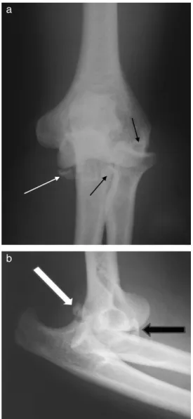
Fig. 1 – Anteroposterior (AP) radiograph of a dislocated left elbow (case 10). White arrow, fragment of the fracture of the coronoid process. Black arrows, fragments of the fracture of the radial head (a). Lateral radiograph of a dislocated left elbow (case 10). White arrow, fragment of the fracture of the radial head. Black arrow, fragment of the fracture of the coronoid process (b).
The patients’ mean age at the time of the treatment was 41 years and four months, with a range from 21 to 66. Nine (60%) were male and six (40%) were female. The dominant side was affected in eleven cases (73.3%) (Table 1).
The trauma mechanism in 10 patients (66.7%) was low-energy (falling to the ground). The others (33.3%) suffered high-energy trauma (falls from a height) (Table 1).
The classification used for the fractures of the coronoid pro-cess was the one proposed by Regan and Morrey.10Thirteen (86.7%) were classified as type I (fractures of the apex of the coronoid process alone) and two (13.3%), as type II (fracturing with fragments, of up to 50% of the height of the coronoid). None of the cases had a fracture classified as type III (frag-ments greater than 50% of the height of the coronoid) (Table 1).
To evaluate the severity of the fractures of the head of the radius, we used Mason’s original classification.11 Two cases (13.3%) were classified as type II (marginal fractures with dis-placement) and 13 (86.7%), as type III (comminuted fractures involving the entire head of the radius). None of the fractures were classified as type I (fissure or marginal fracture without displacement) (Table 1).
With the exception of case 3, which underwent an arthro-scopic procedure, all the cases underwent open operations, by means of the lateral access to the elbow described by Kaplan,12 followed by a medial access.
In 10 cases (66.7%), medial collateral ligament injuries were observed. In five cases (33.3%), this ligament was found to be undamaged. Injuries to the lateral ligament complex were seen in all the cases (Table 1).
The patients underwent closed reduction of the disloca-tion and immobilizadisloca-tion of the elbow with a plaster-cast splint extending from the axilla to the palm, until the surgery was performed. The patients who came to our service with the joint already reduced were immobilized in the same manner. The mean time interval between the trauma and the surgery was eight days, with a range from one to 24 (Table 1).
Regarding the surgical treatment, the fracture of the radial head underwent open reduction and internal fixation in 10 cases (66.7%). In four cases, osteosynthesis was performed only using screws, and in six cases, with a plate and screws. In case 3, the reduction was done by means of arthroscopic view-ing and the fixation was done usview-ing a Herbert screw. In three cases (20%), the radial head was completely resected (cases 13, 14 and 15). In case 12, only the lateral fragment of the radial head fracture was resected (Table 1).
Regarding the fractures of the coronoid process of the ulna, the fracture was reduced as an open procedure and was fixed in accordance with the technique described by Morrey,13in 10 cases (66.7%). In this technique, two sutures with non-absorbable No. 5 thread were performed by passing the thread around the bone fragment (including the anterior joint cap-sule) and then through two bone tunnels to the posterior face of the ulna, where they were tied off, like in the classical pull-out technique (Fig. 2A and B). In one case, the bone frag-ment was resected arthroscopically (case 3), and in four cases (26.7%), the fracture was not dealt with (Table 1).
All the collateral ligament injuries were treated by means of transosseous sutures, without the aid of anchors, with the exception of case 3, in which the injury to the lateral collateral ligament was not repaired.
In no case was residual intraoperative instability observed that would justify the use of transarticular external fixation of the elbow.
In case 8, because of instability of the distal radioulnar joint and injury to the interosseous membrane of the forearm (Essex-Lopresti injury),14 this joint underwent closed reduc-tion and fixareduc-tion with a Kirschner wire at 60◦of supination
of the forearm, which was then maintained for four weeks (Table 1).
To evaluate the range of motion (ROM), we took complete extension to be 0◦ and flexion to be the great degrees of
movement made from this parameter. Deficiency of extension was noted as a negative number (for example, a deficiency of extension of 10◦was noted as
rev
bras
ortop.
2014
;
4
9(3)
:271–278
Table 1 – Clinical data on the patients. Age Sex Dominant
side
Trauma mechanism
Morrey Mason LCL injury
MCL injury
Time interval from trauma to
surgery (days)
Radial head Coronoid Postoperative follow-up (months)
Results
Flexion Extension Pronation Supination Quantitative Bruce
Qualitative Bruce
1 66 F FS 1 3 + + 6 Plate Not fixed 120 130 −35 70 90 86.125 Fair
2 55 F + FS 1 3 + + 2 4 screw Not fixed 12 140 −30 20 80 79.375 Poor
3 28 M + Fall from height
1 2 + 17 Arthroscopic
fixation
Arthroscopic resection
22 130 0 90 90 96.125 Excellent
4 49 F + FS 1 3 + + 9 2 screws Not fixed 94 140 0 90 90 100 Excellent
5 31 M + Fall from height
1 3 + 13 Plate Pull-out 63 140 −15 50 55 81.125 Fair
6 21 M + Fall from height
1 3 + 14 Plate Pull-out 67 140 −10 20 0 61.125 Poor
7 26 M + FS 1 3 + 5 Plate Pull-out 62 140 −10 90 70 94.375 Good
8 42 M Fall from height
1 3 + + 1 4 screws Pull-out 109 130 −30 35 40 72.81 Poor
9 26 M FS 1 3 + + 24 Plate Pull-out 85 130 −5 90 45 83.75 Fair
10 44 M Fall from height
2 2 + + 7 3 screws Pull-out 32 130 −50 70 60 77.375 Poor
11 28 F + FS 2 3 + + 6 Plate Pull-out 24 120 −10 60 55 80 Poor
12 37 M + FS 1 3 + + 10 Resection of
fragment
Pull-out 119 120 −20 90 45 82.0625 Fair
13 64 F + FS 1 3 + 7 Resection of
head
Not fixed 37 130 0 90 80 96.25 Excellent
14 44 M + FS 1 3 + + 4 Resection of
head
Pull-out 66 130 −10 65 75 88.75 Fair
15 59 F + FS 1 3 + + 6 Resection of
head
Pull-out 31 115 −25 90 90 88.625 Fair
Source: SAME – DOT ISCMSP.
r e v b r a s o r t o p .2 0 1 4;4 9(3):271–278
275
Fig. 2 – Intraoperative photograph (left elbow; medial access). White arrow, sutures (No. 5 non-absorbable thread) passing around the fragment of the coronoid process and the anterior joint capsule. Black arrow, percutaneous exit of the threads through the posterior face of the ulna (a). Lateral radiograph of the left elbow (case 10) in the immediate postoperative period. White arrow, bone tunnel for fixation of the fragment of the coronoid process by means of the pull-out technique. Osteosynthesis of the fracture of the radial head using traction screws (b).
were measured from the neutral rotation position of the fore-arm.
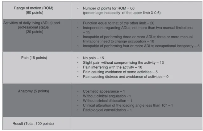
The analysis on the results was based on the score devel-oped by Bruce et al.15(Fig. 3). All the variables were analyzed
statistically by means of Student’s t test, with a significance level of 5%.
Results
With a mean follow-up of 62 months and 24 days (range: 12–120 months), three patients achieved results that were con-sidered to be excellent (20%), one good (7%), six fair (40%) and five poor (33%) (Table 1).
The mean amplitude of elbow flexion was 131◦, with a
range from 115◦ to 140◦; for extension,−16◦, ranging from
−35◦to 0◦; for pronation, 68◦, ranging from 20◦to 90◦; and for supination, 64◦, ranging from 0◦to 90◦. Twelve patients (80%)
attained a minimum flexion-extension ROM of 100◦; 13 (86.7%)
attained a minimum pronation-supination ROM of 100◦
(func-tional arcs of Morrey et al.9) (Table 1). Cases 2, 6 and 8 presented significant deficits of pronation-supination.
In relation to activities of daily living, 13 patients (86.7%) reported that they had recovered the function of the affected limb, in comparison with the contralateral limb. Two patients presented partial limitation of function (Table 1).
Only one patient complained of pain (case 3), but this pain was mild and did not compromise the patient’s activities (Table 1).
All the fractures that were fixed became consolidated, although in case 2, consolidation of the fracture of the radial head was delayed. None of the cases presented joint instabil-ity. Clinical examinations on four patients (26.7%) showed that they presented loading angles greater than 10◦, and in seven
patients (46.7%) there was some angular displacement of the elbow. Nonetheless, all the patients were satisfied regarding the final cosmetic appearance (Table 1).
Range of motion (ROM) (60 points)
• Number of points for ROM = 60
(percentage incapacity of the upper limb X 0.6)
Activities of daily living (ADLs) and professional status
(20 points)
• Function equal to that of the other limb – 20
• Independent regarding ADLs; not more than two manual limitations – 15
• Incapable of performing three or more ADLs; three or more manual limitations; need to change occupation – 10
• Incapable of performing four or more ADLs; occupational incapacity – 5
Pain (15 points) • No pain – 15
• Slight pain without compromising the activity – 13 • Pain interfering with the activity – 10
• Pain causing avoidance of some activities – 5 • Pain causing distress and avoidance of activities – 0
Anatomy (5 points) • Cosmetic appearance – 1 • Without clinical angulation - 1 • Without clinical dislocation – 1
• Clinical alteration of the loading angle less than 10° – 1 • Radiological consolidation – 1
Result (Total: 100 points)
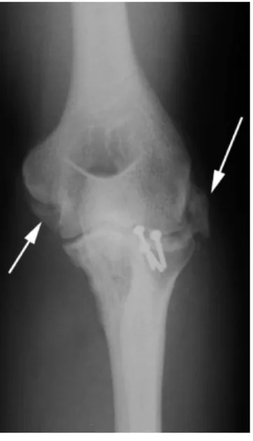
Fig. 4 – Anteroposterior (AP) radiograph of the left elbow (case 10), seven months after the operation. White arrows, heterotopic ossification.
In our series, the mean quantitative Bruce score for the patients affected on the dominant side was 86 points, while for the other group, the value was 80. There was no statistically significant difference between these two groups (p= 0.201).
Regarding the trauma mechanism, the patients who had suffered low-energy trauma had a mean quantitative Bruce score of 88 points. The patients with a high-energy trauma mechanism had a mean of 77.7, without any statistically sig-nificant difference (p= 0.152).
The mean quantitative Bruce score for the patients with fracture of the coronoid process that were classified as Mor-rey type I was 85 points, while for the patients with fractures classified as type II, the score was 78.7 (p= 0.059), also without any statistical difference.
In our sample, 10 cases of fractures of the coronoid pro-cess (66.7%) were fixed and five (33.7%) were not. Among those that were not fixed, the mean quantitative Bruce score was 91.57 points; while for those that were fixed, the mean was 80.9 points. This difference was not shown to be statistically significant (p= 0.056).
Two patients (13.3%) evolved with neuropraxia of the ulnar (cases 1 and 5) and one (6.7%) evolved with heterotopic ossifi-cation (case 10) (Fig. 4). This patient underwent reoperation 32 months after the first surgery in order to gain extension, with went from−50◦to 0◦after anterior and posterior open release. Case 6 had an indication for removal of the synthesis material and anterior release in order to gain supination ROM, but the procedure was not performed, at the patient’s own request. It is important to emphasize that our study evaluated the results before the possible treatment for these complications.
Discussion
Dislocated fractures of the elbow in young patients are often associated with high-energy trauma. These are therefore severe injuries with a high complication rate.2
In our sample, the patients’ mean age was 41 years and four months, and this was seen to be similar to findings from other studies.2–5,16,17Among our patients, 60% were male and 40% were female; these proportions were also found in the literature.2–5,16,17
In relation to the trauma mechanism, there was a discrep-ancy between the findings from our cases (66.7% with low energy) and those of other series, in which high-energy mech-anisms predominated.3–5
The dominant side was affected in 73.3% of our patients, which was greater than what was found in another two studies,4,16 which both found this to be 58%. Like Gomide et al.,4we did not find any statistical correlation between the proportion with the dominant side affected and the result obtained.
The fractures found in TTE cases (coronoid and radial head) have been found to vary in severity. In this regard, certain points relating to their respective classifications need to be borne in mind.
Fractures of the coronoid process classified as Morrey type I occurred in 86.7% of our patients, while type II fractures occurred in 13.3%. A similar relationship was found in a series examined in 2010.5In other series,3,4,16Morrey type I fractures also predominated, but not as clearly. In the series reported by Ring et al.,2in 2002, all the 11 cases were classified as type II fractures. In our study, we did not observe any statistical association between the type of fracture of the coronoid pro-cess and the clinical result, just like Gomide et al.4The latter was the only study in the literature that made a statistical assessment for this comparison.
There is some controversy regarding the need for fixation for Morrey type I fractures. According to some authors, any fracture of the coronoid process associated with dislocation of the elbow is a major marker of instability, regardless of its size.2,3However, these fractures can also be treated conserva-tively, according to other authors.4,5
In our sample, most of the fractures of the coronoid pro-cess were fixed. Among the five patients who did not undergo fixation of the coronoid process, two obtained unsatisfactory results and three, excellent results, according to the qual-itative Bruce score. In our evaluation, we did not find any statistically significant difference between the cases that did and did not undergo fixation of the coronoid process. This result is different from that of other series.2,5In the study by Chemama et al.,5in 2010, the Mayo scores were better among the patients who underwent fixation than among those who did not undergo coronoid fixation, although these authors did not perform any statistical analysis on their results.
Surgical treatment is recommended for type II and III frac-tures of the coronoid process.2–4Among our patients, we did not find any case classified as Morrey type III; in the two cases classified as type II, the fractures were fixed.
Out of all the patients in this study, 13 (86.7%) suffered frac-tures of the radial head that were classified as Mason type III and two as type II (13.3%). In the literature, we found a slight predominance of Mason type III fractures in TTE cases.2–5,16
r e v b r a s o r t o p .2 0 1 4;4 9(3):271–278
277
10 patients who were victims of low-energy accidents (66.7%) were classified as Mason type III. On the other hand, among the five patients with high-energy trauma, two were Mason type II and three were type III. There was no statistical dif-ference in the final results obtained for each group. Gomide et al.,4in 2012, did not find any statistically significant corre-lation between the fracture pattern of the radial head and the clinical result.
The radial head is an important secondary stabilizer against valgus stress and posterior translation of the elbow. In unstable elbows associated with fractures of the coronoid, the stabilizing function of the radial head should be pre-served whenever possible, either by means of reconstruction or through replacement by a prosthesis. Resection arthro-plasty is not recommended in TTE cases, because of the risk of instability and arthrosis.2–4,8,18–26Nonetheless, this was done in three (20%) of our 15 patients, after failure in the attempts to perform osteosynthesis, because of the high degree of com-minution. A prosthesis was not used, because there was none available at our service at that time. However, in analyzing these three cases separately, we observed that they did not evolve with severe complications: two were classified qualita-tively as “fair” and one as “excellent”. Nonetheless, it should be emphasized that if a prosthesis had been available, it would have been used in these cases.
Similar studies have found injuries of the lateral ligament complex in all patients, which were always repaired.3–5In our series, these injuries were also observed in all the cases and were surgically repaired, except in case 3. In this case, after reduction and arthroscopic fixation of the fracture of the radial head, there was no significant residual instability of the elbow and therefore it was decided not to perform ligament repair. It is worth emphasizing that this repair would have been done as an open procedure if it had been necessary.
The protocol most used for managing TTE includes repair of injuries to the coronoid process, fractures of the radial head and injuries to the lateral ligament complex. Explo-ration and repair of the medial collateral ligament are done if there is any residual instability of the elbow.3–5,16However, medial surgical exploration was done in all of our patients and ligament injuries were found in ten cases (66.7%). All of these were repaired. In our opinion, and in agreement with Jeong et al.,27 integrity of the medial ligament of the elbow is important in recovering function after this severe injury (TTE). In this manner, we routinely explored this lig-ament in 100% of our cases and found injuries in 66.7%. Clearly, it can be argued to the contrary that in 33.3% of the cases, the medial route was used unnecessarily. Never-theless, it should be emphasized that several injuries were found without there being any residual instability after con-ventional treatment of the primary injuries. In our opinion, a simple investigative method for preoperative evaluation should for used in order to ascertain in advance whether the medial region of the elbow should be explored surgi-cally in TTE cases. This is a current study objective at our service.
The finding that only 26.7% of the results were satisfactory (good or excellent, according to the Bruce scores) is something that can be debated. Although the results were categorized in this manner, it should be emphasized that 12 patients (80%)
attained a minimum flexion-extension ROM of 100◦ and 13
(86.7%) attained a minimum pronation-supination ROM of 100◦ (arcs of movement of Morrey et al.9). These are consid-ered to be the minimum functional ROMs for the joints that make up the elbow and forearm. These results were similar to those found in other series.2–5,16 However, most authors have used different assessment scores3–5that do not take the pronation-supination ROM into consideration. Thus, there is the possibility of obtaining a good or excellent score even if there are limitations on pronation and supination. The Bruce score takes into account both arcs of movement, i.e. flexion-extension and pronation-supination and may thus expand the spectrum of analyses on the results. Among the five cases that were considered to be qualitatively poor, three had sig-nificant losses of pronation-supination, even though they had a functional arc for flexion-extension (cases 2, 6 and 8).
Other criteria evaluated in the Bruce score are activities of daily living, residual pain and cosmetic appearance. In relation to activities of daily living, 86.7% of the patients reported that they had recovered function in the affected limb, in compari-son with the contralateral limb. Only one patient complained of pain (of mild intensity and without compromising his activ-ities) and all the patients were satisfied with the final cosmetic appearance.
Conclusion
We found that, according to the Bruce score, only 26.7% of the results obtained were good and excellent. Thus, 73.3% of the results were unsatisfactory, despite recovery of Morrey’s functional movement arc in more than 80% of the patients.
Conflicts of interest
The authors declare that there were no conflicts of interest.
r e f e r e n c e s
1. Hotchkiss RN. Fractures and dislocations of the elbow. In: Rockwood CA, Green DP, Bucholz RW, et al., editors. Rockwood and Green’s fractures in adults. 4th ed. Philadelphia:
Lippincott-Raven; 1996. p. 929–1024.
2. Ring D, Jupiter JB, Zilberfarb J. Posterior dislocation of the elbow with fractures of the radial head and coronoid. J Bone Joint Surg Am. 2002;84(4):547–51.
3. Pugh DMW, Wild LM, Schemitsch EH, King GJW, McKee MD. Standard surgical protocol to treat elbow dislocations with radial head and coronoid fractures. J Bone Joint Surg Am. 2004;86(6):1122–30.
4. Gomide LC, Campos DO, de Sá JM, de Sousa MRP, do Carmo TC, Andrada FB. Tríade terrível do cotovelo: avaliac¸ão do tratamento cirúrgico. Rev Bras Ortop. 2011;46(4):374–9. 5. Chemama B, Bonnevialle N, Peter O, Mansati P, Bonnevialle P.
Terrible triad injury of the elbow: how to improve outcome? Orthop Traumatol Surg Res. 2010;96(2):147–54.
6. Armstrong AD. The terrible triad injury of the elbow. Curr Opin Orthoped. 2005;16(4):267–70.
8. Josefsson PO, Gentz CF, Johnell O, Wendeberg B. Dislocations of the elbow and intra-articular fractures. Clin Orthop Relat Res. 1989;(246):126–30.
9. Morrey BF, Askew LJ, Chao EY. A biomechanical study of normal functional elbow motion. J Bone Joint Surg Am. 1981;63(6):872–7.
10. Regan W, Morrey B. Fractures of the coronoid process of the ulna. J Bone Joint Surg Am. 1989;71(9):1348–54.
11. Mason ML. Some observations on fractures of the head of the radius with a review of one hundred cases. Br J Surg. 1954;42(172):123–32.
12. Kaplan EB. Surgical approach to the proximal end of the radius and its use in fractures of the head and neck of the radius. J Bone Joint Surg Am. 1941;23(1):86–92.
13. Morrey BF. Current concepts in the treatment of fractures of the radial head, the olecranon, and the coronoid. Intr Course Lect. 1995;44:175–85.
14. Essex-Lopresti P. Fractures of the radial head with distal radio-ulnar dislocation; report of two cases. J Bone Joint Surg Br. 1951;33(2):244–7.
15. Bruce HE, Harvey JP, Wilson Jr JC. Monteggia fractures. J Bone Joint Surg Am. 1974;56(8):1563–76.
16. Leigh WB, Ball CM. Radial head reconstruction versus replacement in the treatment of terrible triad injuries of the elbow. J Shoulder Elbow Surg. 2012;21(10):1336–41.
17. Forthman C, Henket M, Ring DC. Elbow dislocation with intra-articular fracture: the results of operative treatment without repair of the medial collateral ligament. J Hand Surg Am. 2007;32(8):1200–9.
18. Schneeberger AG, Sadowski MM, Jacob HA. Coronoid process and radial head as posterolateral rotatory
stabilizers of the elbow. J Bone Joint Surg Am. 2004;86(5): 975– 82.
19. Mathew PK, Athwal GS, King GJ. Terrible triad injury of the elbow: current concepts. J Am Acad Orthop Surg.
2009;17(3):137–51.
20. Morrey BF, Chao EY, Hui FC. Biomechanical study of the elbow following excision of the radial head. J Bone Joint Surg Am. 1979;61(1):63–8.
21. Broberg MA, Morrey BF. Results of treatment of
fracture-dislocations of the elbow. Clin Orthop Relat Res. 1987;216:109–19.
22. Moro JK, Werier J, MacDermid JC, Patterson SD, King GJ. Arthroplasty with a metal radial head for unreconstructible fractures of the radial head. J Bone Joint Surg Am.
2001;83(8):1201–11.
23. Fitzpatrick MJ, Diltz M, McGarry MH, Lee TQ. A new fracture model for “terrible triad” injuries of the elbow: influence of forearm rotation on injury patterns. J Orthop Trauma. 2012;26(10):591–6.
24. Morrey BF, Tanaka S, An KN. Valgus stability of the elbow. A definition of primary and secondary constraints. Clin Orthop Relat Res. 1991;(265):187–95.
25. Paccola CA, Defino HL, Barbieri CH. Fraturas da cabec¸a do rádio: resultados tardios após ressecc¸ão ou osteossíntese. Rev Bras Ortop. 1986;21(3):80–6.
26. Motta Filho GR, Motta Filho LAJ, Costa RPA, Mendes HM. Osteossíntese da cabec¸a radial na fratura-luxac¸ão do cotovelo. Rev Bras Ortop. 1998;33(9):709–12.