(Annals of the Brazilian Academy of Sciences)
Printed version ISSN 0001-3765 / Online version ISSN 1678-2690 www.scielo.br/aabc
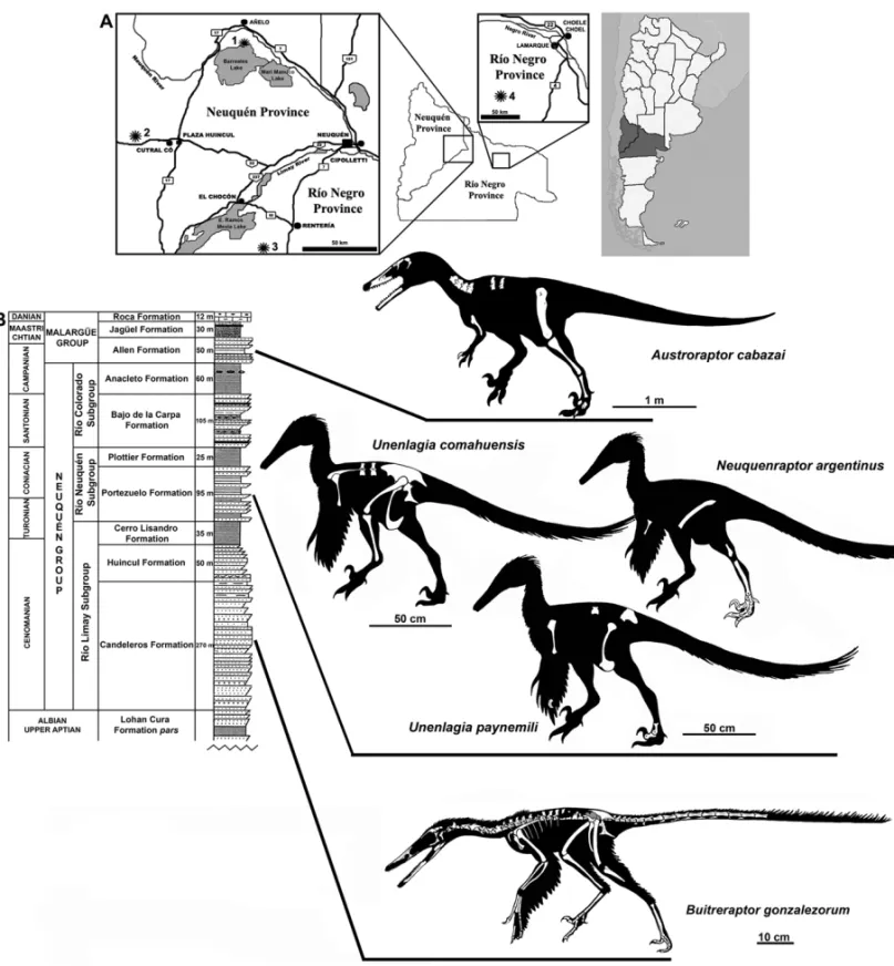
Unenlagiinae revisited: dromaeosaurid theropods from South America
FEDERICO A. GIANECHINI and SEBASTIÁN APESTEGUÍA
CONICET – Área de Paleontología, Fundación de Historia Natural ‘Félix de Azara’ Departamento de Ciencias Naturales y Antropología
CEBBAD, Universidad Maimónides, Hidalgo 775 (1405BDB), Ciudad Autónoma de Buenos Aires, Argentina
Manuscript received on October 30, 2009; accepted for publication on June 21, 2010
ABSTRACT
Over the past two decades, the record of South American unenlagiine dromaeosaurids was substantially increased both in quantity as well as in quality of specimens. Here is presented a summary review of the South American record for these theropods. Unenlagia comahuensis, Unenlagia paynemili, andNeuquenraptor argentinuscome from the
Portezuelo Formation, the former genus being the most complete and with putative avian features. Neuquenraptoris
more incomplete and exhibits pedal features resembling those ofUnenlagia. The earliest and most complete South American dromaeosaurid isBuitreraptor gonzalezorum, whose preserved cranial remains, provides important data in
the characterization of unenlagiines. The most recently described,Austroraptor cabazai, also with cranial remains,
allows further comparisons with Laurasian lineages and a better characterization of unenlagiines. The possible syn-onymy betweenUnenlagiaandNeuquenraptoris discussed. Additional evidences from Brazil and Colombia show that dinosaurs with similar dentition to that of unenlagiines were present in the whole South America. However, it is not possible to discart that these remains may belong to other unknown maniraptoran lineages, considering the increasing number of taxa of this group found in South America.
Key words:Deinonychosauria, Dromaeosauridae, South America, Unenlagiinae.
INTRODUCTION
The Dromaeosauridae is a family of highly derived small to mid-sized theropod dinosaurs characterized by the presence of a raptorial second pedal digit and a tail stiff-ened by the elongated prezygapophyses and chevrons of the medial to distal posterior vertebrae. The manus and pes of these theropods bear sharp trenchant claws and the pubis is generally posteriorly oriented. The majority of phylogenetic analyses found this group of theropods as the closest relatives to Avialae.
After the discovery ofDromaeosaurus albertensis
in Canada (Matthew and Brown 1922) and Velocirap-tor mongoliensisin Mongolia (Osborn 1924) during the
first decades of the twentieth century, and the
descrip-Proceedings of the Third Gondwanan Dinosaur Symposium Correspondence to: Federico A. Gianechini
E-mail: smilodon.80@gmail.com
tion ofDeinonychus antirrhopusfrom the USA (Ostrom
1969), many other new dromaeosaurids have been found in the northern continents, mainly in North America and Asia. The finding of many quite complete specimens with an outstanding preservation in the USA, Canada, Mongolia, and in the last decades in China, prompted a great advance in the knowledge of these theropods. This record suggested that the greatest diversity and dis-tribution of dromaeosaurids was circumscribed to the Laurasian continents.
No-vas and Puerta 1997, which shares some features with avians mainly in the scapula and pelvis (Novas and Puer-ta 1997, Novas 2004). Later, more remains were found in other Argentine localities. New material very simi-lar to that ofUnenlagia comahuensis was attributed to a new species of the same genus, Unenlagia paynemili
Calvo et al. 2004. In 2005, a new and more fragmentary specimen was described, mainly preserving few remains of the hind limbs. It was assigned to a new genus and species,Neuquenraptor argentinusNovas and Pol 2005.
In the same year, the earliest and most complete South American dromaeosaurid to date,Buitreraptor gonzale-zorum (Makovicky et al. 2005), was described. The
available skeletons of this species provide excellent in-formation for a better comprehension of the southern dromaeosaurid anatomy. The most recently described South American dromaeosaurid wasAustroraptor caba-zaiNovas et al. 2009, which bears some unusual
char-acters not observed in Laurasian dromaeosaurids and is therefore important in the characterization of this group of southern theropods. This same group also has a rep-resentative in Madagascar, Rahonavis ostromi (Forster et al. 1998), previously considered an avialan but lately linked withUnenlagiamainly through pelvic characters
(Novas 2004).
Many phylogenetic analyses found these South American dromaeosaurids in a monophyletic clade named Unenlagiinae (Bonaparte 1999, Makovicky et al. 2005). The anatomical differentiation of this group of South American theropods with respect to Laurasian dromaeosaurids could have been due to a vicariant evolution produced after the separation of Pangea into Laurasia and Gondwana during the Late Jurassic (Mako-vicky et al. 2005, Novas and Pol 2005). This separation would have resulted in the isolation of the South Amer-ican dromaeosaurids, allowing a parallel evolution with respect to those of Laurasia and, thus, creating a new South American lineage itself.
A brief overview of the South American dromaeo-saurids and other possible deinonychosaurian taxa is presented here. Since Cretaceous Argentinean fossil re-cord of unenlagiines is the most complete and diverse of South America, the most important features of individ-ual taxa are here characterized according to stratigraphic order, providing some comments and reinterpretations
of certain materials and discussions on the ideas previ-ously exposed by other autors.
INSTITUTIONALABBREVIATIONS
AMNH– American Museum of Natural History, New York, USA.
IGM – Mongolian Institute of Geology, Ulan Baatar, Mongolia.
IVPP– Institute of Vertebrate Paleontology and Paleoan-thropology, Beijing, China.
MCF PVPH– Museo Municipal Carmen Funes, Plaza Huincul, Neuquén, Argentina.
MCZ– Museum of Comparative Zoology, Cambridge, Massachussets, USA.
MML– Museo Municipal de Lamarque, Río Negro, Ar-gentina.
MPCA– Museo Carlos Ameghino, Cipolletti, Río Ne-gro, Argentina.
MUCPv– Museo de Geología y Paleontología de la Uni-versidad Nacional del Comahue, Neuquén, Argentina. UCMP– University of California Museum of Paleontol-ogy, Berkeley, CA, USA.
DROMAEOSAURID THEROPODS FROM ARGENTINA
THEROPODAMarsh, 1881
MANIRAPTORAGauthier, 1986
DEINONYCHOSAURIAColbert and Russel, 1969
DROMAEOSAURIDAEMatthew and Brown, 1922
UNENLAGIINAEBonaparte, 1999
Buitreraptor gonzalezorum
Makovicky, Apesteguía and Agnolín, 2005
Materials: The holotype (MPCA 245) (Figs. 1B, 2B) consists of an almost complete adult skeleton, including a partial articulated skull with both incomplete maxil-lae with teethin situ, left jugal, both postorbitals, both quadrates, right squamosal, both incomplete nasals, both frontals, both parietals, the occipital condyle, and mand-ibular bones, including both dentaries within situteeth,
complete tibia and fibula, metatarsals, several pedal pha-langes, and indetermined fragments of bone. The holo-type ofBuitreraptorwas found in complete articulation
in the field, indicating null or little transporting from the site of death.
A referred specimen (MPCA 238) consists of an almost complete right ilium and pubis, right hindlimb (femur, tibia, astragalus, metatarsals, and phalanges), and sacrum, all preserved in articulation.
Locality and horizon: Both the holotype and referred specimen were found at the fossiliferous locality of ‘La Buitrera’, located in the northwestern Río Negro Province, Patagonia, Argentina, close to the southern shore of the Ezequiel Ramos-Mexía Lake (Fig. 1). Its sedimentary beds correspond to the Candeleros Forma-tion (Cenomanian) and show a high diversity of taxa with a superb preservation of the fossils, which make this fossiliferous locality one of the most outstanding and important of Gondwana. The ‘La Buitrera’ locality presents a peculiar bias towards micro and mesoverte-brates, including remains of sphenodontids, crocodyli-forms, basal limbed snakes, and mammals (including dryolestoids) (Carignano et al. 2002, Apesteguía and Novas 2003, Pol and Apesteguía 2005, Apesteguía and Zaher 2006). Nevertheless, dinosaurs are also present, including carcharodontosaurid and noasaurid theropods, and rebbachisaurid and basal titanosaur sauropods (Cal-vo and Salgado 1995, Coria and Salgado 1995, Gal-lina and Apesteguía 2005, Brissón Egli and Apesteguía 2008).
Main anatomical features and comments: Buitrerap-tor is the earliest dromaeosaurid discovered in South
America to date and the most complete one (Makovicky et al. 2005). It bears some anatomical features, mainly cranial and dental ones, which distinguish it from Lau-rasian dromaeosaurid lineages. The skull is elongated and low (Fig. 2B, a, b), exceeding the femoral length by 25% (Makovicky et al. 2005; synapomorphy of Unen-lagiinae sensu Novas et al. 2009). All the preorbital
bones are very long. Both maxillae are preserved only at mid-length and bear a large maxillary fenestra. This fen-estra is oval in shape, with its major diameter in antero-posterior direction, instead of being small, oval, teardrop-shaped, and dorsally displaced (Fig. 2B, a) as in most
dromaeosaurids (e.g. Colbert and Russel 1969, Ostrom 1969, Sues 1977, Currie 1995, Barsbold and Osmólska 1999, Burnham et al. 2000, Xu and Wu 2001, Burn-ham 2004, Norell et al. 2006, Turner et al. 2007, X. Xu, unpublished data). A maxillary fenestra enlarged and not dorsally displaced is proposed as a synapomorphy of Unenlagiinae by Novas et al. (2009). Posterior to the maxillary fenestra is the antorbital fenestra (inner antor-bital fenestrasensuWitmer 1997), which remains
sepa-rated by a narrow interfenestral bar (Fig. 2B, a) (Mako-vicky et al. 2005). The nasals are very long, flat, and narrow (Fig. 2B, b), suggesting an elongated and nar-row snout, as inVelociraptorandTsaagan(Makovicky
et al. 2005, Norell et al. 2006), as well as in some troodontids likeByronosaurus jaffei(Makovicky et al.
2003). The frontals are also elongated, with laterally projected postorbital rami, resembling other dromaeo-saurids. The jugal is low and the postorbital has the triangular trirradiate form that is common among dro-maeosaurids. The quadrate has an enlarged quadrate foramen (Fig. 2B, c), thus differing from the general condition observed in dromaeosaurids. However, the condition ofBuitreraptorresembles that of troodontids,
in which this character is better represented, as seen in
Troodon formosusandSinovenator changii, (Varrichio
1997, X. Xu, unpublished data).
The dentary is very long and low, with dorsal and ventral parallel rims that run horizontally along the en-tire length. On the lateral side, close to the alveolar margin, there is a deep subalveolar groove that lodges a row of nutrient foramina (Fig. 2B, a), as is frequent in troodontids (Makovicky et al. 2003, 2005). Among the postdentarial bones, only the splenials, left angular, and left surangular are present. The splenials are articu-lated with the medial side of the dentary, but they are not very visible because the jaws are in occlusion and still partially covered by matrix.
suite of dental characters is only observed in Buitre-raptorand can be considered as potentially
autapomor-phic of this species (Gianechini et al. 2009). In most Laurasian dromaeosaurids the teeth are larger than in
Buitreraptor (e.g. Colbert and Russel 1969, Ostrom 1969, Sues 1977, Currie 1995, Xu and Wu 2001, Cur-rie and Varricchio 2004), and they are fewer in number, since most dromaeosaurids bear 11 to 16 dentary teeth (Norell and Makovicky 2004), whereas Buitreraptor
bears up to 20 alveoli (this estimate take into account that many alveoli are not preserved and broken, but due to the length of the dentary, the extension of the den-tal row and the size of the alveoli, it is estimated that more than 20 alveoli were present in the jaw). Among deinonychosaurs, only troodontids have such large den-tal count (Makovicky et al. 2003, Makovicky and Norell 2004). The complete absence of denticles is not a com-mon character acom-mong dromaeosaurids. However, some taxa possess a wide variety of denticle development, ranging from total absence in one border (generally the mesial) to the total absence, like inMicroraptor, Bam-biraptor,Shanag, andSinornithosaurus(Burnham et al. 2000, Xu et al. 2000, Hwang et al. 2002, Burnham 2004, Hwang 2005, Turner et al. 2007, X. Xu, unpub-lished data). Nevertheless, the total absence of denti-cles in some of these taxa is not characteristic of all teeth, as is conversely observed in Buitreraptor
(Gia-nechini et al. 2009), Austroraptor(Novas et al. 2009)
and avialans, as will be discussed later. The presence of grooves on the sides of the crown is strange among dromaeosaurids. Moreover, the grooves observed in the teeth ofSinornithosaurus(Xu and Wu 2001, X. Xu,
un-published data) and some isolated teeth assigned with doubts toDromaeosaurus(Sankey et al. 2002) are very different from those ofBuitreraptor, both in morphol-ogy, density, and location. Accordingly, the eight-shaped basal section is not common among dromaeosaurid teeth, and only is recorded in some taxa such as Saurornit-holestes(Currie et al. 1990, Sankey et al. 2002), Tsaa-gan (Norell et al. 2006), and Pyroraptor (Allain and
Taquet 2000, S. Apesteguía, personal observation). Nev-ertheless, the teeth of these taxa are not as labiolingually compressed as those ofBuitreraptor.
The cervical vertebrae (Fig. 2B, d) have low neu-ral spines and small epipophyses, in contrast to the large
epipophyses observed on the cervical vertebrae of
DeinonychusandVelociraptor(Ostrom 1969, Norell et
al. 2006). Carotid processes are present in the poste-rior cervical centra as in some dromaeosaurids, such as Microraptor and Tsaagan, and also in troodontids and alvarezsaurids (Hwang et al. 2002, Makovicky and Norell 2004, Norell et al. 2006). A characteristic fea-ture ofBuitreraptoris the presence of low ridges on the ventrolateral corners of the last cervical centrum, which terminate posteriorly as small tubers (Makovicky et al. 2005). The dorsal vertebrae (Fig. 2B, e) have tall and rectangular neural spines, as is common among dro-maeosaurids, without a distal transverse expansion as a spine table. Hypapophyses are present on the ven-tral side of the anterior dorsal centrae, as in the dro-maeosauridsVelociraptor, Saurornitholestes, Deinony-chus,Luanchanraptor,Bambiraptor,Sinornithosaurus, Microraptor, and Rahonavis (Ostrom 1969, Forster et
al. 1998, Norell and Makovicky 1999, Burnham 2004, Lü et al. 2007, X. Xu, unpublished data), and also in troodontids, oviraptorosaurs, alvarezsaurids, ornithomi-mosaurs, Ornitholestes hermanni, and basal avialans such as hesperornithiforms andIchthyornis(Osmólska
et al. 1972, Kurzanov 1981, Perle et al. 1994, Norell and Makovicky 1999, Barsbold et al. 2000, Norell et al. 2000, Currie and Dong 2001, Makovicky et al. 2003, Clarke 2004). The dorsal vertebrae ofBuitreraptorbear
stalked parapophyses, considered as a common feature among Dromaeosauridae, despite also being observed in alvarezsaurids (Novas 1997, Norell and Makovicky 1999) and in the basal bird Confuciusornis sanctus
(Chiappe et al. 1999). The caudal vertebrae (Fig. 2B, f) are low and elongated as in other dromaeosaurids, and the distal ones are devoid of neural spines. Unlike Lau-rasian lineages, the prezygapophyses of these vertebrae are not anteriorly extended forming bony rods, and they only overlap up to half of the preceding vertebra (Mako-vicky et al. 2005). Some chevrons are present, which are dorsoventrally compressed and bifids at their ante-rior and posteante-rior ends, as in other paravians (Makovi-cky et al. 2005).
The pectoral girdle of Buitreraptor is
←−
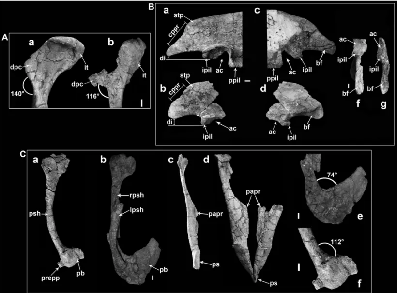
Fig. 2 –A: holotype ofAustroraptor cabazai(MML-195); a-g: skull bones; a: left maxilla; b and c: left lacrimal, in lateral and ventral views; d and e: right frontal, in dorsal and lateral views; f: right postorbital, in lateral view; g: left dentary, in lateral view; h: right humerus, in lateral view; i-l: presacral vertebrae; i and j: cervical 8th?, in cranial and left lateral views; k and l: dorsal 4th?, in cranial and left lateral views; m: left femur, in lateral view; n: right tibia, in cranial view. Scale bars: 5 cm. B:Buitreraptor gonzalezorum; a-k: holotype (MPCA 245); a and b: skull, in left lateral and dorsal views; c: close-up of the right quadrate; d: cervicodorsal vertebrae, in ventral view; e and f: mid-dorsal and distal posterior vertebrae, in lateral view; g: furcula, in posterior view; h: scapulocoracoid, in lateral view; i: right humerus, in lateral view; j: left femur, in anterior view; k: right ilium and ischium, in lateral view; l-m: referred material (MPCA 238); l and m: right metatarsus, in plantar and lateral view. Scale bars: 5 cm, except in c (1 cm).Abbreviations: ac, acetabulum; an, angular; aof, antorbital fenestra; bf, brevis fossa; cor, coracoid; cp, carotid process; ct, coracoid tubercle; czapa, contact zone of the ascendent process of the astragalus; dg, dentary groove; dn, dentary; dp, diapophysis; dpc, deltopectoral crest; ep, epicleidum; fh, femur head; fpl, fan-shaped process of the lacrimal; fr, frontal; gl, glenoid; ifb, interfenestral bar; ipp, pubic process of the ischium; it, internal tuberosity; jppo, jugal process of the postorbital; ju, jugal; lf, lacrimal foramina; mf, maxillar fenestra; mr, medial ridge; mt II, III, IV, metatarsals II, III and IV; mx, maxilla; na, nasal; ns, neural spine; oc, occipital condyle; op, obturator process; pa, parietal; pdp, proximodorsal process; pf, pneumatic foramen; plf, posterolateral flange of metatarsal IV; po, postorbital; pof, postorbital process of the frontal; pp, parapophysis; prez, prezygapophysis; pw: postantral wall; qu, quadrate; rpm, anterior process of the maxilla; rrsf, anterior rim of the supratemporal fenestra; sc, scapula; sppo, squamosal process of the postorbital; sq, squamosal; st, spinal table.
2B, g) is stout, curved, with a very rudimentary hypo-cleidum, and is nearly U-shaped in form. Moreover, the furcula ofBuitreraptoris pneumatic, being hollow and reinforced by internal trabeculae (Makovicky et al. 2005). The strap-like scapular blade (Fig. 2B, h) is curved close to the glenoid, with a triangular acromion that resembles that of other dromaeosaurids such as
Sinornithosaurus and Velociraptor, as well as that of Archaeopteryx(Ostrom 1976b, Norell and Makovicky
1999, Xu et al. 1999, X. Xu, unpublished data). The cora-coid (Fig. 2B, h) is bent in an approximately straight angle between the glenoid portion and the ventral re-gion, the latter being expanded as a blade-like structure. The anterior surface of the coracoid has a prominent tu-bercle (“biceps tutu-bercle” or “biceps tuber” for Ostrom 1974, Xu et al. 1999, Burnham 2004, Makovicky et al. 2005, and other authors) at the point of flexion of the coracoid. The glenoid appears to be laterally directed, as inArchaeopteryx(Ostrom 1976b, Wellnhofer 1992,
Paul 2002). In many dromaeosaurids the coracoid ac-quires a L-shape, reaching the dorsal part that articu-lates with the scapula at a 90◦ angle with respect to the ventral part, as inBuitreraptor. This flexion allows
the ventral portion of the coracoid to articulate with the anterolateral margin of the sternum, as occurs in Bam-biraptor (Burnham et al. 2000, Burnham 2004), and
brings the glenoid into a lateral position. In this way,
the general form of the scapulocoracoid is reminiscent of that of Asian dromaeosaurids, such asMicroraptorand
Sinornithosaurus, and also of that of basal birds includ-ing Archaeopteryx andConfuciusornis (Ostrom 1975, 1976a, b, 1986, Wellnhofer 1992, 1993, Chiappe et al. 1999, Norell and Makovicky 1999, Xu et al. 1999, 2000, Hwang et al. 2002, X. Xu, unpublished data).
The forelimb ofBuitreraptoris proportionally very
long, as inSinornithosaurusandMicroraptor(Xu et al.
1999, 2000, X. Xu, unpublished data). The humerus (Fig. 2B, i) has a well-developed deltopectoral crest, similar in form to that ofUnenlagia comahuensis
The pelvis shares several features with other dro-maeosaurids, such as Sinornithosaurus, Microraptor,
andRahonavis, but also with early birds as Archaeopte-ryx(Ostrom 1976b, Wellnhofer 1992, 1993, Forster et al.
1998, Xu et al. 1999, Xu et al. 2000, Hwang et al. 2002, X. Xu, unpublished data). The ilium (Fig. 2B, k) shows a slightly convex dorsal rim, with a posteriorly curved postacetabular blade bearing a posterior concave rim due to the posterior expansion of the brevis shelf (character 227, Turner et al. 2007), resembling other unenlagiines (an issue that will be discussed later). A peculiar fea-ture is a strong lateral torsion of the dorsal rim of the iliac blade at the level of the ischiadic peduncle, so that dorsal margins of both iliac blades diverge later from the sagittal axis at this zone. A similar eversion of the dorsal margin of the iliac blade is also observed in Sinovena-tor(Xu et al. 2002, X. Xu, unpublished data), whereas
inVelociraptorandArchaeopteryxthe dorsal margin of
the ilium, above the ischiac process, is thickened and slightly laterally everted, and the blade is laterally con-cave (Wellnhofer 1974, Norell and Makovicky 1997), thus resembling the ilium ofBuitreraptor.
The acetabulum is medially partially occluded, a derived avian feature also present inUnenlagia coma-huensis(Novas and Puerta 1997). A supracetabular crest
is present, similar to that of Rahonavis (Forster et al.
1998), and the brevis shelf is laterally projected and pos-teriorly extended beyond the posterior end of the ver-tical lamina (Makovicky et al. 2005). However, in the referred material, the brevis shelf is slightly more lat-erally projected and more ventrally curved at the pos-terior end, so some small differences can be observed between the two specimens, which seem to be a case of intraspecific variation. The pubis is preserved in the referred material, and is vertically oriented with a pos-teriorly curved distal half. Unfortunately the distalmost portion of the pubis is not preserved, and therefore it is not possible to observe the form and degree of fusion at the symphysis. The ischium (Fig. 2B, k) is platelike, as in other paravians, with an anteroposteriorly short iliac process and an anteriorly projected ischiadic pro-cess, which is dorsoventrally expanded. The ischium ta-pers distally to a posteroventrally oriented pointed end. The obturator process is long, pointed, and anteroven-trally projected, as in Microraptor, Sinornithosaurus,
andRahonavis(Forster et al. 1998, Hwang et al. 2002,
X. Xu, unpublished data). A posterodorsal process is present, similar to the proximodorsal process of Bambi-raptor,Sinornithosaurus, Microraptor, andRahonavis
among dromaeosaurids, the one of the troodontid Sinove-nator, and of early birds such asArchaeopteryx, Enan-tiornithes,Iberomesornis, andConfuciusornis (Ostrom 1976b, Forster et al. 1998, Chiappe et al. 1999, Xu et al. 1999, 2000, Burnham et al. 2000, Hwang et al. 2002, Burnham 2004, X. Xu, unpublished data). On the lateral side, a sharp ridge is extended in dorsoventral direction, approximately at the middle of the shaft, dividing the latter into anterior and posterior halves.
The femur (Fig. 2B, j) is a slender bone, 25% shorter than the skull, with a ventrolaterally oriented head, and without a distinct neck (Makovicky et al. 2005). The shaft is strongly anteriorly bowed, as in some non-avian theropods and avialans (Ostrom 1976b, Norell and Makovicky 1999, X. Xu, unpublished data). The fourth trochanter is poorly developed, as common among dromaeosaurids, and a conspicuous lateral ridge (linea intermuscularis lateralissensuHutchinson 2001) is present, which extends from the base of the posterior trochanter towards the cranial edge of the proximal tip of the lateral condyle, often fading away near midshaft. This is a character also observed, for example, in Veloci-raptor,Microraptor, andSinornithosaurus(Norell and
ornithomi-mids, and troodontids (Holtz Jr. 2004, Makovicky and Norell 2004, Makovicky et al. 2004). However, the proximal most portion of the metatarsus of Buitrerap-tor is not preserved, so it is not possible to discern if
metatarsals II and IV were proximally in contact to each other. Therefore, although metatarsal III is pinched proximally, it cannot be asserted if it was completely hidden beneath metatarsals II and IV in anterior view, as it was stated by Hu et al. (2009). The metatarsus (Fig. 2B, l, m) is long, ca. 70% of the femoral length and ca. 67% of the tibial length (see Table I). By contrast, in
Velociraptor, the metatarsus is only about 40% and
35% of the femoral and tibiotarsal length, respectively, while inDeinonychusthese ratios are ca. 49% and ca.
45%, respectively (Ostrom 1969, 1976c, Norell and Makovicky 1999). Accordingly, the metatarsal length of Buitreraptor, in comparison with the femoral and
tibial lengths, is similar to that observed in Sinornitho-saurus,Microraptor, andBambiraptor(Xu et al. 1999, 2000, Burnham et al. 2000, Hwang et al. 2002, Burn-ham 2004, X. Xu, unpublished data). There is also sim-ilarities with troodontids, such as Sinovenator, Sinor-nithoides, and Saurornithoides(Xu et al. 2002, X. Xu,
unpublished data), and with basal birds such as Archaeo-pteryx (Wellnhofer 1974, 1992, X. Xu, unpublished
data), in which the length of the metatarsus, in compar-ison with the femoral length, varies between 70% and 80% approximately. Metatarsal III is the longest ele-ment of the metatarsus, whereas metatarsal II is slightly shorter than the IV. Metatarsals II and IV have a diam-eter similar to each other, unlike the condition observed in derived troodontids, in which metatarsal IV is the most robust (Xu et al. 2002, Makovicky et al. 2003). Metatarsals II and III have a partially developed ging-lymoid distal articulation. The distal anterior surface of metatarsal III is slightly transversely expanded over the anterior surfaces of metatarsals II and IV. On the other hand, the distal posterior portion of metatarsals II and IV are expanded over the posterior surface of metatarsal III. The posterolateral surface of metatarsal IV is porteri-orly projected as a sharp ridge (Fig. 2B, l,m), resembling
Microraptor,Sinornithosaurus, andArchaeopteryx(Xu
et al. 1999, 2000, Hwang et al. 2002, Paul 2002, X. Xu, unpublished data). The pedal phalanges are simi-lar to those of other dromaeosaurids, with phalanx II-2
with a proximoventral flexor heel and a distal articula-tion dorsoventrally expanded. However, this phalanx is shorter and with a shaft not as constricted at mid-length. Additionally, the ventral heel is less developed, when it is compared with more derived dromaeosaurids such asDromaeosaurus,Velociraptor,Deinonychus, and
Saurornitholestes (Matthew and Brown 1922, Colbert and Russel 1969, Ostrom 1969, Currie 1995, Norell and Makovicky 1997, Longrich and Currie 2009). Thus, it is morphologically more similar to the phalanx II-2 of
Rahonavis,Microraptor,Sinornithosaurus, Gracilirap-tor, and some primitive troodontids (Forster et al. 1998,
Xu and Wang 2000, Xu et al. 2002, X. Xu, unpub-lished data).
Unenlagia comahuensisNovas and Puerta, 1997
Materials: The holotype of Unenlagia comahuensis
(MCF PVPH 78) (Figs. 1B and 3A) consists of an in-complete skeleton found in partial articulation, which includes three presacral vertebrae considered as the 8th, 10th, and 13th dorsals (the latter articulating to the sacrum), sacrum, dorsal ribs, two proximal haemal arches, left scapula and incomplete humerus, ilia, pubes, right ischium, right femur (found in direct association with the other elements), and left tibia (Novas and Puerta 1997). This theropod is medium-sized, approx-imately 2 meters in length.
Locality and horizon: Portezuelo Formation (late Tu-ronian-early Coniacian), Sierra del Portezuelo region, Neuquén Province, Argentina (Fig. 1B, A). This geo-logical sedimentary unit is composed of continental sed-iments, bearing frequent paleosol tops (Leanza et al. 2004). Apart from this species, this unit has provided other theropod species such as Patagonykus puertai
Novas 1996, Megaraptor namunhuaiquii Novas 1998, a big undescribed coelurosaur (Coria et al. 2001), frag-ments of an abelisauroid (Novas 1996), and fragfrag-ments of an undescribed Neornithes.
Main anatomical features and comments: Unenlagia comahuensis has been considered as one of the
mani-raptoran theropods most closely related to birds, sharing many anatomical features with basal birds, mainly with
Archaeopteryx(Novas and Puerta 1997, Novas 2004).
>
>
>
>
>
.
<
.
<
.
<
.
>
.
<
.
>
characterized by the presence of hyposphenes and pleu-rocoels on the dorsolateral surface of the centrum. The first of these vertebrae (considered as the 8th dorsal) has an anteroventral heel on the centrum, similar but less de-veloped than that present on the last cervicals and first dorsals of some dromaeosaurids such as Deinonychus
(Ostrom 1969). The other dorsals preserved (the 10th? and the 13th) (Fig. 3 A, e) ofU. comahuensishave tall neural spines, which are anteroposteriorly expanded at its distal ends, being that of the anterior sacral more vertically elongated. A peculiar feature of the anterior sacral is the presence of deep lateral pits at the base of the neural spines (Fig. 3A, e) (Novas and Puerta 1997). The sacrum consists of six fused vertebrae, although the ilia extend spanning the last two dorsal vertebrae an-teriorly and the first caudal vertebra posan-teriorly (Novas and Puerta 1997). Six sacral vertebrae are also observed in dromaeosaurids such as MahakalaandVelociraptor
(IGM 100/986) (Norell and Makovicky 1999, Turner et al. 2007), and troodontids such asSaurornithoidesand
Troodon(Forster et al. 1998, Rauhut 2003, Makovicky and Norell 2004, Norell and Makovicky 2004, Turner et al. 2007, Norell et al. 2009, P.J. Makovicky, unpub-lished data). On the other hand, the number of sacral vertebrae is five in the dromaeosaurids Saurornitho-lestes,Sinornithosaurus, and Microraptor (Norell and
Makovicky 1997, Hwang et al. 2002, Rauhut 2003, X. Xu, unpublished data), in the basal troodontids Mei long,SinovenatorandSinusonasus magnodens(Xu and
Norell 2004, Xu and Wang 2004, X. Xu, unpublished data), and inArchaeopteryx(Ostrom 1975, 1976b,
Well-nhofer 1974, 1992).
In the original description ofU. comahuensis, No-vas and Puerta (1997) observed several avian traits in this dromaeosaurid, especially in the pectoral girdle, fore-limb and pelvic bones. The general shape of the scapula (Fig. 3 A, a,b) is certainly quite similar to that of Ar-chaeopteryxandBuitreraptor, with a twisted shaft,
be-ing strap-like in dorsal aspect and curved in lateral view (Novas and Puerta 1997, Novas 2009). Furthermore, the glenoid ofUnenlagiawas interpreted as laterally
ori-ented, resembling the condition observed in birds (Novas and Puerta 1997, Norell and Makovicky 1999). This is in contrast to the more posteroventrally faced glenoid present in some dromaeosaurids, such asDeinonychus
antirrhopus(Ostrom 1969, 1974). Nevertheless, a
later-ally oriented glenoid has also been observed in other dro-maeosaurids, including, for example,Velociraptor mon-goliensis(Norell and Makovicky 1999), Sinornithosau-rus millenii(Xu et al. 1999, X. Xu, unpublished data),
Bambiraptor feinbergi(Burnham et al. 2000, Burnham 2004),Tsaagan mangas(Norell et al. 2006),Rahonavis
(Forster et al. 1998), and Buitreraptor(Makovicky et al. 2005), among others. The presence of a laterally oriented glenoid would have permitted a forelimb fold-ing involvfold-ing rotation of the humerus and also a more marked humeral abduction. Additionally, it would allow an almost vertical position of the forelimb during max-imum upstroke, resembling the avian movements of the forelimb (Novas and Puerta 1997). However, Carpenter (2002) considered that the anatomical orientation of the scapula proposed by Novas and Puerta (1997) is wrong, i.e. the scapula ofUnenlagiahas been interpreted with
its widest surface horizontally oriented, so the scapular blade results in a dorsoventrally compressed bone, in-stead of laterally compressed as predominantly observed in theropods (Carpenter 2002). If the scapula is oriented like a laterally compressed bone, with the costal surface medially oriented as in some articulated specimens of dromaeosaurids and other non-avian theropods, then the glenoid is oriented posteroventrally, not facing laterally (Carpenter 2002, Paul 2002, Senter 2006). On the other hand, inRahonavis (Forster et al. 1998), a non-avian
theropod with close anatomical affinities with Unenla-gia(Novas 2004), the scapula might have been dorsally
positioned on the ribcage, with the costal surface ven-trally faced and lateral to the vertebral column, as occurs in avialans. This scapular position results in a more lat-erally facing glenoid. However, a lateralized position of the scapula and less dorsalized must be taken into ac-count because the curvature of the scapula would match the curvature of the ribcage, and so the glenoid takes a more posteroventrally facing position. Additionally, the presence of a dorsally positioned scapula leads us to assume a large and flexed coracoid, like that observed inBuitreraptor, and the necessary presence of a
ster-num inUnenlagia, though one was not found. Unen-lagiaprobably possessed a sternum, as many other
data), just based on its close phylogenetic position to Avialae. Nevertheless, placing the scapula in Buitrerap-tor as Carpenter (2002) explained, the coracoid takes
an anatomically incorrect position, as its posteroventral portion pierces the thoracic cavity (Novas 2009). De-spite the described situation, Unenlagia does not pre-serve coracoids. Therefore, the position of scapula and shape, ubication, and mode of articulation of the cora-coid with the sternum (if the latter exists) are hypothet-ical. Nevertheless, the similarity among the scapulae ofArcheopteryx,BuitreraptorandUnenlagiashould be
considered when analyzing the position of the scapula and the subsequent location of the glenoid. A more de-tailed analysis about the scapulocoracoid position and the subsequent orientation of the glenoid is beyond the objetive of this paper.
The humerus (Fig. 3A, c, d) has a well-developed and laterally projected deltopectoral crest, very similar to that ofBuitreraptor, and also shows a large internal tuberosity that is proximodistally extended.
Recently, Novas (2004) considered further ana-tomical features with avian trends inUnenlagia, espe-cially concerning features of the iliac morphology. In general shape, the ilium of Unenlagia (Fig. 3A, f, g)
is anteroposteriorly elongated, although the postacetab-ular iliac blade is short and with the dorsal margin in-flected, being convex anteriorly and concave posteri-orly in lateral view (Novas and Puerta 1997). This il-ium differs from that of other dromaeosaurids in some characters, and on the other hand presents similar fea-tures with the ilium ofRahonavisand early birds, such
asArchaeopteryxandConfuciusornis(Novas 2004). As
stated by Novas (2004), a preacetabular blade with an anteriorly expanded rounded border beyond a “pendant process” (Norell and Makovicky 1997), situated at the anteroventral corner of the ilium, is shared with Raho-navis, Archaeopteryx, Confuciusornis, and Patagopte-ryx(Novas 2004). By contrast, in other
dromaeosau-rids, the preacetabular blade is anterodorsally projected with respect to the pendant process, as inDeinonychus
(Ostrom 1969, 1976c, Novas 2004) and Achillobator
(Perle et al. 1999), or it is not anteriorly expanded, as inBambiraptor(Burnham et al. 2000, Burnham 2004).
A supratrochanteric process is observed on the dorsal edge of the ilium (Fig. 3A, g) above the acetabulum,
in the form of a prominence that continues with a very shallow ridge that connects with the dorsal edge of the acetabulum. This process is also observed inRahonavis
(Forster et al. 1998), Archaeopteryx, Confuciusornis
(Chiappe et al. 1999), and other early birds, but also in some dromaeosaurids such asMahakala omnogovae
and Hesperonychus elizabethae (Turner et al. 2007, Longrich and Currie 2009). The brevis fossa of Unen-lagiais more reduced, transversely narrower, and
an-teroposteriorly shorter (Fig. 3A, f, and 4B, f) than in
Buitreraptorand other dromaeosaurids, resembling the
condition observed in early birds (Novas 2004). The ilium ofU. comahuensis includes an inflection on the
dorsal border of the iliac blade (Fig. 3A, g) close to the supratrochanteric process, and also a dorsally concave postacetabular blade, the latter feature shared also with
Rahonavis andBuitreraptor(Forster et al. 1998,
No-vas 2004, Makovicky et al. 2005), while in other dro-maeosaurids the postacetabular blade is convex (e.g. Ve-lociraptor,Bambiraptor,Deinonychus, andMahakala) (Ostrom 1969, 1976c, Norell and Makovicky 1997, 1999, Burnham et al. 2000, Burnham 2004, Turner et al. 2007). A similar inflection of the dorsal border of the postacetabular process is observed in Microraptor
andHesperonychus(Hwang et al. 2002, Longrich and
Currie 2009), but, in both taxa, the postacetabular blade between the inflection point and the tip of the blade is not concave, but straight. A medially constricted acetab-ulum is present (Novas and Puerta 1997, Novas 2004), as inHesperonychusandBuitreraptor(Makovicky et al.
2005, Longrich and Currie 2009). Another feature is the anteroventral inclination of the pubic peduncle, but this feature is widely observed in many dromaeosaurids including Adasaurus, Velociraptor, Microraptor, Ma-hakala,Hesperonychus, and also Rahonavis(Barsbold 1983, Norell and Makovicky 1997, 1999, Forster et al. 1998, Hwang et al. 2002, Turner et al. 2007, Lon-grich and Currie 2009). By contrast, inDeinonychusand
some early birds (i.e., Archaeopteryx, Confuciusornis,
and enanthiornitines), this peduncle has a posteroven-tral inclination (Ostrom 1969, 1976c, Wellnhofer 1993, Chiappe et al. 1999, Novas 2004). In Unenlagia the
supracetabular crest is prominent, particularly its ante-rior portion, resembling the condition ofBuitreraptor,
like that seen inMicroraptor,Velociraptor, and Deino-nychus (Ostrom 1969, 1976c, Norell and Makovicky
1997, 1999, Hwang et al. 2002). In addition, the an-terior rim of the acetabulum ofU. comahuensisis
later-ally projected, in similar way to that ofHesperonychus
(Longrich and Currie 2009).
The pubis ofUnenlagia comahuensis (Fig. 3A, i, and 4C, b, d) is long, slightly shorter than the femur, and ventrally projected, resembling the condition of Deino-nychus, Rahonavis, Buitreraptor, and Archaeopteryx
(Ostrom, 1976c, Forster et al. 1998, Makovicky et al. 2005), but differing from the more posteroventraly ori-ented pubis of many maniraptorans. The pubic shaft is straight and medially expanded forming the pubic apron, which extended approximately along ¾ of the length of the bone (Fig. 4C, d). It differs from that of Velocirap-tor, Bambiraptor, Microraptor, and Sinornithosaurus,
in which it is extended about half the length of the pu-bis (Norell and Makovicky 1997, Burnham et al. 2000, Xu et al. 2000, Hwang et al. 2002, Burnham 2004, X. Xu, unpublished data). Distally, the pubes are fused to a symphysis, and a transversely compressed pubic boot is present (Fig. 3A, i). It is slightly inclined posteriorly and short in anteroposterior direction, being devoid of an anterior process and tapering posterodorsally to end in a blunt tip, thus resembling that ofBambiraptor
(Burn-ham et al. 2000, Burn(Burn-ham 2004).
The ischium (Fig. 3A, h) is poorly preserved when compared with the rest of the pelvic bones. It is plate-like and short, as commonly observed in maniraptorans, and it bears an anteriorly projected and pointed distal obturator process, resemblingSinornithosaurusand Bui-treraptor(Xu et al. 1999, Makovicky et al. 2005, X. Xu, unpublished data). A proximodorsal process, similar to that ofBuitreraptor, is also present.
The femur ofUnenlagiais elongated and slender, as inMicroraptor,Bambiraptor, andBuitreraptor. It has
a small proximal head and lacks the fourth trochanter, as occurs in SinornithosaurusandMicroraptor(Burnham
et al. 2000, Burnham 2004, Novas 2004, Makovicky et al. 2005, X. Xu, unpublished data). Both femoral fea-tures are also observed inRahonavisandArchaeopteryx
(Forster et al. 1998, Novas 2004). The anterior troch-anter is proximally projected similarly to that of Bui-treraptorandRahonavis(Novas 2009). The tibia is also
a slender bone, longer than the femur (see Table I), with a transversely expanded distal articular portion.
Unenlagia paynemiliCalvo, Porfiri and Kellner, 2004
Materials:The holotype ofUnenlagia paynemili
(MU-CPv-349) (Fig. 1B; 3B; and 4C, a, c) consists of a left femur and a left pubis. Referred specimens are a dor-sal vertebra (MUCPv-416) (Fig. 3B, c-g), the posterior end of a right ilium (MUCPv-409) (Fig. 4B, b, d, g), one pedal phalanx (MUCPv-415), and a manual claw (MUCPv-343) (Fig. 3B, h, i).
The materials belonging to the holotype were found disarticulated but in close association. The ilium was collected 23 meters from the pubis, but with the same bone color, kind of preservation and size of the holo-type. The phalanges and the ungual were also found isolated and between 5 and 15 meters from the pubis. The dorsal vertebra was found next year following the original discovery of the holotype, by surface collecting, so it is interpreted as it has been washed out from the quarry during the flooding of Barreales Lake (Calvo et al. 2004).
Locality and horizon: Futalognko site, placed at Cos-ta Dinosaurio Locality, northern coast of the Barreales Lake, northeastern Neuquén Province (Calvo et al. 2004) (Fig. 1A). The fossil-bearing beds of this locality are in-cluded in the Portezuelo Formation (Fig. 1B), the same geological unit of procedence ofUnenlagia comahuen-sis. Among other tetrapods found at this locality are
titanosaurian sauropods, theropods, ornithopods, turtles, osteichthyan fishes, crocodylomorphs, and pterosaurs (Calvo et al. 2004). The holotype ofUnenlagia payne-miliwas discovered during a fieldtrip in 2002, and
ad-ditional remains in 2003 also from continental deposits from the top of the Portezuelo Formation (Calvo et al. 2004).
Main anatomical features and comments: Several anatomical similarities are certainly found between the theropod from the Futalognko site andU. comahuensis
(see Fig. 4). However, the former also presents some minor traits that support the specific differentiation given by the authors (Calvo et al. 2004). In general parame-ters, the bones of U. paynemili are more gracile than
Fig. 4 – Osteological comparison betweenU. comahuensisandU. paynemili; those differences discussed by Calvo et al. (2004) and those discussed in this paper are included.A: comparison of the proximal part of the left humerus ofU. comahuensis(a) andU. paynemili(b) (observe the eroded surface of the deltopectoral crest); the angle between the deltopectoral crest and the shaft is marked. B: comparison of the posterior part of the ilium ofU. comahuensis(a, c, e) andU. paynemili(referred material: MUCPv-409) (b, d, f); a and b: lateral views; c and d: medial views; e and f: ventral views.C: comparison between the pubes ofU. comahuensis(b, d, e) andU. paynemili(holotype: MUCPv-349) (a, c, f); a: left pubis ofU. paynemili, in lateral view (the supposed prepubic process is marked) ; b: pubes ofU. comahuensis, in left lateral view; c and d: left pubis ofU. paynemiliand pubes ofU. comahuensis, in posterior view; e: pubic boot of the right pubis ofU. comahuensis, in medial view; d: pubic boot of the left pubis ofU. paynemili, in lateral view. The angle between the pubic boot and the shaft of the pubis is marked. Scale bars: 1 cm.
Abbreviations: inAthe same of Figure 2. InBthe same of Figure 3, except: cppr, concavity of the posterior rim of the postacetabular blade; di, distance between the tip of the postacetabular blade and the tip of the ischiadic process of the ilium.C: lpsh, left pubic shaft; papr, pubic apron; pb, pubic boot; prepp, prepubic process; ps, pubic symphysis; psh, pubic shaft; rpsh, right pubic shaft.
(Fig. 3B, a, b) is almost complete, but its proximal and distal ends are poorly preserved. The proximal end of this bone is deflected relative to the longitudinal axis of the shaft, in a similar way to that in U. comahuensis. Nevertheless, the differences between U. comahuensis
andU. paynemili exposed by Calvo et al. (2004) are the smaller size of the humerus (about 20% smaller), a smaller angle between the ventral margin of the
del-topectoral crest and the humeral shaft (116◦inU.
pay-nemili; 140◦ U. comahuensis), and the presence in U.
comahuensisof a ridge on the dorsal margin posterior
to the deltopectoral crest, not observed inU. paynemili
dorsal margin posterior to the deltopectoral crest is dif-ficult to ascertain with certainty, because the surface of the crest is severely eroded (F.A. Gianechini, personal observation).
The pubis of U. paynemili (Fig. 4C, a, c) is a slender bone, with the distal half of the shaft curved posteroventrally, resembling the common condition of Dromaeosauridae. Distally, both pubes are fused along the midline forming a pubic boot, which is broken at the posterior end. Anteriorly, the pubic boot has a small projection, which was considered to be a prepubic pro-cess (Fig. 4C, a, f). This propro-cess has its dorsal and ventral surfaces broken, but a continuity between the lat-eral and medial surfaces of the pubic boot and these sur-faces of the process is observed (F.A. Gianechini, per-sonal observation). Therefore, it is possible to consider it as a true prepubic process. Another difference from
U. comahuensisis a greater angle between the shaft and
the proximal dorsal rim of the posterior process of the pubic boot (Fig. 4C, e, f). In U. comahuensis there is a more pronounced angle in this sector, with a pu-bic boot very inclined posterodorsally forming a pupu-bic cup, as inBambiraptor(Burnham et al. 2000, Burnham
2004). The pubic apron of the left pubis extends medi-ally (Fig. 4C, c), but without contact with the oppos-ing pubic apron, despite the fact that both pubes would have fused distally at the zone of pubic boot similarly to the pubes ofU. comahuensis. Nevertheless, the
pu-bic apron of U. paynemiliextends from about half the
length of the pubic shaft to the distal part, thus start-ing more distally with respect toU. comahuensis.
More-over, the proximal part of the shaft is narrower and more slender than in U. comahuensis (Fig. 4C, c, d). Another difference arises in the angle between the dor-sal border of the pubic apron and the medial border of the pubic shaft, which is greater in U. paynemili(Fig. 4C, c). Although in general appearence the pubes ofU. paynemiliandU. comahuensisare very similar to each
other, it is not possible to certainly assert if the pubis ofU. paynemiliwas vertically oriented, as inU. coma-huensis, because the entire preacetabular portion of the
ilium is not preserved. However, in Bambiraptor the
pubis is similar to that of Patagonian genera, being pos-teroventrally inclined (Burnham et al. 2000, Burnham 2004). The posteroventral curvature of the distal part
of the pubic shaft was considered as an autopomorphy of Unenlagiaby Novas and Puerta (1997), but a later
contribution indicated that the pubis ofU. comahuen-sis is also posteroventrally curved as in U. paynemili
(Calvo et al. 2004) (Fig. 4C, b), and this character is also present in Buitreraptor. This curvature is better observed in the left pubis of U. comahuensis because the right one is broken and the distal part is displaced from its original position. Another similarity between both taxa is the presence of a sigmoid lateral border of the pubic shaft in posterior view, with a proximal part slightly concave while the distal third has a convex border (Fig. 4C, c, d). This character is more pronounc-ed inU. paynemiliand is considered synapomorphic of
the genusUnenlagia (Calvo et al. 2004). The pubic
boot inU. paynemiliwas interpreted similarly to that of U. comahuensis(Calvo et al. 2004, Fig. 12, pag. 555),
but its distal portion is missing.
The postacetabular blade of the ilium ofU. payne-mili(Fig. 4B, b, d) has an acuminate end like in other Maniraptora. The dorsal margin, posterior to the ac-etabulum, has a marked inflection and the posterodorsal border behind the inflection is concave as inU. coma-huensis,Buitreraptor, andRahonavis. In the inflection
zone there is a rugose area, which may correspond to the supratrochanteric process observed inU. comahuensis
(Novas and Puerta 1997), and was interpreted as a mus-cle attachment zone (Calvo et al. 2004). Such inflec-tion in the margin of the postacetabular blade, a putative synapomorphic character ofUnenlagia, is also present
in some microraptorine dromaeosaurids such as Micro-raptorandHesperonychus, and also (though less
con-spicuous) inVelociraptor(Norell and Makovicky 1997), but the dorsal rim is straight posterior to the inflection point, and not concave as is observed inUnenlagia. On the other hand, in Deinonychus, Bambiraptor, Luan-chuanraptor, Tianyuraptor, Mahakala, in troodontids
such asSinovenatorandAnchiornis, and in basal birds
such asArchaeopteryx,Jeholornis, andConfuciusornis,
2003. Burnham 2004, Lü et al. 2007, Turner et al. 2007, Hu et al. 2009, Xu et al. 2009, Zheng et al. 2010, X. Xu, unpublished data). Furthermore, inMicroraptor, Hesperonychus, andArchaeopteryxa supratrochanteric
process is also present (Ostrom 1969, 1976b, Wellnhofer 1974, 1992, Elzanowski 2002, Hwang et al. 2002, Lon-grich and Currie 2009). InU. paynemili, in compari-son withU. comahuensis, the end of the postacetabu-lar blade is more rounded, and the postacetabupostacetabu-lar entire blade is less posteroventrally inclined, so that its poste-rior end is situated above the level of the ventral end of the ischiadic peduncle (Fig. 4B, a, b). By contrast, in
U. comahuensisthe posterior end of the postacetabular
blade is situated at almost the same level of the ventral end of this peduncle. Moreover, the posterior portion of the acetabulum is more open inU. paynemili, and it is not
separated from the brevis fossa by a ridge, as occurs in
U. comahuensis(Calvo et al. 2004). Furthermore, inU. paynemilithe brevis fossa shallower and its medial shelf is less developed with respect to that ofU. comahuensis
(Fig. 4B, c, d).
The dorsal vertebra (MUCPv-416) (Fig. 3B, c-g), corresponding to the referred material, consists of a cen-trum and the base of the neural arch. This vertebra has a lateral pit close to the base of the neural spine, on each side (Fig. 3B, d, g), a feature also present in the pos-terior dorsal vertebrae of U. comahuensis (Novas and
Puerta 1997, Calvo et al. 2004). On the dorsolateral portion of the centrum and close to the base of the neural arch, on each side, there is a pleurocel, and pos-teriorly to this pleurocel and slightly ventrally, a small depression is ubicated (Fig. 3B, d, f). On the ventral side of the centrum there is a shallow longitudinal groove, followed by a small foramen (Calvo et al. 2004). The parapophyses are laterally projected, a common feature among the Dromaeosauridae, and are situated on the neural arch (Calvo et al. 2004).
The remaining referred materials include one pedal phalanx (MUCPv-415) and an ungual manual phalanx (MUCPv-343). The pedal phalanx MUCPv-415 was in-terpreted as the first phalanx of the right digit I (Calvo et al. 2004), but in a later contribution (Porfiri and Cal-vo 2007) the same phalanx was correctly reinterpreted as the first of the pedal digit II. It has a ginglymoid distal articulation, with asymmetric condyles, being the
lateral one slightly larger, separated by a deep groove. For its part, the proximal articulation is formed by two asymmetric concave surfaces, being the lateral surface slightly larger, and both are separated by a ridge (Calvo et al. 2004). The general form of this phalanx resem-bles that of the phalanx II-1 of other dromaeosaurids, such asVelociraptor(Norell and Makovicky 1997). It has a proximal end with asymmetric medial and lateral concave surfaces, being the medial surface larger than the lateral one, and both are divided by a blunt ridge (Calvo et al. 2004). The ventral rim of this end is slightly more proximally projected than the dorsal rim, thus forming a small tongue-like process, which in Velo-ciraptor overlaps the trochlea of metatarsal II (Norell
and Makovicky 1997).
There is a ridge extended from this tongue, which reaches another ridge present in the trochlea of the dis-tal articulation, and which was considered as lying on the dorsal surface by Calvo et al. (2004). The shaft is dorsoventrally constricted close to the distal articulation. The latter is distinctly ginglymoid, almost circular in lat-eral view, and dorsally extended, with two large medial and lateral trochleae separated by a deep median groove. The form of the distal articulation confers a wide angle of movement to the phalanx II-2, with a great degree of extension of the latter (Ostrom 1969). InVelociraptor,
the ridge on the ventral surface of the phalanx II-1 ex-tends from the proximal articulation to the medial distal condyle, a condition that is considered here as present in
U. paynemili. The lateral distal condyle is transversely
wide and shows a sub-circular fossa on the lateral side for attachment of the collateral ligaments. The medial condyle is narrower and presents a much less developed fossa on the medial side. The pits for the collateral liga-ments are usually dorsally displaced in dromaeosaurids, and a slight displacement is observed in U. paynemili. An asymmetric development of the condyles of the dis-tal articulation, with a narrower medial condyle and a wider lateral one, is also observed inSinornithosaurus
andMicroraptor(Xu et al. 1999, 2000, X. Xu,
unpub-lished data).
prox-imal end below the articular facets (Calvo et al. 2004). On each side, there is a groove extending through the medial part of this phalanx from the flexor tubercle, but not from the proximal edge. This groove follows the curvature of the ventral rim of the claw and reaches the dorsal margin close to the tip (Calvo et al. 2004). Two additional grooves in the proximal region, on one side (lateral or medial undetermined), have been observed by Calvo et al. (2004). This kind of trait is also observed inSinornithosaurusandTroodon(Russel 1969, X. Xu,
unpublished data). Following the overall morphology of the claw, Calvo et al. (2004) related this element to the ungual phalanx of digit I ofRahonavis. However,
more recently, Porfiri and Calvo (2007) attributed this to a manual ungual, an assignment that is followed here. In this regard, the poor extension of the articular facets of this claw indicates that it is a manual ungual pha-lanx. The manual claws are also very curved and have a strong flexor tubercle, but contrasting with the pedal claws, the proximal articular surface has a minor dorso-ventral extension, more dorsally situated, and less con-cave and defined (Novas et al. 2005, Senter 2007). In addition, the flexor tubercle of the manual claws is more ventrally projected, as observed in the claw ofU. payne-mili(Colbert and Russel 1969, Ostrom 1969, Sues 1978,
Kirkland et al. 1993, Forster et al. 1998, Norell and Makovicky 1999, Xu et al. 1999, 2000, Allain and Taquet 2000, Burnham et al. 2000, Burnham 2004, Longrich and Currie 2009, X. Xu, unpublished data).
Neuquenraptor argentinusNovas and Pol, 2005
Materials: the holotype ofNeuquenraptor argentinus
(MCF PVPH 77) (Fig. 1B, and 5A, B) consists of a fragmentary cervical vertebra, dorsal ribs, haemal arches, left proximal radius, right femur, left distal tibia, left proximal tarsals, and an almost complete left foot. The total length of the holotype has been estimated in ca. 2 m. All of these materials were found partially articulated and associated with sauropod bones.
Additional materials referred to this taxon were found more recently, which consists in an articulated left foot (MUCPv-1163) comprising a complete meta-tarsus and digits, together with undetermined fragments of bone (Porfiri et al. 2007).
Locality and horizon: Portezuelo Formation
(Conia-cian), Sierra del Portezuelo, Neuquén Province, Argen-tina (Fig. 1B). The additional materials were found at Baal quarry, north Coast of Barreales Lake, Neuquén Province, and also come from the Portezuelo Formation. The geological features and paleontological records of this unit have already been discussed above, in the sec-tion of locality and horizon of provenance ofU. coma-huensisandU. paynemili.
Main anatomical features and comments: The only forelimb bone recovered is the proximal portion of a left radius (Fig. 5A, a, b). This bone is slender and long, as in other deinonychosaurs, and it has a proximal ar-ticular surface triangular in contour (Fig. 5A, b), as is the case in Saurornitholestes langstoni, Deinonychus, Bambiraptor, andBuitreraptor(Ostrom 1969, Burnham
2004, Makovicky et al. 2005, Novas and Pol 2005). The femur is incomplete and the proximal and dis-tal articular ends are absent (Fig. 5A, c, d). Neverthe-less, Novas and Pol (2005), from the preserved portion of the shaft, interpreted that this bone is proportionally short and robust, resembling the condition of Sauror-nitholestes, but differing from the longer and slender femur of most deinonychosaurs and avialans, includ-ingU. comahuensis(Novas and Puerta 1997, Novas and
Pol 2005). However, Makovicky et al. (2005, supple-mentary information) considered that, even similar in size and robustness, there are no substancial differences between the femora ofU. comahuensis and Neuquen-raptor. Nevertheless, the preservation of the femur of Neuquenraptor is quite poor, limiting any attempt of
comparison with the femur ofU. comahuensis. Only
the distal ends of the left tibia and fibula are preserved (Fig. 5A, e, f). The fibula is distally splint-like, similar to that ofBuitreraptor(Makovicky et al. 2005). Prox-imal left tarsals are present (Fig. 5A, e, f), with a cal-caneum being transversely compressed. The astragalus has preserved only part of the ascending process (Novas and Pol 2005).
A subarctometatarsal condition is observed in the pes ofNeuquenraptor(see Fig. 5A, g, i), as occurs in
several dromaeosaurids such asBuitreraptor, Microrap-tor zhaoianus,Microraptor gui,Bambiraptor, Gracili-raptor, Sinornithosaurus, and Sinovenator (Xu et al.
Makovicky et al. 2005, X. Xu, unpublished data). In this regard, the proximal portion of metatarsal III of
Neuquenraptoris slightly transversely pinched between
metatarsals II and IV, and the distal portion expands over the anterior surfaces of metatarsals II and IV (Fig. 5A, g, i). On the other hand, the distal posterior surface of metatarsal III is covered by lateral and medial projec-tions of metatarsals II and IV, respectively (Novas and Pol 2005) (Fig. 5A, g, i). The lateral expansion of metatarsal II over the posterior surface of metatarsal III was considered as an autopomorphy ofNeuquenraptor
(Novas and Pol 2005). However, Buitreraptor shares
this character withNeuquenraptor(Novas 2009).
Meta-tarsals II and IV are sub-equal in length, both with the distal end approximately at the same level (Fig. 5A, g, i), but metatarsal II is transversely wider than meta-tarsal IV, as occurs inGraciliraptor,Sinornithosaurus,
andMicroraptor(Xu et al. 1999, 2000, Hwang et al.
2002, Makovicky and Norell 2004, X. Xu, unpublished data), differing from the condition observed in derived troodontids, in which metatarsal IV is more robust than metatarsal II. The distal portion of metatarsal II is gin-glymoid, as is common among dromaeosaurids, whereas metatarsal III has an incipient ginglymoid distal articu-lation (Novas and Pol 2005). The lateral and medial condyles of metatarsal III are not well developed and separated by a shallow groove, thus resembling the con-dition ofBuitreraptor, Sinornithosaurus, Microraptor, Graciliraptor, Rahonavis and Sinovenator (Forster et
al. 1998, X. Xu, unpublished data), and differing from the better developed distal ginglymoid of metatarsal III present in more derived dromaeosaurids, such as Velo-ciraptor and Deinonychus (Ostrom 1969, Norell and Makovicky 1997). Metatarsal IV is characterized by the presence of a well-developed and sharp longitudi-nal flange ubicated in the posterolateral surface of the shaft, which is posteriorly projected, extending approxi-mately from the mid-length of the shaft to near the distal articulation of the bone (Fig. 5A, h, i). This flange is also present inMicroraptor,Sinornithosaurus,Buitreraptor,
and in the troodontidsSinornithoides andSinovenator
(Xu et al. 1999, 2000, Xu and Wang 2000, Hwang et al. 2002, Makovicky et al. 2005, Novas and Pol 2005, X. Xu, unpublished data). Metatarsal III of Neuquen-raptoralso has an extensor sulcus on the anterior
sur-face, as inSinovenatorandBuitreraptor(Makovicky et
al. 2005, X. Xu, unpublished data), which could corre-spond to the extensor distal fossa. The metatarsus/femur ratio among Neuquenraptor, Buitreraptor, and
micro-raptorines (see Table I) is similar, indicating the pres-ence of an elongated metatarsus forNeuquenraptorand the other taxa.
The phalanges of Neuquenraptor (Fig. 5A, j-m; 5B) show general features widespread among deinony-chosaurs, especially those of the second pedal digit. Thus, phalanx II-1 shows a proximal articulation surface formed by two lateral and medial depressions. It also shows a ridge between them, and a distal ginglymoid articulation very expanded dorsoventrally (Fig. 5A, k; 5B). The shaft of this phalanx is slender and dorsoven-trally constricted close to the distal end. Vendorsoven-trally, a ridge is extended from the proximal articulation through the medioventral surface until the medial condyle of the distal articulation (Fig. 5B, h). The medial condyle pres-ents a lesser developed collateral ligament pit than the lateral condyle (Fig. 5B, e). The morphology of the phalanx II-1 of Neuquenraptor is almost identical to that of Unenlagia paynemili (Makovicky et al. 2005,
supplementary information) (see Fig. 5B). Phalanx II-2 bears a proximoventral heel, but not developed in the same degree observed in most derived dromaeosaurids (Longrich and Currie 2009), with a shaft constricted dorsoventrally and a distal ginglymoid dorsoventrally expanded (Fig. 5A, k). The constriction of the shaft is narrower than in basal dromaeosaurids, including Sinor-nithosaurus,Microraptor, andGraciliraptor(Xu et al.
1999, 2000, Hwang et al. 2002, X. Xu, unpublished data), but it does not reach the constriction grade of more derived dromaeosaurids, such as the Velocirapto-rinae and DromaeosauVelocirapto-rinae. The proximoventral heel is triangular both in dorsal and ventral views, and more robust on the medial side, as usually occurs among deinonychosaurs. Phalanges II-1 and II-2 are subequal in length, while in Unenlagia paynemili the phalanx
←−
Fig. 5 –A: holotype ofNeuquenraptor argentinus(MCF PVPH 77); a and b: proximal part of the left radius, in lateral and proximal articular views; c and d: right femur, in lateral and anterior views; e and f: distal portion of the left tibia, and tarsus, in anterior and lateral views; g-i: left metatarsus, in anterior (g), lateral (h), and posterior (i) views; j-m: pedal digits of the left pes, all in medial view; j: digit I; k: digit II; l: digit III; m: digit IV. Scale bars: 1 cm for radius, tibiotarsus, and digits; 5 cm for femur and metatarsus. B: comparison between phalanges II-1 ofU.
paynemili(MUCPv-415) andN. argentinus(MCF PVPH 77), in lateral (c, g), medial (a, e), dorsal (b, f), and ventral views (d, h).Abbreviations: as, astragalus; ca, calcaneum; clp, collateral ligament pit; dlc, distal lateral condyle; dmc, distal medial condyle; fi, fibula; fsh, femoral shaft; icg, intercondylar groove; lclp, lateral collateral ligament pit; mclp, medial collateral ligament pit; mt I, metatarsal I; PH, phalanx; pvh, proximoventral heel; pvp, posteroventral process; rsh, radial shaft; ti, tibia; vr, ventral ridge. Metatarsus: the same abbreviations of Figure 2.
one situated more dorsally than the medial one. How-ever, it does not exhibit the strong asymmetric disposi-tion observed in derived dromaeosaurids, with the lat-eral groove dorsally displaced and the medial more ven-tral, such as in Saurornitholestes, Velociraptor, Dei-nonychus, and Utahraptor (Ostrom 1969, Sues 1978,
Kirkland et al. 1993, Norell and Makovicky 1997, 1999, Longrich and Currie 2009). The pedal ungual pha-langes III-4 and IV-5 resemble those of other deinony-chosaurs, being less curved than the claw of the second digit, without lateral and medial grooves and without strong flexor tubercles.
The additional materials from Barreales Lake pres-ent a number of features shared with the holotype, like phalanges 1 and 2 subequal in length, a subarctometa-tarsal condition with metasubarctometa-tarsal III pinched proximally between metatarsals II and IV, metatarsal IV with a pos-terolateral flange, an extensor sulcus on the proximal half of metatarsus, distal end of metatarsal III with a poorly developed ginglymoid compared with that ob-served in Laurasian dromaeosaurids, and metatarsal II with a lateral expansion over the posterior surface of metatarsal III (Porfiri et al. 2007). Except by the sube-qual length of phalanges 1 and 2 of the second digit, the remaining characters are also present in Buitrerap-tor. Moreover, some variations have been indicated in
the proportions of the phalanges of the digit II, which have been attributed to a probable subadult stage of the specimen from Barreales Lake (Porfiri et al. 2007). Ac-cordingly, the assignment of this material to Neuquen-raptoris considered here as tentative.
Austroraptor cabazai
Novas, Pol, Canale, Porfiri and Calvo, 2009
Materials:the holotype (MML-195) (Fig. 1B, and 2A)
includes both cranial and postcranial remains. The cra-nial material consists in a right frontal and postorbital, both lacrimals, both maxilla and dentaries within situ
teeth, right surangular and prearticular. The postcra-nial remains consist of cervicals 3, 5, 6, 7 and 8, dor-sals 2 and 4, isolated ribs and gastralia, right humerus, manual ungual of digit III, left pubic shaft, left femur, and right tibia, astragalus, calcaneum, metatarsal III and pedal phalanges I-2, II-2, III-4 and IV-2. The length of the holotype has been estimated in ca. 5 m.
A new specimen has been recently reported (MML-220) from the same locality of the holotype (Paulina Carabajal et al. 2009). This specimen preserves bones not recorded in the holotype, consisting of a fragmen-tary maxilla, isolated teeth, posterior vertebrae, rib frag-ments, humerus, radius, ulna, one metacarpal (although the position of this element is not specific), four manual phalanges, two possible pedal phalanges, and metatarsals II-IV (it is not known if they are right or left elements).
Locality and horizon: Bajo de Santa Rosa, about 90 km southwest of Lamarque town, Río Negro Prov-ince, Argentina, in sediments of the Allen Formation (Campanian-Maastrichtian) (Fig. 1A). This formation is composed of continental sedimentary facies of mostly fluvial and lacustrine environments (Leanza et al. 2004). Among the dinosaur fauna recorded in this unit are ti-tanosaurian sauropods (Salgado and Coria 1993, Sal-gado and Calvo 1999, SalSal-gado and Azpilicueta 2000, J. Powell, unpublished data), and hadrosaurid and an-kylosaurian ornithischians (Powell 1987, Salgado and Coria 1996). The theropod dinosaurs are represented by the abelisaurid Quilmesaurus curriei (Coria 2001,