Iranian Journal of Basic Medical Sciences
ijbms.mums.ac.ir
Chronopharmacological effects of growth hormone on the
executive function and oxidative stress response in rats
Carlos K B Ferrari
1, Eduardo L França
1, 3*, Luciane A Monteiro
2, Bruno L Santos
2, Alfredo
Pereira-Junior
3, Adenilda C Honorio-França
1, 31 Federal University of Mato Grosso (UFMT), Mato Grosso, Brazil 2 University Center of Araxá (UNIARAXA), Minas Gerais, Brazil 3 São Paulo State University (UNESP), São Paulo, Brazil
A R T I C L E I N F O A B S T R A C T
Article type: Original article
Objective(s): To investigate the chronopharmacological effects of growth hormone on executive function and the oxidative stress response in rats.
Materials and Methods: Fifty male Wistar rats (36-40 weeks old) had ad libitum access to water and food and were separated into four groups: diurnal control, nocturnal control, diurnal GH-treated, and nocturnal GH-treated animals. Levels of Cu, Zn superoxide dismutase (Cu,Zn-SOD), and superoxide release by spleen macrophages were evaluated. For memory testing, adaptation and walking in an open field platform was used. GH-treated animals demonstrated better performance in exploratory and spatial open-field tests.
Results: The latency time in both GH-treated groups was significantly lower compared with the latency time of the control groups. The diurnal GH treatment did not stimulate superoxide release but increased the CuZn-SOD enzyme levels. The nocturnal GH treatment did not influence the superoxide release and CuZn-SOD concentration. GH treatment also resulted in heart atrophy and lung hypertrophy.
Conclusion:Growth hormone treatment improved the performance of executive functions at the cost of oxidative stress triggering, and this effect was dependent on the circadian period of hormone administration. However, GH treatment caused damaging effects such as lung hypertrophy and heart atrophy.
Article history: Received: Apr 18, 2016 Accepted: Jun 30, 2016
Keywords:
Chronopharmacology Executive function Growth hormone Superoxide anion Superoxide dismutase
►
Please cite this article as:Ferrari CKB, França EL, Monteiro LA, Santos BL, Pereira-Junior A, Honorio-França AC. Chronopharmacological effects of growth hormone on the executive function and oxidative stress response in rats. Iran J Basic Med Sci 2017; 20:17-22; http://dx.doi.org/10.22038/ijbms.2017.8087
Introduction
There is evidence in neuropsychology literature linking growth hormone (GH) deficiency with cognitive impairment. Patients with GH deficit are at risk of declining cognitive quality (1, 2); those with GH decline
usually have an abnormalcognitive function (e.g. poor
concentration and declining mental drive), tiredness, reduced physical stamina, irritability, decreased vitality,
andproblems in coping with stressful situations which
can result in social isolation (1,2). The age-related
spontaneous decrease in serum GHcould, therefore, be
related to the impairment of cognitive performance. These findings suggest that GH may play an important role in the age-related decline of cognitive performance (3-5).
Prospective studies should be performed in order to better explain and understand the effects of GH on cognitive function. Those studies can answer a lot of questions regarding case-control studies. Although there have been several prospective studies
regard-ing the effect of GH on cognitive function (6-11), no consensus could be established on whether GH treatment is really useful for cognitive function.
A meta-analysis by Arwert et al (10) considered the
relationship between GH treatment and cognitive function, concluding that GH treatment had no beneficial effect. However, this conclusion was based on only four scientifically robust studies and did not consider all available data. On the other hand, another
meta-analysis performed by Falleti et al (12) suggested
that cognitive impairment caused by GH deficiency can be enhanced with GH replacement therapy. It is also possible that neurocognitive benefits of GH
replacement could be applicable to other conditions (12).
It has been established that GH levels decline with age in both rodents and humans (13-15). It has been suggested that human GH secretion declines as much as 14% with each decade beyond 20 years of
age (16). Additionally,ithasbeenproposedthatGH
*Corresponding author:Eduardo Luzía França. Biomedical Research Group, Institute of Biological and Health Sciences (ICBS), Federal University of Mato
deficiency has been associated with progressive loss of body mass, especially of the lean body mass, with an increment of adiposity and metabolic risk events that are both age-related (17-18). GH treatment of elderly people improves body mass by decreasing both the adiposity and the bone density loss rate and increases the subjective well-being (19). However, other authors informed that treatment with GH induces smaller improvements in bone density but potentially enhances adverse effects such as carcinogenesis risk (16).
Several experimental models have been identified having elucidative roles of GH in aging animals (20), but the role of GH in the executive function is still controversial. The aim of this paper was determination of the chronopharmacological roles of GH in executive function and macrophage activation in rats.
Materials and Methods
Animals and operation procedures
Fifty male Wistar rats weighing (300–310 g) living
in controlled light (photoperiod L: D 12:12) and
temperature (25 ˚C) conditions with free access to
water and food, were divided into four groups: diurnal control, nocturnal control, diurnal GH-treated, and nocturnal GH-treated. Diurnal and nocturnal GH groups received four intramuscular GH injections at 0.07 mg/kg of body weight (21), in the morning and night, respectively. The GH injection was administrated on
days 1, 4, 7, and 14. Animals of the control group
received a saline volume equivalent to 0.9%, following the same experimental protocol.
On the morning of the 14th day, diurnal GH-treated
animals were submitted to the probe test. Nocturnal GH-treated animals were submitted to the same test at night periods. Before the test probe, both animal groups were deprived of food for a 12 hr period.
On the 16th day, animals were anesthetized with
pentobarbital (50 mg/Kg body weight, IP) and submitted to the surgical procedure, which removed the organs for biometric analyses. The spleen was placed in phosphate buffered saline (PBS) solution for macrophage isolation and superoxide release determination. The blood was collected for superoxide dismutase (SOD) determination. All experimental animal procedures followed ethical standards and the study was institutionally approved.
Obtaining the plasma
Animal blood was collected using an anticoagulant and centrifuged for 15 min at 160 g with separation of the plasma from the red blood cells. Plasma samples
were stored in individual tubes (1 ml) at -20 ˚C for
determination of the Cu,Zn-SOD enzyme.
Isolation, purification, and identification of macro-phages
After maceration of the spleens in PBS, cells were fractioned by centrifugation (160 g, 30 min) by gradient density (1,077 g/liter) using Ficoll-Paque
(Pharmacia, Sweden). Then, the isolated macro-phages were independently re-suspended in serum-free medium 199 (Gibco, USA) which was subse-quently washed two times. Using a Newbauer hemocytometer, the macrophage levels were adjusted to a final
concentration of 1 x 10⁶ cells/ml (21, 22) and used in
superoxide release test. Light microscopy was used to characterize macrophages populations and the cell viability was checked using the trypan blue exclusion assay (21).
Superoxide anion releasing
As previously described in the literature (21-23) the macrophage release of superoxide anion was measured by the cytochrome C (Sigma) reduction assay. Briefly, macrophages were centrifuged twice at 160 g, for 10
min at 4 ˚C and 500 ml of phagocytic cells was
re-suspended in PBS buffer containing 2.6 mM CaCl2, 2.0
mM MgCl2 and cytochrome C (2 mg/ml). Macrophages
were stimulated with μl Phorbol Myristate Acetate
(PMA, Sigma) at a concentration of 10-7M (21, 22).
μl cell suspensions were incubated for 60 min at
37 ˚C on cell culture plates. The rates of reactions were
measured by absorbance at 450 nm, and the results
expressed as nmol/O2-. Duplicate or triplicate
experiments were done.
CuZn-Superoxide Dismutase Determination (CuZn-SOD)
SOD enzyme was measured in plasma samples using the nitroblue tetrazolium test (NBT, Sigma) and was spectrophotometrically read at 560 nm (24).
0.5 ml aliquots of plasma were placed in test tubes; and 0.5 ml aliquots of standard hydro-alcoholic solutions (1:1; v/v) were prepared in other pipes. The experimental tubes and standard solutions were added to 0.5 ml of chloroform-ethanol solution (1:1 ratio) followed by 0.5 ml of NBT and EDTA (diaminoethane tetra acetic acid) solution at a 1.0:1.5 ratio (v/v). pH was raised to 10.2 using 2.0 ml of carbonate buffer plus hydroxylamine (25-26).
The test pipes stayed at room temperature for 15 min and were read using spectrophotometry at 560 nm. Superoxide dismutase was calculated as follows: SOD = (Abs standard - Abs sample / Abs standard) x 100 = % of NBT reduction / SOD. The results were expressed in international units (IU) of CuZn-SOD (22, 24).
Open-field test
brought to explore the apparatus again for another 10 min and the same measures were recorded to evaluate their performance in the maze (27-28).
Statistical analysis
Weight, SOD release, SOD values, and latency time were analyzed by the variance analysis (ANOVA), the statistical F and significant differences minimum were calculated by the Tukey method (29). Results were expressed as mean±standard deviation (SD).
The statistical significance was considered when
P-value was less than 0.05.
Results
Effects of GH on biometrical evaluation
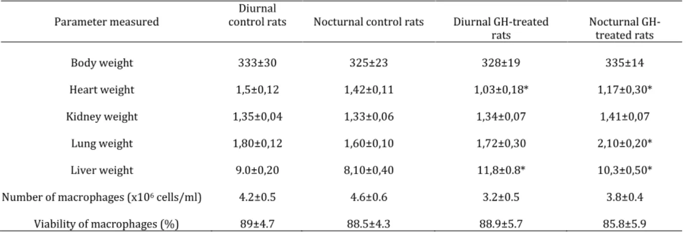
There was no difference in body weight of the GH-treated group compared to the control group. Biometrical measurement of organs showed that GH effects were not observed in the kidney, independently
of administration periods. Heart weight was
significantly decreased in both groups treated with GH, whereas the lung and liver weights of nocturnal
GH-treated rats were significantly (P<0.05) enhanced as
compared with the weights of those of the nocturnal control rats (Table 1).
Effect of GH on oxidative stress
Neither diurnal nor nocturnal GH treatment significantly changed the quantity or quality (viability) of the macrophages (Table 1). GH treatment, independently of administration phases, did not significantly stimulate superoxide release by spleen macrophages. However, when stimulated with PMA macrophages, released more superoxide in both groups (Figure 1).
The diurnal GH-treatment influenced serum CuZn-SOD concentration in rats. A significant rise was observed in SOD levels in diurnal GH-treated rats
(83.5±13.1) compared with the diurnal control rats
Figure 1. Effects of GH on macrophages superoxide release of control and diurnal GH-treated rats. PBS (unstimulated cells) and PMA (107M –stimulated cells). Results represent the mean and SD
of 10 experiment with different cells. *P<0.05 comparing the superoxide release PMA cells stimulated with PBS, considering the same experimental group
(16.2±4.3) (Table 1). Nocturnal GH administration did not significantly change the CuZn-SOD concentrations when compared to the control group (29.0±10.5 x 26.4±12.5, respectively) (Figure 2).
Figure 2. CuZn-Superoxide Dismutase Serum of control and GH-treated rats in different phases (diurnal and nocturnal). The results represent the mean and SD of 10 experiments of animals. *P<0.05 comparing the controls groups with the GH-treated rats groups
Table 1. General characteristics of GH-treated rats and control rats
Parameter measured
Diurnal
control rats Nocturnal control rats Diurnal GH-treated rats
Nocturnal GH-treated rats
Body weight 333±30 325±23 328±19 335±14
Heart weight 1,5±0,12 1,42±0,11 1,03±0,18* 1,17±0,30*
Kidney weight 1,35±0,04 1,33±0,06 1,34±0,07 1,41±0,07
Lung weight 1,80±0,12 1,60±0,10 1,72±0,30 2,10±0,20*
Liver weight 9.0±0,20 8,10±0,40 11,8±0.8* 10,3±0,50*
Number of macrophages (x106 cells/ml) 4.2±0.5 4.6±0.6 3.2±0.5 3.8±0.4
Viability of macrophages (%) 89±4.7 88.5±4.3 88.9±5.7 85.8±5.9
The results represent mean±SD of experiments with 5 different animals
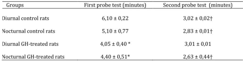
Table 2. Open-field test performance of GH-treated and control rat groups
Groups First probe test (minutes) Second probe test (minutes)
Diurnal control rats 6,10 ± 0,22 , ± , †
Nocturnal control rats 5,10 ± 0,77 ,8 ± , †
Diurnal GH-treated rats 4,05 ± 0,40 * 3,01 ± 0,01
Nocturnal GH-treated rats 4,40 ± 0,51* ,6 ± , †
The results represent mean±SD of experiments with 5 different animals
* P<0.05 (ANOVA) comparison of control groups with GH-treated groups, considering the same periods' probe test
† P<0.05 comparing the first test probe with the second test probe, considering the same group
Effects of GH-treatment on executive function
In the first test, it was observed that GH-treated groups had a significant decrease in latency times when compared with the control groups (Table 2).
In the second test, both control groups (diurnal and nocturnal) had a significant decreased on the latency time. The second latency time in the diurnal GH-treated group tended to decrease, but this difference was not statistically significant. Comparing with the first latency time the nocturnal GH-treated group had a significantly decreased second latency time.
Discussion
The present study experimentally assessed the chronopharmacological roles of GH in executive function and oxidative stress response, which could be implicated in pathophysiological changes of rats.
Several studies have suggested that GH affects neuronal networks and psychological processes. GH hormone deficiency in children may cause sleep disturbances, immaturity and deviant personality development (30-31). In adults GH deficiency has been associated with subjects who are single and unemployed (32), and who had lowered subjective well-being (33, 34).
Our data shows that GH-treated rats displayed improved performance in a task that requires spatial orientation, implementation of a strategy, and working memory, all these capacities are included in the executive function category. The GH treated animals spent less time to accomplish their exploratory and spatial tasks in the open-field test. The time latency in both GH-treated groups was significantly lower compared to the control groups.
Several studies have suggested that GH also affects pathophysiological processes.
In patients with heart failure, it was suggested that GH-treatment restored heart contraction and relaxation (35). In GH-deficient patients, GH treatment improved both heart muscle mass and heart function (36). Studies regarding GH and heart failure have reported that hormone effect is dose-dependent. Lower doses exert beneficial effects,
enhancing antioxidant defense and reducing
oxidative stress, whereas higher GH doses exert detrimental effects related to mitochondrial energy
metabolism and oxidative stress (37). GH
overproduction reduces life span and enzymatic antioxidant defenses, and increases the production of pro-inflammatory cytokines, tissue inflammatory markers, cardiac apoptotic rates, and also increases the burden of oxidative stress, which decisively contributes to aging, cardiac dysfunction, and neurodegeneration (38-45).
In the current work, GH-treated rats presented a decrease in heart weight and an increase in lung weight which reinforces the pathological effects of supraphysiological levels of GH. In addition, another model of excessive GH production is represented by acromegaly patients. In acromegaly, a condition in which the patient is exposed to higher levels of GH, mortality due to diabetes, hypertension, stroke, congestive cardiac failure, and ischemic heart disease were associated with the excess of blood GH values (46). This data confirms findings from acromegalic patients that had decreased cardiac performance and heart vascular impairment despite left ventricular hypertrophy (47).
In our experiment, GH treatment exerted beneficial effects by enhancing antioxidant defenses in the nocturnal phase. Antioxidant defense system includes both enzymatic and non-enzymatic pathways. Among the enzymatic antioxidant mechanisms, various studies have reported that CuZn-SOD has an important role (48, 49), whereas it has been previously reported that SOD activity displays no difference in GH-treated animals (37). The rise of the SOD concentration occurred in the nocturnal GH-treated group showing
that GH may be both dose- and phase-dependent.
On contrary, there was no change detected in the quantity or viability of spleen macrophages in either GH-treated or control groups. The capacity of activation of those cells evidenced by superoxide release, showed no significant differences either, indicating that GH-treatment did not alter those cells. When those cells were stimulated by PMA we observed an increase in superoxide release in both GH-treated and control groups.
human patients as proposed by many authors (5, 11, 12, 50, 51).
Conclusion
We suggest that GH administration may enhance executive functions according to the period of the day, at the cost of increasing the risk of oxidative stress. Administration in the diurnal period increased oxidative burst, while in the nocturnal period GH is more likely to contribute to the reduction of oxidative stress.
Acknowledgment
The results reported in this paper were part of a student thesis. The study was partially supported by the Araxa Univesity Center, Minas Gerais, Brazil and Brazilian CNPQ agency.
Conflict of interest
There is no conflict of interest since this study was not supported by third parties.
References
1. Aleman A, Vries WR, Haan EHF, Samson MM,
Koppeschaar HP. Age-sensitive cognitive function, growth hormone and insulin-like growth factor I
plasma levels in healthy older men.
Neuropsychobiology 2000; 4:73-78.
2. Lichtenwalner RJ, Forbes ME, Bennett SA, Lynch
CD, Sonntag WE, Riddle DR. Intracerebroventricular infusion of insulin-like growth hormone factor-I ameliorates the age-related decline in hippocampal neurogenesis. Neuroscience 2001; 107:603-613.
3. Maruff P, Falleti M. Cognitive function in growth
hormone deficiency and growth hormone
replacement. Hormone Res 2005; 64:100–108.
4. Landi F, Capoluongo E, Russo A, Onder G, Cesari
M, Lulli P, et al. Free insulin-like growth factor-I and
cognitive function in older persons living in
community. Growth Horm IGF Res 2007; 17:58–66.
5. Sathiavageeswaran M, Burman P, Lawrence D,
Harris AG, Falleti MG, Maruff P, et al. Effects of GH on
cognitive function in elderly patients with adult-onset GH deficiency: a placebo-controlled 12-month
study. Eur J Endocrinol 2007; 156:439–447.
6. Deijen JB, de Boer H, Blok GJ, van der Veen EA.
Cognitive impairments and mood disturbances in
growth hormone deficient men.
Psychoneuroendo-crinology 1996; 21:313-322.
7. Baum HB, Katznelson L, Sherman JC, Biller BMK,
Hayden DL, Schoenfeld JC, et al. Effects of
physiological growth hormone (GH) therapy on cognition and quality of life in patients with adult-onset GH deficiency. J Clin Endocrinol Metab 1998; 83:3184-3189.
8. Pavel ME, Lohman T, Hahn EG, Hoffman M.
Impact of growth hormone on central nervoussystem activity, vigilance and tiredness after short term therapy in growth hormone-deficient adults. Horm Metab Res 2003; 35:114-119.
9. Oertel H, Schneider HJ, Stalla GK, Holsboer F,
Zihl J. The effect of growth hormone substitution on
cognitive performance in adult patients with hypopituitarism. Psychoneuroendocrinology 2004;
29:839–850.
10. Arwert LI, Deijenb JB, Mqllerb M, Drenta ML.
Long-term growth hormone treatment preserves GH-induced memory and mood improvements: a 10-year follow-up study in GH-deficient adult men.
Horm Behav 2005; 47:343–349.
11. van Dam PS. Neurocognitive function in adults
with Growth Hormone deficiency. Horm Res 2005; 64:109-114.
12. Falleti MG, Maruff P, Burman P, Harris A. The
effects of growth hormone (GH) deficiency and GH replacement on cognitive performance in adults: A
meta-analysis of the current literature.
Psychoneuroendocrinology 2006; 31:681-691.
13. Corpas E, Harman SM, Blackman S. Human
growth hormone and human aging. Endocr Rev 1993; 14:20-39.
14. Zadik Z, Chalew SA, McCarter RJ, Meistas M,
Kowarski AA. The influence of age on the 24-hour integrated concentration of growth hormone in normal individuals. J Clin Endocrinol Metab 1995; 60:513-516.
15. Xu X, Bennett SA, Ingram RL, Sonntag WE.
Decreases in growth hormone receptor signal transduction contribute to the decline in insulin- like
growth factor I gene expression with age.
Endocrinology 1995; 136:4551-4557.
16. Rosen CJ, Conover C. Growth hormone/insulin-like
growth factor-1 axis in aging: a summary of a National Institutes of Aging-Sponsored Symposium. J Clin Endocrinol Metab 1997; 82:3919-3922.
17. Rudman D, Feller AG, Nagraj H, Gergans GA,
Lalitha PY, Goldberg AF, et al. Effects of human growth
hormone in men over 60 years old. N Engl J Med 1990; 323:1-6.
18. Attanasio AF, Mo D, Erfurth EM, Tan M, Ho KY,
Kleinberg D, et al. Prevalence of metabolic syndrome in
adult hypopituitary growth-hormone (GH)-deficient patients before and after GH replacement. J Clin Endocrinol Metab 2010; 95:74-81.
19. Kinneya BA, Coschiganob KT, Kopchickb JJ, Stegera
RW. Evidence that age-induced decline in memory retention is delayed in growth hormone resistant GH-R-KO (Laron) mice. Physiol Behav 2001; 72:653-660.
20. Santos LOM, Simões MLPB, Machado APB,
Matioski Filho GR, Endo PC, Gruen GR, Cipriani VR, Mesquita LD. Effect of somatotropin on skin wound healing in rats. Acta Cir Bras2002; 17:220-224.
21. Honorio-França AC, Carvalho MP, Isaac L, Trabulsi
LR, Carneiro-Sampaio MM. Colostral mononuclear phagocytes are able to kill enteropathogenic escherichia coli (EPEC) opsonized by colostral IgA. Scand J Immunol 1997; 46:59-66.
22. França EL, Feliciano ND, Silva KA, Ferrari CK,
Honorio-França AC. Modulatory role of melatonin on superoxide release by spleen macrophages isolated from alloxan-induced diabetic rats. Bratisl Lek Listy 2009; 110: 517-522.
23. Pick E, Mizel D. Rapid microassays for the
24.Novelli EL, Rodríguez NL, França EL, Gebra LMN, Ribas BO. High Dietary Carbohydrate and Panceatic
lesion. Braz J Med Biol Res 1993; 26:31-36.
25.Ledig MM, Paria JR, Mandel P. Superoxide
dismutase activity in rat brain during acute and
chronic alcohol intoxication. Neurochem Res 1981;
6:385-390.
26.Endo H, Nito C, Kamada H, Yu F, Chan PH.
Reduction in oxidative stress by superoxide dismutase over expression attenuates acute brain injury after subarachnoid hemorrhage via activation of Akt/glycogen synthase kinase-3beta survival signaling. J Cereb Blood Flow Metab 2007; 27:975-982.
27.Barros D, Amaral O, Izquierdo I, Geracitano L, do
Carmo Bassols Raseira M, Henriques AT, et al.
Behavioral and genoprotective effects of vaccinium
berries intake in mice. Pharmacol Biochem Behav 2006; 84:229-334.
28.Mello PB, Benetti, F, Cammarota M, Izquierdo I.
Effects of acute and chronic physical exercise and stress on different types of memory in rats. An Acad Bras Ciênc 2008; 80:301-309.
29.Zar JH. Bioestatical Analysis. Englewood cliffs:
Prentice-Hall International Editions, 1984.
30.Rotnem D, Genel M, Hintz RL, Cohen DJ.
Personality development in children with growth hormone deficiency. J Am Acad Child Psychiatr 1977; 16:412-426.
31.Hayashi M, Shimohira M, Saisho S, Shimozawa K,
Iwakawa Y. Sleep disturbance in children with growth hormone deficiency. Brain Dev 1992; 14:170-174.
32.Dean HJ, McTaggart TL, Fish DG, Friesen HG. The
educational, vocational, and marital status of growth hormone-deficient adults treated with growth hormone during childhood. Am J Dis Child 1985; 139:1105-1110.
33.Mitchell CM, Joyce S, Johanson AJ, Libber S,
Plotnick L, Migeon CJ, et al. A retrospective evaluation
of psychological impact of long-term growth
hormone therapy.Clin Pediatr 1986; 25:17-23.
34.McGauley GA, Cuneo RC, Salomon F, Sonksen PH.
Psychological well-being before and after growth hormone treatment in adults with growth hormone
deficiency. Horm Res 1990; 33:52–54.
35.Libera LD, Ravara B, Volterrani M, Gobbo V,
Barbera MD, Angelini A, et al. Beneficial effects of
GH/IGF-1 on skeletal muscle atrophy and function in experimental heart failure. Am J Physiol Cell Physiol 2004; 286:C138-C144.
36.Isgaard J, Arcopinto M, Karason K, Cittadini A. GH
and the cardiovascular system: an update on a topic at heart. Endocrine 2015; 48:25-35.
37.Seiva FR, Evadí GM, Castro AV, Okoshi K,
Nascimento A, Rocha KK, et al. Growth hormone and
heart failure: Oxidative stress and energetic
metabolism in rats. Growth Horm IGF Res 2008;
18:275-283.
38.Brown-Borg H, Bode AM, Bartke A. Antioxidative
mechanisms and plasma growth hormone levels: potential relationship in the aging process. Endocrine 1999; 11:41-48.
39.Brown-Borg H, Johnson WT, Rakoczy S, Romanick
M. Mitochondrial oxidant generation and oxidative damage in Ames dwarf and GH transgenic mice. J Am Aging Assoc 2001; 24:85-96.
40.Coschigano KT, Holland AN, Riders ME, List EO,
Flyvbjerg A, Kopchik JJ. Deletion, but not antagonism, of the mouse growth hormone receptor results in severely decreased body weights, insulin, and insulin-like growth factor 1 levels and increased life span. Endocrinology 2003; 144:3799-3810.
41.Ferrari CKB. Total Antioxidant Capacity: a
biomarker in biomedical and nutritional studies. J Cell Mol Biol 2008; 7:1-15.
42.Ferrari CKB., França EL, Honorio-França AC.
Nitric Oxide, Health and Disease.J Appl Biomed 2009;
7:163-173.
43.Coschigano KT, Wetzel AN, Obichere N, Sharma A,
Lee S, Rasch R, et al. Identification of differentially
expressed genes in the kidneys of growth hormone transgenic mice. Growth Horm IGF Res 2010; 20:345-355.
44.Bogazzi F, Russo D, Raggi F, Bohlooly-Y M, Tornell
J, Sardella C, et al. Cardiac extrinsic apoptotic
pathway is silent in young but activated in elderly mice overexpressing bovine GH: interplay with the intrinsic pathway. J Endocrinol 2011; 210:231-238.
45.Masternak MM, Bartke A. Growth hormone,
inflammation and aging. Pathobiol Aging Age Relate Dis 2012; 2:17293.
46.Varadhan L, Reulen RC, Brown M, Clayton RN. The
role of cumulative growth hormone exposure in determining mortality and morbidity in acromegaly: a single centre study. Pituitary 2016; 19:251-261.
47.Colao A, Spinelli L, Cuocolo A, Spiezia S, Pivonello
R, di Somma C, et al. Cardiovascular consequences of
early-onset growth hormone excess. J Clin Endocrinol Metab 2002; 87:3097-3104.
48.França EL, Bitencourt RV, Fujimori M, Morais TC,
Calderon IMP, Honorio-França AC. Human colostral phagocytes eliminate enterotoxigenic Eschechia coli opsonized by colostrums supernatant. J Microbiol Immunol Infec 2011; 44:1-7
49.Morceli G, França EL. Magalhães VB, Damasceno
DC, Calderon IMP, Honorio-França AC. Diabetes-induced immunobiochemical changes in human colostrum Acta Paediat 2011; 100:550-556.
50.van Dam PS, Aleman A, de Vries WR, Deijen EA,
van der Ven EA, de Haan EH, et al. Growth hormone,
insulin-like growth factor I and cognitive function in
adults. Growth Horm IGF Res 2000; 10:69S–73S.
51.Glisky EL, Kong LL. Do young and older adults
rely on different processes in source memory tasks? A neuropsychological study. J Exp Psychol 2008;