Departamento de Biologia Animal
Faculdade de Ciências
The role of microRNAs in
X-Linked Myotubular Myopathy
Mariana Miranda Fontes
Mestrado em Biologia Humana e Ambiente
Departamento de Biologia Animal
Faculdade de Ciências
The role of microRNAs in
X-Linked Myotubular Myopathy
Mariana Miranda Fontes
Mestrado em Biologia Humana e Ambiente
2009
Alan H Beggs, PhD, Children´s Hospital Boston, Harvard Medical School
Maria do Mar Oom, PhD, Faculdade de Ciências da Universidade de Lisboa
First and foremost I would like to thank my advisor and tutor, Prof. Dr. Alan Beggs, for all the motivation and scientific inspiration during this past year. I am truly grateful and thankful for all the opportunities you created to me and for all the excitement and enthusiasm you put on your support to my ideas and dreams. It definitely couldn’t have made my experience more pleasant. Thank you for your sensitivity and care, contagious passion for research and knowledge.
I also would like to specially thank Prof. Dr. Vandana Gupta for all the motivation in the everyday learning experience, for all the incredible scientific ideas, unequal patience and critic point of view that made this thesis project such an exciting challenging journey. Thank you for your unstoppable dedication and continuous help!
To my parents and grandma for all the patience and support since the early first day I decided to do my way in this project until the last minute, and I’m sure the minute after. It is unbelievable for me how you can always be there, believe in me so hard and pull me up so easily. Your help and positive attitude is so fascinating and inspiring to me.
To my best and forever friends Ines Freitas, Ana Queiros, Matthew Raphael, Joana Reis, Joao Cerveira, Joao Delfim and Pedro Fonseca for being there and sharing with me their fun times and frustrations. A particular thank you to the first three ones for this amazing year filled with incredible wonderful times together, strong friendship, and constant support in most stressful moments.
To my advisor and friend Prof. Dr. Maria do Mar Oom for the constant connection, care and excitement about my career progression. Thank you.
And finally I would like to thank my master’s coordinators Prof. Dr. Deodalia Dias e Prof. Dr. Ana Crespo for making this experience possible and supporting the development of my master thesis outside of Portugal.
X-linked myotubular myopathy (XLMTM) is a congenital neuromuscular disorder characterized by profound hypotonia and severe skeletal muscle weakness in the affected newborn males. The pathology is associated with mutations in the MTM1 gene leading to loss of function of the resulting encoded protein, myotubularin. Myotubularin is a phosphoinositol lipid phosphases known to be involved in endosome trafficking and membrane remodeling, however, the molecular mechanisms underlying myotubular myopathy are not yet clear.
MicroRNAs (miRNAs) are post transcriptional modulators of gene expression and play an important role in many developmental processes and diseases. To identify functional miRNA-protein networks that may be dysregulated in myotubular myopathy, we performed miRNA as well as mRNA expression profiling of skeletal muscle of Mtm1 knockout mice. Bioinformatic analysis and real-time RT-PCR validation resulted in identification of 12 miRNAs that showed significantly differential expression in
Mtm1 mice. The functional targets of these miRNAs in myotubular myopathy were identified by a
combinatorial approach in which computationally predicted targets genes of these 12 miRNAs were matched with statistically altered genes obtained by mRNA profiling of skeletal muscle tissues from
Mtm1 mice. Ontological classification of target genes revealed genes primarily belonging to skeletal
muscle development and maintenance, regulation of cell cycle and differentiation of muscle fibers.
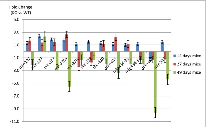
Expression analyses of miRNA-target genes identified from this study were also performed during earlier developmental time points (2 and 4 weeks) in Mtm1 mice for a better comprehensive insight of miRNA-mRNAs in the progression of the disease. We observed that an increase in the severity of XLMTM is associated with an increase in the fold change of several miRNAs and their target genes, suggesting their crucial role in pathology of myotubular myopathy. We hope understanding the molecular pathways involving these miRNA-mRNA networks, which are disrupted in myotubular myopathy, will contribute to uncovering the mechanisms of muscle development and maintenance and the development of new therapies for myotubular myopathy.
Miopatia miotubular associada ao cromossoma X (XLMTM) é a forma mais severa de um grupo de doenças musculares congénitas denominado miopatias centronucleares. XLMTM é caracterizada por uma marcada redução de tónus muscular e profunda debilitação do músculo esquelético em neonatais do sexo masculino. Como consequência, a maioria dos doentes falece nos primeiros oito meses de vida, devido a insuficiência respiratória. XLMTM tem uma estimada incidência de 1:50.000 neonatais, estando associada a mutações no gene MTM1 que conduzem à perda de função da proteina por este codificado, miotubularina. A miotubularina é o membro prototipico de uma família evolucionariamente conservada de fosfatases de fosfatidilinositois (PtdIns), envolvida na regulação de tráfico endossomal e remodelação membranar. As vias bioquímicas reguladas por miotubularina estão fundadamente reconhecidas, no entanto o modo como a deficiência em miotubularina e consequentemente uma alteração nos níveis de fosfatidilinositol 3-P e fosfatidilinosital (3,5)P2 conduzem a um fenótipo musculo-esqueletico especifico é ainda indeterminado.
Estudos recentes atribuem à miotubularina um papel crucial no manutenção dos tubulos transversais (tubulos T). Os túbulos T são parte integral das triadas que constituem a região subcelular responsável pelo mecanismo de excitação e contração muscular. Os túbulos T são estruturas membranes que requerem constantes oscilações dos níveis de PtdIns, regulados por miotubularina. Segundo este modelo, uma deficiência em miotubularina conduz a uma descoordenação dos mecanismos de contração muscular, resultando na atrofia muscular observada nos doentes de miopatia miotubular.
Neste projecto pretendemos clarificar as vias afectadas pela ausência de miotubularina e compreender o envolvimento dos microRNAs na patofiosiologia da miopatia miotubular. Os microRNAs (miRNAs) são moléculas recentemente descobertas que modulam a expressão génica a nível pos-transcripcional. Após transcrição, transporte para o citoplasma e vários ciclos de processamento mediados pelas RNAses III Drosha e Dicer, os miRNAs funcionam como moléculas guia, que reconhecem por complementaridade as regiões 3’UTR dos mRNA alvo e promovem o seu silenciamento. Elevados graus de complementaridade miRNA:mRNA geram degradação do mRNA alvo enquanto que ligações com mismatch conduzem a repressão da tradução. A importância dos microRNAs no desenvolvimento muscular e em estados patológicos foi previamente demonstrada através da formação de Dicer knockouts condicionais e reportada em várias doenças neuromusculares.
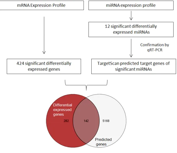
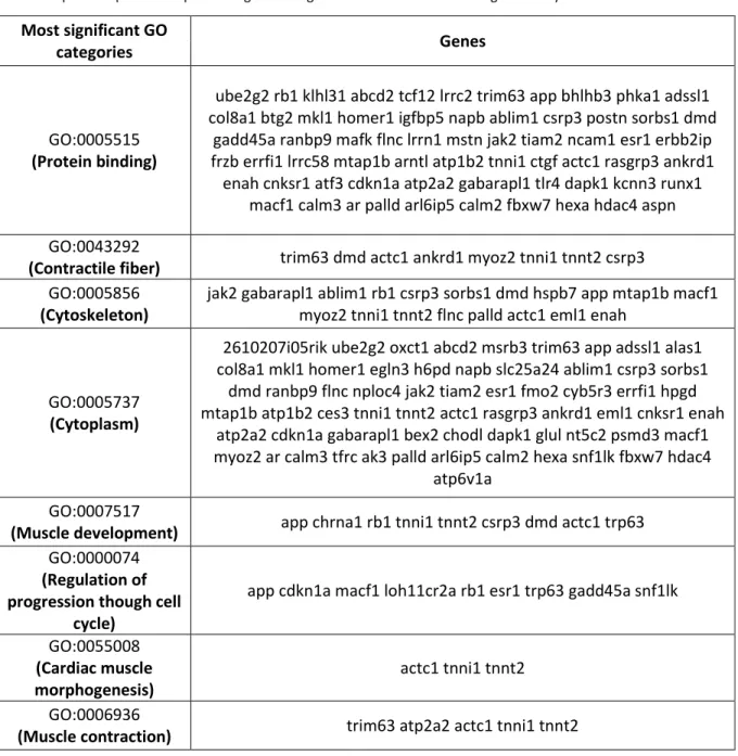
mRNAs foi primariamente efectuado e potenciais interações miRNA-target identificadas. miRNA profilling e subsequente validação por real time PCR revelaram expressão diferencial de miRNAs em ratinhos deficientes em miotubularina quando comparados com os controlos, demonstrando o seu envolvimento na miopatia miotubular. Na tentativa de decifrar redes miRNA/mRNA alvo relevantes para o desenvolvimento de XLMTM, o perfil de expressão de mRNAs foi comparado com a lista de potenciais alvos dos microRNAs obtidos como significantes nesta experiência por abordagens bioinformaticas. A combinação dos resultados de mRNA profiling (com 424 genes significantes) para a identificação de potenciais alvos dos 12 microRNAs obtidos como significantes nas análises de miRNA microarrays resultou em 142 genes alvos diretos de microRNAs. Este grupo de genes potencialmente regulados por miRNAs e diferencialmente expressos no contexto da patologia foram subsequentemente classificados em grupos funcionais sobre-representados usando o software GOstat. As categorias funcionais de contração muscular, regulação da progressão através do ciclo celular, morfogénese do músculo cardíaco, assim como desenvolvimento do músculo esquelético e componentes do sarcomero foram identificadas.
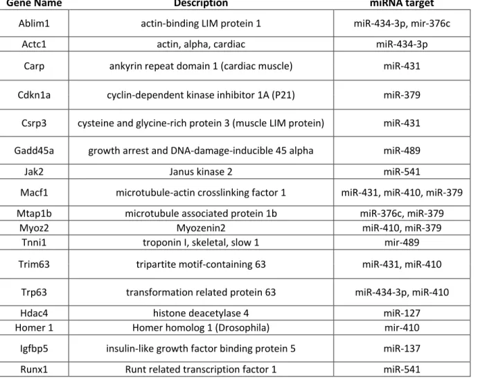
Dada a relevância biológica dos grupos funcionais obtidos, estudos posteriores basearam-se em genes presentes nestas categorias. Apenas genes que apresentaram um padrão de expressão diferencial contrário ao dos seus potenciais miRNAs foram considerados uma vez que os microRNAs regulam negativamente a expressão dos seus mRNAs alvo. Subsequente validação da expressão dos genes selecionados por qRT-PCR array permitiu a formulação de hipóteses quanto às vias moleculares envolvidas na XLMTM.
Estes estudos permitiram verificar que a atrofia muscular observada em pacientes de miopatia miotubular é dependente da expressão de FoxO1/Murf1/Mafbx. FoxO1 é um membro crucial da via de sinalização celular Akt/Pi3k que conduz a atrofia muscular como resposta a uma redução da actividade desta via. FoxO1 promove um aumento de expressão de componentes chave da via de ubiquitinacao-proteossoma. Em particular, Murf1 e Mafbx (moléculas sobreexpressas em XLMTM como foi detectado por análises de microarrays) foram identificados como marcadores necessários para a activação de atrofia muscular. Igualmente várias proteínas integrais do sarcomero previamente reportadas como envolvidas no processo de atrofia muscular foram encontradas sobre-expressas e potencialmente alvo de miRNAs sub-expressos na miopatia miotubular.
forma, usando adenovirus para sobre-expressar estes miRNAs (sub-expressos em condições de atrofia muscular) poderá ser possível reduzir os níveis de proteínas implicadas no processo de atrofia e consequentemente atenuar o progresso da miopatia miotubular, entre outras doenças musculares. Adicionalmente foi também observada uma persistente sub-expressão de microRNAs alvo de genes inibidores de proliferação celular e respectiva sobre-expressão dos respectivos mRNA. Entre este os genes supressores de tumores p21, Gadd45a e Dusp4 representam alvos candidatos de microRNAs sub-expressos nesta doença. Igualmente, marcadores de diferenciação celular foram consistentemente detectados sobre-expressos.
Por outro lado, foi observado um aumento de expressão de genes (regulados por miRNAs) envolvidos no controlo da manutenção da população de células estaminais em ratinhos mutantes para miotubularina. Estas observações despoletaram o desenvolvimento de hipóteses explicativas da incapacidade do músculo esquelético de responder a estímulos regenerativos. É postulado que a incapacidade do músculo de ratinhos mutantes de promover hipertrofia muscular poderá estar correlacionada com um defeito no potencial proliferativo das células estaminais. De uma forma cooperativa vários miRNAs pertencentes a um mesmo cluster genomico (Dlk1-Dio3 cluster no cromossoma 12 em Mus musculus) poderão ser responsáveis pela sobre-expressão de genes inibidores do ciclo celular e igualmente genes indutores da diferenciação celular.
Estas evidências sugerem que uma redução na capacidade de proliferação celular acoplada a um aumento de proteinas de diferenciação nos ratinhos knockout deficientes em miotubularina, resultam na produção de apenas um pequeno pool de satellite cells activadas (células estaminais progenitoras de tecido muscular), insuficientes para uma eficiente reparação da injuria muscular. Esta incapacidade regenerativa poderá ser um dos mecanismos básicos fundamentais a contribuir para o desenvolvimento de atrofia muscular. Os genes responsáveis por estas duas vias (activação das células estaminais precursoras de mioblastos e indução de atrofia muscular) são regulados por vários microRNAs que formam uma rede altamente coordenada levando a que mesmo um pequeno defeito no circuito resulte no desenvolvimento de severa atrofia, incluindo associada às manifestações de miopatia miotubular.
Estudos de expressão génica realizados para diferentes tempos de desenvolvimento do ratinho usando a tecnologia de highthroughput qRT-PCR array permitiram demonstrar a relevancia do grupo de genes regulados por miRNAs selecionados. 26 genes envolvidos nas vias de diferenciação/proliferação
clinico da doenca. Desta forma, a hipotese de estas moléculas representarem apenas representarem um artefacto experimental ou apenas serem inespecificamente induzidas como consequência do estado geral de doença foi refutada. Os microRNAs encontrados diferencialmente expressos na XLMTM e identificados como responsáveis pelo estabelecimento de várias das vias funcionais desreguladas neste sistema constituem candidatos preferenciais para estudos futuros de terapia genética.
1) Introduction - p.1
1.1) X-linked myotubular myopathy - p.1
1.2) Myotubularin and the etiology of myotubular myopathy - p.2 1.3) Skeletal muscle development – p.5
1.4) microRNAs biogenesis and cellular function - 7 1.5) miRNAs in skeletal muscle – p.11
1.6) Mouse model of XLMTM – p.12
1.7) Study description and biological impact – p.13
2) Methods – p.15
2.1) Samples preparation – p.15 2.2) RNA extraction – p15 2.3) miRNA Array analysis – p.15
2.4) mRNA microarray hybridization – p.16
2.5) Principal Component Analysis (PCA), Box-Whisker plot, Hierarchical Clustering – p.16 2.6) Statistical Analysis of microarray data – p.16
2.7) miRNA and mRNA Real Time PCR (qRT-PCR) – p.17 2.8) miRNA target prediction and functional analysis – p.17
3) Results/Discussion – p.19
3.1) miRNAs are differentially expressed in XLMTM – p.19
3.2) Validation of differentially expressed miRNAs by qRT-PCR analysis – p.22 3.3) miRNAs genomic location – p.23
3.4) Biological relevance of identified miRNAs – p. 26
3.8) Functional categories enriched in potential miRNA-regulated genes correlate with disease phenotype- p.31
3.9) Biological pathways modulated by miRNAs relevant in the etiology of XLMTM – p. 35
3.9.1) Muscle atrophy molecular pathways are upregulated in XLMTM – p. 35 3.9.2) Structural sarcomeric proteins implicated in atrophy are
overexpressed in XLMTM – p.36
3.9.3) Inhibition of proliferation and induction of differentiation in satellite cells – p.39
3.9.4) Overexpression of type I myofiber genes - p.42
3.10) Differential expression of candidate miRNA-regulated genes during development -p.44
4) Concluding remarks – p.46
5) Bibliography – p.48
1) Introduction
1.1) Linked Myotubular Myopathy
linked myotubular myopathy (XLMTM) is the most severe form of a group of congenital muscular disorders named centronuclear myopathies, and presents an estimated incidence of 1 in 50,000 newborn males. (D'Amico and Bertini, 2008) In contrast to the heterogeneous patterns found in the majority of congenital myopathies, XLMTM has a relatively homogeneous clinical presentation, where males are born with marked hypotonia and generalized muscle weakness, with respiratory difficulties often requiring ventilation. Most XLMTM patients die within 4-8 months as a consequence of respiratory failure. Even though some of them are able to survive several years (approximately 15%), and some achieving independent respiration, so far, it is not possible to accurately predict the severity of the phenotype at birth. (Jungbluth et al., 2008)
The morphological hallmark in muscle biopsies of XLMTM patients is the presence of numerous rounded hypotrophic fibers with a higher than expected occurrence of centrally placed nuclei in hematoxylin/eosin stained sections. This pattern resembles the structure of fetal myotubes, whereas in healthy individuals nuclei usually occupy a peripheral location. The percentage of these “myotubes” in a sample can vary widely among cases with a reported range between 2% to 60%. Based on this canonical characteristic this pathology previously was thought to be due to an arrest in muscle development at the myotube stage. (Sarnat, 1990) Subsequent studies, however, refuted this hypothesis and suggested instead that myotubular myopathy represents a defect in skeletal muscle maintenance. Typically found surrounding these nuclei in central locations is a clear perinuclear zone containing glycogen and mitochondria deposits, which can be observed in histochemical stains PAS and NADH-TR, respectively. (Tronchere et al., 2003)
Disease diagnostic results from an integrative approach compiling pathological findings, age of onset and, if available, genetic testing. The definitive diagnosis, however, is based on genetic screening for mutations in the MTM1, the only gene associated with the development of the pathology. (Laporte et al., 1996) So far, more than 200 different loss-of-function mutations have been identified throughout the entire coding sequence of MTM1, in more than 300 unrelated families. (Laporte et al., 2000) The MTM1 gene spans approximately 100Kb at the genomic level, contains 15 exons and is located in Xq28 locus. MTM1 mRNA is expressed as of 3.9 Kb long transcript, ubiquitously expressed in all human tissues as idenfied by northern blot analysis. Interestingly, in skeletal muscle and testis, an additional smaller 2.5 Kb tissue-specific transcript is detected, resulting from the use of a different polyadenylation site. The biological significance of
this mechanism of gene regulation for MTM1 remains unknown, pointing out the importance of further studies in MTM1 expression control. (Pierson et al., 2005)
1.2) Myotubularin and the etiology of myotubular myopathy
The protein product of the ubiquitously expressed MTM1 gene is myotubularin, a phosphatidylinositol (PtdIns) lipid phosphatase of 603 amino acids. Myotubularin contains an active phosphatase domain (PTP) and a prixomal PH-GRAM domain that binds phosphoinositides. Phosphoinositides (PI) are lipid second messengers that play key roles in signal transduction, trafficking and cellular homeostasis through the recruitment of effectors proteins to their subcellular target sites. The selection of the PI specie to be formed (in a set of seven) is dependent on the phosphorylation/ desphosphorylation balance in the position 3, 4 or 5 of their inositol sugar rings. These interconversions are controlled by their respective PI kinases and phosphatases. Myotubularins are 3’-phosphatases specific for PtdIns3P and PtdIns(3,5)P2, PIs involved in the endosomal–lysosomal pathway. (Robinson and Dixon, 2006)
Myotubularin is the archetypical member of a protein tyrosine phosphatase (PTP) superfamily of 14 closely related genes that share high levels of homology, present in a wide spectrum of eukaryotic organisms from yeast to mammals. (Clague and Lorenzo, 2005) In the myotubularin family only 8 members were defined has containing phosphatase activity. Other myotubularins that have an inactive phosphatase seems to be crucial for the stability and activation of the other catalytic active members. Most of the myotubularin family members are able to form homodimers or heterodimers with other members of the family by direct protein-protein interactions. The interaction between the inactive MTMR9 and active MTMR6 form reflects this essential function by leading to an increase in the 3-phosphatase activity of MTMR6 up to 6-fold. (Zou et al., 2009) Mutations in MTMR13 or its binding partner MTMR2 have been found in human patients of Charcot-Marie-Tooth disease further strengthing the importance of their interactions.
The biochemical functions of myotubularin have been intensively described, although, the biological pathways and regulatory mechanisms that when disrupted lead to myotubular myopathy, still remain uncertain. To address these questions several knockout/overexpression experiments were recently developed. Using siRNA-mediated technology it was demonstrated that knocking down the levels of myotubularin results in an increase of 60% to 120% of endogenous PI3K that accumulates on early endosomes. The sequential waves of PI(3)P synthesis
and degradation, regulated by the interplay of PI3 kinases and phosphatases and controlled by the activation of Rab GTPases, has been postulated to be the mechanism for signal transduction and receptor sorting necessary for acute recruitment, release or activation of the trafficking machinery components, during endocytosis. (Cao et al., 2008) A dysregulation in the orchestration of these antagonistic proteins seems to lead to impairment cellular metabolism, contributing to the development of several pathologies, including XLMTM.
Likewise MTM1, several MTM related genes (MTMR) have been established as involved in different types of neuromuscular disorders (e.g. MTMR2 and MTMR13 mutations are associated with Charcot-Marie-Tooth type 4B1 and 4B2 neuropathy, MTMR14 mutations result in autosomal centronuclear myopathy, MTMR1 splicing variants are associated with myotonic dystrophy and MIP/Mtmr14 mutations lead to muscular weakness and fatigue). (Azzedine et al., 2003); (Houlden et al., 2001); (Shen et al., 2009)
It is remarkable to note that ubiquitously expressed PI phosphatases acting in the same biological pathway and with similar catalytic active profiles are involved in pathologies affecting different specific tissues. As noted above, MTMR2 gene that shares 65% sequence identity with MTM1 affects primarily Schwann cells in peripheral nerves instead of skeletal muscle as MTM1 mutations. The reason for large amounts of apparently functional redundant PI proteins seems to be directly correlated with their specific spatiotemporal expression pattern and subcellular location. Both MTM1 and MTMR2 desphosphorylate pools of Ptdns(3)P and Ptdns(3,5)2P, however, MTM1 primary acts both on early and late endosomes, whereas MTMR2 is exclusively active on late endosomes. (Nicot and Laporte, 2008)
Although displaying specific subcellular functions, at least some MTM-related genes are able to compensate for the lack of expression of a particular family member, demonstrating a fine efficient mechanism of gene regulation, to restore cell homeostasis. MTM1 is ubiquitously expressed, although XLMTM is a muscle specific disorder. A possible justification for this phenomenon is the ability of MTMR1 and MTMR2 to compensate the loss of myotubularin in all but skeletal muscle tissue, where MTM1 is the main 3’-phosphatase. (Dowling et al., 2009)
The overexpression of myotubularin promotes formation of fillapodia-like structures and disrupts protein transport from late endosomes to lysosomes. (Tsujita et al., 2004) To deeper define the subcellular function of myotubularin in skeletal muscle, overexpression experiments were performed using adeno-associated virus (AAV) vectors expressing myotubularin. The formation of packed membrane assemblies and the presence of vacuoles positives for T-tubules
and plasma membrane markers were observed. Subcellular staining of myotubularin revealed the protein associates with sarcolemma and triads. Triads are highly specialized junctions where the control of calcium concentration, and therefore, regulation of excitation-contraction occurs. These structures are constituted by transverse T-tubules (plasma membrane invaginations) and terminal cisternae of sarcoplasmic reticulum. Myotubularin is suggested to be important for the maintenance of triads, remodeling of longitudinal T-tubules to become transverse T-tubules and plasma membrane homeostasis. The failure of these processes would lead to impaired excitation-contraction coupling and consequently generate muscular atrophy. (Buj-Bello et al., 2008); (Al-Qusairi et al., 2009)
In vivo studies using morpholino antisense technology to knockdown myotubularin,
provides additional functional evidences supporting this hypothesis. Knockdown of myotubularin in zebrafish results in impaired motor function and histopathologic changes in skeletal muscle, resembling clinical XLMTM patient observations. Also, tubule-reticular structural abnormalities were found in MTM1-morpholino treated zebrafish and human patient biopsies, displaying disorganized and irregular patterns. T-tubule integrity is required for proper force generation, muscle contraction and more specifically for the process of excitation-contraction coupling. Defects in T-tubule structure and localization (T-tubule were found concentrated around the abnormal located nuclei in XLMTM patients) were also associated with T-tubule functional defects, as was demonstrated by abnormal mechanisms of excitation-contraction observed in skeletal muscle. (Dowling et al., 2009)
The physiological implications of T-tubules dysfunction in the etiology on myotubular myopathy can be hypothesized: T-tubules are membrane invaginations whose biogenesis and maintenance requires continuous recycling of membrane components, including phosphoinositides. If MTM1 function is disrupted, membrane recycling does not occur efficiently. Consequently, T-tubule function is impaired, leading to abnormal excitation-contraction mechanism, necessary to produce muscle force. Mutations of MTM1, causing loss of function, deregulate this process, and subsequently promote muscle wasting and atrophy, characteristics present in skeletal muscle of myotubular myopathy patients. (Dowling et al., 2009)
Recent findings, originated through investigation of MIP/MTMR14 mutant mice, remarkably elucidate the crucial function of MTMR14 in control of PtdInsP levels in establishing calcium homeostasis and muscle performance. Several PtdInsP were identified to directly bind and activate ryanodine receptors (RyR1) - the skeletal muscle calcium release channel in sarcoplasmic
reticulum, crucial for excitation-contraction coupling. These studies strongly help to understand how loss of myotubularin family proteins that are responsible for regulation of PtdInsP function lead to muscle contraction deficiencies. (Shen et al., 2009) (Treves et al., 2005)
Although considerable progress has been made in the understanding the pathogenesis of XLMTM, many critical aspects still remain mainly uncovered, in particular concerning the translation of the identified metabolic defects, into the skeletal muscle phenotype found in the disease.
In this project we will try to bring new insights to address these questions by studying gene expression control in the myotubularin-deficient mouse, model of XLMTM, using microarray analysis of mRNA as well as miRNA (endogenous modulators of gene expression) expression profiles. We aim to contribute to dissect primary molecular pathways disrupted in myotubular myopathy through the identification of miRNA-mRNA regulatory interactions and differential expression of relevant genes in the context of the disease, in knockout mice when compared to wild-type littermates.
1.3) Skeletal muscle development
The knowledge of the networks underlying muscle development and the regulatory mechanisms responsible for the homeostasis and maintenance of muscle integrity are a pivotal basis of muscle biology we hope to improve with our findings.
Skeletal muscle results from muscle progenitor cells derived from mesoderm. In humans, at approximately 3 weeks of gestation, these muscle stem cells generate myoblasts which fuse to generate myotubes at 7 weeks. The myotube stage is characterized by long multinucleated filaments with central located nuclei sharing the same basal lamina. Along the development, myotubes start to produce significant amounts of contractile components required for muscular function. Also, each nucleus acquires an individual basal lamina and become innervated. At this step, mature myotubes are called myofibers and possess peripheral located nuclei and contractile apparatus located in the central region. The determination of cell fate and coordination to myogenic lineage specific seems to be upstream regulated by Pax3 and Pax7. (Maroto et al., 1997) (Seale et al., 2000) Pax3 acts mainly during primary myogenesis while Pax7 during the later stages of muscle growth.
After the initial phase of embryonic muscle development, muscle mass can be produced, according to functional needs, by mechanisms such as hypertrophy (the most common adult
muscle growth) and hyperplasia (primary occurs during embryonic development). During skeletal muscle growth through hypertrophy, myofibers increase their volume by increasing the amount of contractile components, requiring the incorporation of additional nuclei. Satellite cells are mononucleated cells able to fuse and increase the growing capacity of myofibers. (Sanger, 2004) Satellite cells are the major muscle precursor cells and are involved in maintenance and repair of skeletal muscle. These stem cell-like structures derive from the dermomyotomal population (the dorsal compartment of the somites, where the embryonic myogenesis starts to take place), located in a specific niche between the basal lamina and the sarcolemma. Typically, there is a small pool of mitotically quiescent satellite cellswhich are activated in response to external stimuli that produce stress such as need for growth by hypertrophy or muscular injuries. Activated satellite cells proliferate and generate myogenic precursor cells (myoblasts), which undergo multiple rounds of cell division until they experience terminal differentiation. The resulting cells undergo fusion, leading to the formation of multinucleated myofibers.
The molecular mechanisms that underlie the regulation of the satellite cell pool and their self-renewal are still poorly understood. Myogenesis requires co-expression of myogenic regulatory factors (MRFs) such as Myf5, MyoD, Myogenin and MRF4 which promote cell cycle arrest and subsequent terminal differentiation into contractile muscle fibers. Concomitant with this process is the down-regulation of the levels of pax7. (Buckingham and Montarras, 2008) Several studies indicated pax7 as a key molecule in adult muscle cell remodeling pointing out its action as a negative regulator of MyoD and Myogenin and consequently delaying the successful adoption of the myotube phenotype. It has also been recently elucidated that myostatin controls self-renewal of satellite cells and their state of activation, through negative regulation of pax7 via the Erk1/2 pathway. (Bryson-Richardson and Currie, 2008; McFarlane et al., 2008)
Skeletal muscle development is organized by evolutionarily conserved networks of transcription factors that coordinate the expression of muscle-specific genes responsible for muscle growth, differentiation and contractibility. MADS-box transcription factors MEF2 (myocyte enhancer factor 2) and SRF (serum response factor), in combination with MyoD and Myogenin (basic helix-loop-helix factors) play a central role in the regulation of myogenesis through the activation of fine subsets of muscle specific genes according to developmental stages. (Chen et al., 2009)
1.4) microRNAs biogenesis and cellular function
Recently, an additional layer of complexity in skeletal muscle gene regulatory circuits started to be revealed. MicroRNAs (miRNAs) are pos-transcriptional gene expression modulators. Through their ability to coordinately regulate networks of genes, they enable a fast and precise cellular reaction to developmental, physiologic and pathologic signals in skeletal muscles. (Williams et al., 2009)
The recognition of fundamental catalytic activities carried out by RNA molecules on gene expression dates back to 1961 when Jacob and Monod proposed the role of RNAs in the inhibition of operons expression, through Watson-Crick base-pairing interactions with the operator sequence. (Jacob and Monod, 1961) However, the steeply growing knowledge and interest about these tiny molecules was triggered by the discovery of RNA interference mechanism in 1998 by Craig Mello’s group. (Fire et al., 1998) By silencing the expression of specific genes in specific spatial-temporal limits this phenomenon of gene silencing generated by small non-coding RNAs (and effector proteins) is implicated in innumerous crucial cellular and biological processes and has been associated with human pathologies. MicroRNAs are defined as ssRNAs of ~22 nucleotides in length generated by the RNAse-III enzyme Dicer from an endogenous transcript containing a local hairpin structure. (Ambros et al., 2003) Bioinformatics methodologies have predicted that almost 30% of mammalian mRNAs of protein coding genes are regulated by these molecules, revealing their remarkable biological significance. (Bartel, 2004)
The first experiments highlighting the existence of miRNAs were conducted by the Ambros and Ruvkun labs. It was reported that short lin-4 RNA directly down-regulate lin-14 gene product by binding to specific repetitive sequences on the 3’UTR of the lin-14 messenger. This functional interaction promotes the progression from the first nematode larval stage to the second. (Lee et al., 1993) (Wightman et al., 1993)
Despite the fact that it is only seven years the next miRNA was discovered (let-7) (Reinhart et al., 2000) at the date of writing, the official miRNA database - miRBase (release 14.0, Sep 2009) - reports 10581 mature miRNAs in 115 species, including more than 700 human miRNAs. (http://www.mirbase.org) For a long period of time, miRNAs were thought exclusive to multicellular organisms, as a vehicle for the transition to a more complex organism design. Recent studies have identified microRNAs in Chlamidomonas reinhardtii, a unicellular alga, demonstrating the evolutionarily conserved nature of this mechanism of gene regulation. (Molnar et al., 2007)
With only one unique exception detected so far (Bao et al., 2004) microRNAs function at the post-transcriptional level. MicroRNAs regulate gene expression either by blocking mRNA translation or decreasing mRNA stability in the cytoplasm. New perspectives expect microRNAs to operate in almost every cellular process, including regulating pre-mRNA processing in the nucleus, acting as chaperones that modify mRNA structure or modulating mRNA-protein interactions. (Filipowicz et al., 2008)
The genomic localization and transcription process used for microRNAs is not deeply understood yet, however, different mechanisms seem to be involved. Initial studies have pointed out that the majority of microRNAs were encoded in intergenic regions (> 1kB away from the annotated/predicted gene) although the presence of microRNA codified in intronic regions in the
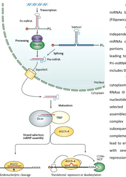
Figure 1: Schematic representation of miRNAs biogenesis and their mode of action. (Filipowicz, 2008)
miRNAs can either be transcribed as independent transcriptional units producing pri-miRNAs or as a result of splicing of intronic portions of protein-coding genes (mirtrons) leading to the direct formation of pre-mRNAs . Pri-miRNAs are processed by a complex that includes Drosha giving rise to pre-miRNAs.
These molecules are transported to the cytoplasm through exportin5 and cleaved by the RNAse III type Dicer to yield approximately 20 nucleotide miRNA duplexes. One of the strands is selected to form the mature miRNA being assembled into a miRNA-induced silencing complex (miRNP) while the other strand is subsequently degraded. High levels of complementarity between miRNA:mRNA target lead to endonucleolytic cleavage and sequences with several mismatches guide translational repression of mRNA targets.
sense or antisense orientation were quickly reported: the notion of microRNAs as autonomous transcription units had emerged. Since more than 50% of the miRNAs are in close proximity with each other, and it was experimentally proven that they can be transcribed by their own promoters as a unique unit of clusters of pri-miRNAs, there is sufficient data to demonstrate that microRNAs might be transcribed as mono or polycistronic transcription units.
Their biogenesis is usually catalyzed in two major steps, processed by two RNAse III family proteins, namely Drosha and Dicer. (Figure 1) Once the precursors miRNAs (pri-miRNAs) are transcribed, typically by RNA Pol II (besides some exceptions reported (Cai et al., 2004)) they fold in a long hairpin-like structure containing an imperfectly base-pairing stem, often including several sequences for different miRNAs. Pri-miRNAs are processed by the endonuclease Drosha, complexed with a dsRNA-binding protein DiGeorge syndrome critical region gene 8 (DGCR8), leading to the release of ~70 nucleotide hairpins known as pre-miRNAs. As an RNAse III endonuclease, this enzyme cuts the RNA duplex at both strands of the stem near the base of the primary stem loop, resulting in a stem with 5’ phosphate and 2 nucleotides 3’ overhang. An alternative pathway that circumvents the requirement of Drosha-DGCR8 machinery was found in C. elegans, D. melanogaster and mammals and enable a subset of pre-miRNAs (mirtrons) to be generated from introns, by cooperative actions of spliceosome and lariat-debranching enzyme (LDBR). (Berezikov et al., 2007; Ruby et al., 2007)
In animals the pre-miRNAs are actively transported to the cytoplasm by the nuclear transport receptor exportin-5, in a cooperative complex with Ran-GTP, which after hydrolysis permits the release of the cargo. Once there, pre-miRNAs are subsequently subject to the second processing step by Dicer (associated with TAR RNA binding protein), generating the final ~22 nucleotides miRNA duplex product. This short duplex is incorporated into the functional miRNA-Ribonucleoprotein complex (miRNP), where the mature miRNA preferentially remains assembled in the complex.
The criterion for the selection of the strand to be matured seems to be the thermodynamic stability of the 5’ end, with the less stable end to be generally elected and the other degraded. The key components of miRNP or miRISC (microRNA- induced silencing complex) are the Argonaute family proteins (AGO). This family comprises four AGO proteins with only AGO2 protein having the ability to participate in the RNAi-like mechanism developed by this complex. After assembly, the mature miRNA strand functions as the guide molecule to the complementary mRNA target to be silenced. The recognition of the exact mRNA to be targeted seems to be the
more controversial and critical issue in understanding the function of microRNAs in mammals. In plants the interactions between microRNA:mRNA are nearly perfect but when this relation is transposed to metazoans, miRNAs usually bind to mRNA by imperfect base-pairing, yielding variable degrees of miRNA-target mismatches. Consequently, this leads to large difficulties in the prediction of their target mRNAs. (Winter et al., 2009)
Although the mechanistic details of this interaction are poorly understood it is accepted that a stringent requirement must be present for a functional and productive binding: a contiguous and perfect base-pairing on the 2-8 nucleotides of the 5’end of the miRNA with the mRNA, denominated seed region. (Lewis et al., 2005) Further studies have also mentioned the importance of secondary structures on the 3’ untranslated region (UTR) surrounding the target site (seed) in the target mRNA and the ability of complementarity at the 3’ end of the cognate miRNA to compensate for imperfect seed matching. (Brennecke et al., 2005)
The molecular basis of this interaction is directly correlated with the selected pathway in miRNA-mediated gene regulation. If miRNA:mRNA-target interaction presents high affinity and nearly perfect complementary, the mRNP complex will trigger endonucleolytic mRNA cleavage by an RNAi-like process. On the other hand, when this match is imperfect, the most common event in mammals, miRNA lead to translation repression and possibly destabilization of the mRNA target. (Carthew and Sontheimer, 2009)
Despite the consensus about the role of microRNAs in promoting translation repression of mRNA targets, the mechanism through which miRNAs interfere with active translation, and, in particular, the step of translation inhibited, is a matter of controversy. Perhaps, the paramount open question is if miRNAs modulate gene expression by single or multiple mechanisms. Thus far, the model of miRNA gene regulation interfering with translation initiation stage has received increasing support by in vivo and in vitro studies. However, repression mechanisms starting after the initiation of translation mediated by miRNAs were also identified when miRNAs were found associated with actively translating polysomes. (Filipowicz et al., 2008)
miRNA regulatory pathways can not only affect the translation of cognate mRNAs but also might directly decrease their amounts, through the promotion of molecule destabilization. miRNAs control transcript levels by recruiting the machinery involved in mRNA decay. miRNPs recruit GW182 (a protein of the P-body structure), which subsequently recruits proteins involved in 3’ poly-A cleaving. As a result, an unstable transcript is generated, susceptible to 3’-5’ exonucleotidic activity, and vulnerable to decapping proteins with subsequent 5’-3’ exonucleases
exposure. (Behm-Ansmant et al., 2006) P-bodies are highly dynamic cytoplasmatic structures, enriched in translation repressor molecules and mRNA-catabolizing enzymes. By conducting mRNA targets to P-bodies, miRNAs can promote the cleavage or storage of the repressed mRNAs, making miRNA regulation a versatile and wide ranging system. (Rana, 2007)
Furthermore, Vasudevan and colleagues have illustrated this versatility when they reported that miRNAs complexed with Ago2 and FXR1 (Fragile X mental retardation protein 1) can trigger upregulation of mRNAs target, in specific cellular conditions. MicroRNAs can switch from repression to activation according to cell cycle state: in proliferating mammalian cells they repress translation whereas in G1/G0 arrest, which usually precedes differentiation, they can potentially promote activation of gene expression. (Vasudevan et al., 2007)
As trans-acting molecules, miRNAs act in the maintenance of cellular homeostasis and development in cooperation with transcription factors. It is now clear that miRNAs have highly cell type-specific expression profiles and mutations in specific miRNAs that lead to the development of severe diseases.
1.5) miRNAs in skeletal muscle
The importance of the role of miRNAs in skeletal muscle development was identified through the generation of conditional Dicer null alleles under the control of MyoD regulatory elements (a muscle specific marker). The resulting Dicer-deficient mice show skeletal muscle hypoplasia associated with perinatal lethality. C2C12 experiments demonstrate increased apoptosis of cultured embryonic myoblasts. (O'Rourke et al., 2007)
Further studies revealed the involvement of particular muscle specific miRNAs (MyomiRs) in skeletal muscle proliferation and differentiation. (Chen et al., 2009; Williams et al., 2009) miR-1 and miR-206 were identified as enhancers of myogenesis and mir-133 was ascribed as a critical factor for myoblast proliferation and repression of myoblast differentiation. miR-1 promotes muscle cell differentiation by targeting histone deacetylase 4 (hdac4). Hdac4 is a main repressor of Mef2c, which in turn constitutes an essential muscle related transcription factor. Besides belonging to the same miRNA polycistron and being transcribed together, mir-133 triggers an opposite effect to mir-1. miR-133 stimulates myocyte proliferation, to a certain extent, by reducing the levels of serum response factor (SRF), a crucial regulator of muscle cell differentiation (Chen et al., 2006) Similarly to mir-1, mir-206 promotes myoblast differentiation. miR-206 has been shown to inhibit electrical coupling between myofibers via gap junctions through the
suppression of gap-junction protein connexin 43 (Cx43), a required step after fusion of skeletal myoblast during myogenesis (Anderson et al., 2006) Also, mir-206 is responsible for the suppression of cell proliferation by the repression of p180 subunit of DNA polymerase-alpha and inhibition of genes encoding follistatin-like 1 and utrophin in skeletal muscle. (Kim et al., 2006); (Rosenberg et al., 2006)
A surprising evidence of the connection of miRNAs to skeletal muscle disorders came from a direct genetic link, observed in a Texel sheep detected with exceptional muscularity. Fine mapping identified a mutation in the 3´-UTR of the myostatin gene (GDF8), a transforming growth factor family B (TGF-B) member, responsible for inhibition of muscle growth. This mutation creates an illegitimate microRNA target site for miR-1 and miR-206, muscle-specific miRNAs, that lead to specific translation repression of myostatin, and consequently promote muscular hypertrophy. (Clop et al., 2006)
As of late, miRNA overexpression or downregulation was associated with several primary muscular disorders, including muscular dystrophy and nemaline myopathy, contributing to progress in the understanding of these pathologies. (Eisenberg et al., 2007) Nevertheless, these studies have not been conducted for X-linked myotubular myopathy.
1.6) Mouse model of XLMTM
Given that XLMTM is a rare disorder, we decided to use a knockout mouse (KO) model, which reproduces the major features of the human disease, to facilitate sample collection and improve statistical relevance. The knockout mouse Mtm14 was generated for Mtm1, through
homologous recombination followed by selection of mice with exon 4germline excision, by Professor Jean-Louis Mandel´s group. This excision causes absence of myotubularin protein production by creating a frameshift mutation that induces an early codon stop. Despite this deletion generating myotubularin-deficient viable mice, some degree of pre or neonatal lethality was observed with only 16.6% of the litter born, instead of the 25% expected. The resulting male mice recapitulate the histopathology of human XLMTM, showing morphological variation in muscle fiber size and accentuated hypotrophy (predominantly in type 1 fibers), with centrally located nuclei present in a higher proportion in the affected animals. A perinuclear halo containing ER glycogen and mitochondria accumulations was also detected, as well as myofibrillar disorganization with sarcomeric disarray and Z-line streaming. In accordance with the observed pathology in humans, Mtm14 mice manifest a muscle-specific disorder. These data corroborate
the tissue specific impact of myotubularin dysfunction in muscle integrity and the inability of other MTMRs to compensate for this defect in muscle. (Buj-Bello et al., 2002)
Although this model reproduces the human disease, some clinical differences are found and consequently should be considered when extrapolating conclusions. Mtm1 KO mice present with variable and progressive muscular weakness with associated severe reduced life expectancy following four characterized stages, based on distinctive clinical manifestations. Mtm1 (-/-) mice are
born asymptomatic (phase I) manifesting a disease onset around 4-5 weeks of life with accentuated decrease in muscular hindlimb strength (phase II) that reaches forelimbs at 5~7 weeks and is associated with the manifestation of kyphosis (phase III). A reduction of 64% in muscle strength is observed in the later phase IV and leads to complete hindlimb paralysis in addition to severe respiratory difficulties that often result in cachexia and respiratory insufficiency, promoting death at 59 19 days. The human disease is mainly nonprogressive, contrasting with the clinical evolution reported in the knockout. Different times of myogenesis may be the reason for these discrepancies since mouse muscle differentiation is completed much later during development (at birth) as compared with humans (which ends around 16 weeks of gestation). Analysis of Mtm1-deficient mice enabled identification of muscle weakness in XLMTM as a consequence of atrophy rather than hypoplasia, as well as demonstrated that the observed muscle fiber defects are not due to an arrest in myogenesis, but perhaps attributable instead to a defect in the maintenance of muscle structure. (Buj-Bello et al., 2002)
1.7) Study description and biological impact
To decipher the molecular mechanisms underlying XLMTM, it is crucial to improve the knowledge about MTM1 gene regulation expression, and in particular, the process involved in its translation. In this project we will try to develop new insights about gene expression control in XLMTM by bioinformatic analysis of mRNA as well as miRNA expression profiles, using qRT-PCR for confirmation of obtained data.
Gene expression profiles of XLMTM patients were previously investigated by Nishino group. (Noguchi et al., 2005) Using custom cDNA microarrays, the molecular signature of eight XLMTM patients was characterized. An upregulation of extracellular/sarcolemmal proteins and cytoskeletal components was observed (in particular, actin-interacting proteins). On the other hand, proteins involved in muscle contraction and energy metabolism were found downregulated. These findings implicate myotubularin as a key regulator of membranous cytoskeletal actin
remodeling and suggest that a defect in myotubularin function contributes to disorganization of actin filament architecture, giving rise to the characteristic phenotype of XLMTM myofibers, with altered cell morphology and abnormal intracellular organelle distribution. (Noguchi et al., 2005)
Although the study was the first of its kind aimed at the understanding of the pathomechanisms of myotubular myopathy, the project was performed using a custom cDNA microarray with only 4,200 selected genes – muscle specific genes and others expected to be part of the dysregulated pathways in XLMTM. Furthermore, the patient group consisted primarily of infants was not well matched in age with the controls who were significantly older.
In the current investigation we developed a microarray-based approach, using oligonucleotide microarrays able to detect whole mice transcriptome, for an unbiased study of the molecular mechanisms involved in myotubular myopathy. Also, in a combinatorial methodology we analyzed simultaneous significant changes in mRNA and miRNA expression to recognize potential miRNA-based regulatory circuits in myotubular myopathy. mRNA and miRNA profiling by microarrays were evaluated in endstage mice (7 weeks), given that this is the mouse developmental disease time point that better recapitulated human pathology. However, for an accurate extrapolation of conclusions to human disease, final significant results should be experimentally repeated for confirmation in human skeletal muscle biopsies. Additionally, for a deeper understanding of the relevance of statistically significant identified miRNAs in the disease context, we analyzed the expression levels of these miRNAs and also several of their candidate cognate target genes, by qRT-PCR in two other developmental stages (14 and 27 days mice).
Through the study of differential regulated pathways in myotubular myopathy, as well as the identification of specific miRNAs potentially responsible for the development of the disease, we hope to dissect fundamental molecular features of disease and open new avenues for the generation of new therapies. The identification of miRNA signatures in diseases and their specific characteristics, such as small size and ability to affect the expression of several genes present in a given pathway, makes miRNA targeting a promising approach for pathology treatment. New approaches, such as locked nucleic acids (LNAs) and antagomiRs, which are synthetic complementary sequences able to inhibit the function of miRNAs, represent exciting new potential therapies to be used when a particular miRNA is upregulated and identified as promoter of a disease phenotype. Adeno-associated viruses (AAV) represent a very efficient delivery method to introduce miRNAs into cells of patients where these molecules are downregulated, potentially restoring health status. (Brown and Naldini, 2009); (Eisenberg et al., 2009); (Saunders and Lim,
2009) Thus, the findings achieved in this investigation constitute a comprehensive foundation for future research into microRNAs-mediated gene regulation in X-linked myotubular myopathy.
2) Methods
2.1) Samples preparation
Gastrocnemeus muscle from 14, 27 and 49 days mice were dissected under Rnase free conditions and immediately frozen using dry ice. Eight samples from 7 week-old wild-type (WT) mice and Mtm1 KO mice (Mtm14) were collected for microarray expression profiling. Four WT
and four 4 week old mice, as well as 4 WT and 4 KO 2 week old mice gastrocnemeus samples were used for additional qRT-PCR experiments. All the protocols for mice experimentation were performed in compliance with IACUC (Institute of animal care and use committee) guidelines.
2.2) RNA extraction
Total RNA was isolated using mirVana isolation kit (Ambion, Austin, TX) accordingly to manufacturer´s instructions.
The extraction was made
according to the manufacturer’s instructions. Yield and purity of RNA was examined using ND1000 Nanodrop (NanoDrop Technologies) and 2100 Bioanalyzer (Agilent Technologies).2.3) miRNA Array analysis
miRNA profilling was performed by Asuragen Services (Austin, TX) applying a custom-manufactured Affymetrix® GeneChip from Ambion – DiscovArrayTM. RNA samples were processed according to the standard operating procedures of the company. (Shingara J, 2005) The miRNA-enriched fraction was obtained by total RNA fractionation through a flashPAGE Fractionator apparatus (Ambion) followed by clean-up and concentration using the flashPAGE Reaction Clean-Up Kit (Ambion). Subsequently, the resulted purified fraction was hybridized to the miRNA DiscovArray containing 623 human and 361 mouse probes plus additional species and >12,000 exploratory probes. GenePix 4200AL scanner (Molecular Devices) was used to scan processed arrays. Microarray signal processing included substraction of estimated background levels based on the median signal of a set of G-C-matched anti-genomic controls.
Raw data was also normalized through variance stabilization and normalization (VSN) method by Asuragen services. For each mature miRNA sequence a minimum of at least two
probes were available. After data processing, Asuragen designated the two probes as A and B, according to their hybridization performance and signal quality. Only probes A values were selected for subsequent statistical analyses.
2.4) mRNA microarray hybridization
mRNA expression profiles of Mtm1 KO mice and controls were evaluated by hybridization of the isolated total RNA with GeneChip® Mouse Gene 1.0 ST Arrays (Affymetrix) containing approximately 27 probes for each of the 28,853 genes, covering the whole mouse transcriptome. The chip was processed by the Microarray Core Facility at Children’s Hospital Boston, MA, according to the company standard protocol. Raw data was also further normalized using the VSN method using GeneSpring software.
2.5) Principal Component Analysis (PCA), Box-Whisker plot and Hierarchical Clustering
PCA was constructed using normalized scaling and mean centered values. Box whisker plot was created based on Pearson correlation. Hierarchical unsupervised 2D-clustering was processed for non-averaged significant entity lists on WT/MTM1 conditions via Euclidean distance metric and average linkage. GeneSpring GX 10.0.2 allowed these analyses.
2.6) Statistical Analysis of microarray data
For statistical analysis a combinatorial strategy was used. miRNAs were only considered statistical significant if both GeneSpring statistics and SAM identified the same miRNA as significant.
GeneSpring GX was used to apply the non-parametric unpaired Mann-Whitney U test with a cut-off p-value<0.05. The resulted mRNAs were subject to a filter of fold change> 1.5 and the genes still present in the final list considered significant by this statistic approach. In parallel the additional Statistical Analysis of Microarrays (SAM) method was also run. A False Discovery Rate (FDR) of 18% combined with a bootstrap of 5000 randomization of samples was selected to test T-statistic.
miRNAs matching both statistical criteria detected on GeneSpring and SAM analyses were selected for further research.
2.7) miRNA and mRNA Real Time PCR (qRT-PCR)
miRNA and mRNA expression were independently assed by real time PCR, using Taqman microRNA assays (containing specific primers to generate miRNA cDNA) and Taqman mRNA expression assays respectively (Applied Biosystems, Foster City, CA).
miRNA expression was evaluated using 5ng of initial RNA sample and 3 replicates per sample in a 7300 ABI Real Time PCR system. The relative amount of each miRNA was normalized to snRNA U6 endogenous control and the fold change calculated as described by the equation 2-ΔΔCt, where ΔΔCt = (CtWt miRNA - CtWt U6) – (CtKO miRNA – CtKO U6).
mRNA expression was quantified by qRT-PCR using 48.48 Dynamic array (BioMark System, Foster City, CA). For each sample 3 technical replicates were examined with 100 ng of initial RNA sample used. Gapdh was select as the endogenous control and similarly fold change calculated accordingly to the 2-ΔΔCt method.
2.8) miRNA target prediction and functional analysis
To predict miRNA target genes a multi-step sequential approach was performed.
The list of significant miRNAs obtained from the match of SAM and Genespring analysis was individually searched against the Argonaute database to identify already described and validated mRNA targets of the miRNAs in the study. ( http://www.ma.uni-heidelberg.de/apps/zmf/argonaute/) The previously described miRNA targets found in this database were compared with the experimental mRNA expression profiles. Only mRNAs with differential expression were considered as potential miRNA target genes.
Statistically significantly changed miRNAs were subjected to in silico target prediction using the public TargetScan 4.2 algorithm incorporated in the GeneSpring software. (http://www.targetscan.org) Matching of subset gene lists obtained for statistically significant miRNAs predicted genes and mRNAs profiled by microarrays allowed the identification of most probable functional miRNA:mRNA interactions. Predicted genes, that didn´t show an inverse differential expression when compared with their associated miRNAs, were excluded from the group of genes of interest.
Genes targeted by several miRNAs differentially expressed in miRNA microarray analysis and overrepresented in significant functional categories were preferentially kept for further research.
To characterize gene ontology categories overrepresented in the knockout mouse model of XLMTM, GOstat software associated with the MGI (mouse genome informatics) GO database was used. The minimum of 3 genes per functional class and an associated p-value<0.02 were used as inclusion criteria. (http://gostat.wehi.edu.au) (Beissbarth and Speed, 2004)
The precise genomic location and nucleotide sequence of each miRNA was determined using miRBase (Wellcome Trust Sanger Institute; http://microrna.sanger.ac.uk). miRBase and BLAST allowed search for evolutionary conservation of microRNAs (http://blast.ncbi.nlm.nih.gov/Blast.cgi). (Griffiths-Jones et al., 2006)
3) Results/Discussion
In this work we have used a genome wide microarray based approach to identify miRNA-mediated regulatory networks in XLMTM. miRNA expression profile was evaluated using a Mtm1 mouse model to assess miRNAs potentially contributing to myotubular myopathy. In parallel, transcriptional profiles were generated to investigate the occurrence of miRNA/predicted target mRNA pairs anti-correlations. The resultant subset of target genes and associated enriched pathways might represent significant miRNA-regulated circuits in the pathophysiology of myotubular myopathy.
3.1) miRNAs are differentially expressed in XLMTM
In an attempt to identify miRNAs expression changes in myotubular myopathy, microarray experiments were performed for 8 Mtm1 (49 days) mice as well as 8 age-matched WT control mice using Affymetrix DiscoVarry chips. These chips were based on miRBase 9.2 that contained >13000 miRNAs and representated by all miRBase organisms including Mus musculus (mmu) and
Homo sapiens. In our studies, only results of hybridization of RNA samples with mmu probes were
considered for analysis. Each miRNA was represented by two probes on the chip and depending on the efficiency of hybridization, they were classified in to two groups: A and B. Among these two groups, probes A (108 miRNAs) provided superior reliability and were further used for data analysis by Genespring Software.
Quality control analyses were done to identify potential outliers among 8 Mtm1 and 8 WT control samples by performing Principal Component Analysis (PCA) and Pearson Correlation. All samples showed a good correlation by these approaches except two control samples: WT2 and WT5. Moreover, these two samples also showed showed a distinct pattern of expression when compared with the other samples of the same class by Hierarchical classification so were further excluded from the experiment. (Appendix Fig.1 and Fig.2)
After removing the outlier samples, the 3D-PCA aggregated samples in two different clusters which corresponding to two conditions in this study: Mtm1 mice and WT control muscle groups. The clear separation of muscle samples in two different classes on PCA plot illustrated the unique miRNA expression profile in myotubular myopathy disease vs normal condition. (Fig.2)
These samples were then normalized using variance stabilization method. Normalized samples were analyzed by a box whisker plot to compare homogeneity among all samples. In each box plot, the box represents the main body of the data and the whiskers show the extreme values.
The variability is indicated by the size of the box and the length of the whiskers. A box whisker plot of all normalized samples showed a low variability among them suggesting high quality of hybridization data. (Fig. 2)
Different kind of test statistics may be applied to microarray data. All of these tests are aimed at identifying p-values that help assess the likelihood that particular genes are regulated. Tests may be parametric or nonparametric. Parametric tests are applied to data sets that are assumed to follow a normal (Gaussian) distribution. Common parametric test for comparing two groups includes the t-test. Nonparametic tests do not make assumptions about the distribution of data. They rank the outcome variable (gene expression) from low to high and analyze the ranks. Mann-Whiteny and Wilcoxon tests are the most commonly used among nonparametric tests.
We analyzed our miRNA microarray data using both parametric as well as nonparametric statistical analysis methods to improve the confidence of obtained results. Only those miRNAs that were identified as significant in both methods were selected for further experimental validations. In nonparametric statistical analysis, GeneSpring GX software (version 10.0.2, Agilent Technologies) was employed to perform the Mann-Whitney test using a cut-off p-value<0.05. In parametric analysis SAM was used to perform t-statistics test with a FDR threshold of 18% to minimize the number of false positives. A high FDR value was selected to avoid exclusion of true
Figure 2: miRNA microarray data analysis for endstage mice. a) 3D-PCA analysis shows a robust distinction of the two classes (WT and KO) and reveals small variance intra-populations. b) Box Whisker Plot reflects a consistent homogeneity of the samples after normalization, as it is illustrated by the predominance of the values around 0.
hits (Appendix Fig.3). A filter of fold change >1.5 was applied to the subset of list of significant results in both statistical methods.
27 miRNAs were obtained as significant in SAM and 16 miRNAs were identified using Genespring. 12 miRNAs matched both lists and therefore were chosen for subsequently studies (miR-127; miR-137; miR-337; miR-376a; miR-376c; miR-379; miR-434-3p; miR-434-5p; miR-410; miR-431; miR-489 and miR-541). (Table 1)
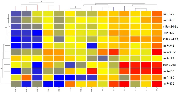
A hierarchical 2D-clustering constructed for these 12 statistically significant miRNAs using the Euclidean measure enabled to additionally investigate the clustering of the distinct patterns of expression of both groups. In agreement with the PCA comparison, the heatmap assorts miRNAs in mainly up or downregulated in knockout mice versus wild-type animals. Despite this observation, three mutant samples (MTM1-3, MTM1-5 and MTM1-8) showed an intermediate level of expression being separated from the remaining mutated samples on the clustering tree. (Fig.3)
Upregulated miRNAs q-value (%) Downregulated miRNAs q-value (%) Upregulated miRNAs p-value Downregulated miRNAs p-value
mmu-miR-137 17,97 mmu-miR-434-5p 0 mmu-miR-137
0,0201 mmu-miR-127 0,002 mmu-miR-324-5p 17,97 mmu-miR-127 0 mmu-miR-329 0,010 mmu-miR-469 17,97 mmu-miR-376a 0 mmu-miR-337 0,020 mmu-miR-546 17,97 mmu-miR-541 0 mmu-miR-376a 0,020 mmu-miR-429 17,97 mmu-miR-434-3p 0 mmu-miR-376c 0,020 mmu-miR-344 17,97 mmu-miR-410 0 mmu-miR-379 0,002 mmu-miR-290 17,97 mmu-miR-431 8,66 mmu-miR-410 0,028 mmu-miR-367 17,97 mmu-miR-379 8,66 mmu-miR-411 0,010 mmu-miR-542-5p 17,97 mmu-miR-383 8,66 mmu-miR-431 0,039 mmu-miR-199b 17,97 mmu-miR-337 8,66 mmu-miR-434-3p 0,002 mmu-miR-465 17,97 mmu-miR-489 12,6 mmu-miR-434-5p 0,002 mmu-miR-464 17,97 mmu-miR-376c 12,6 mmu-miR-466 0,020 mmu-miR-295 17,97 mmu-miR-483 0,010 mmu-miR-224 17,97 mmu-miR-489 0,028 mmu-let-7d 17,97 mmu-miR-541 0,005
SAM GeneSpring statistical Analysis
Table 1: Genespring and SAM results of miRNA microarrays data analysis for endstage mice. miRNAs highlighted in green were identified as significant using both statistical methods.
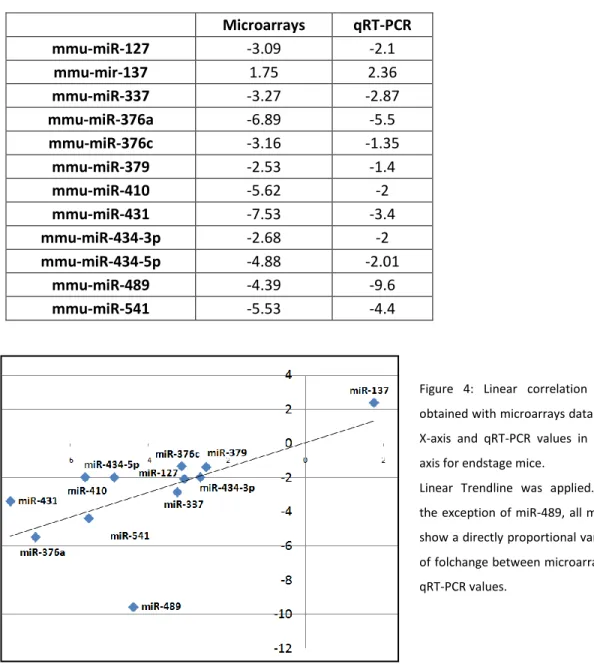
3.2) Validation of differentially expressed miRNAs by qRT-PCR analysis
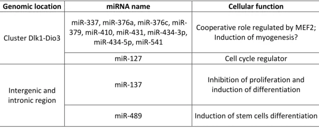
To validate the results obtained in microarray data analysis, qRT-PCR experiments were developed for 12 significantlly altered miRNAs identified from the match between SAM and statistical analysis using GeneSpring GX. (Table 2) All tested miRNAs, with the exception of miR-489, showed a good correlation between microarray and qRT-PCR results. (Figure 4) miR-24 and snRNA U6 were tested to use as internal control miRNAs. miR-24 showed significant up-regulation in mutants demonstrating not to be an appropriate reference miRNA for skeletal muscle expression experiments. snRNA U6 expression levels in mutant and control samples were indistinguishable. Thus, snRNA U6 was adopted for data normalization in qRT-PCR experiments.
To check the accuracy of our hypothesis that only those miRNAs that are common between two statistical analysis are significant, we also performed RT-PCR on few miRNAs that were excluded by parametric test, but considered significant by nonparametric and vice versa. No significant expression differences were observed for mir-411 (fold difference: significant by nonparametric) or miR-295 (fold difference: only identified as significant in parametric) between WT and Mtm1 KO samples, corroborating the efficiency of the approach applied.
Figure 3: Hierarchical 2D-clustering produced for non-averaged significant miRNA lists via Euclidean distance metric and average linkage. The miRNA clustering tree is shown on the left and the sample clustering tree is shown on the top. The colored range represents the differential expression of miRNAs with red denoting <0 and blue >0.