Universidade de Aveiro Departamento de Química 2018
Alexandra Moreira Pais Catabolismo muscular associado à quimioterapia: estudo dos mecanismos moleculares subjacentes
Chemotherapy-induced muscle wasting: exploring the underlying molecular mechanisms
Universidade de Aveiro Departamento de Química 2018
Alexandra Moreira Pais Catabolismo muscular associado à quimioterapia: estudo dos mecanismos moleculares subjacentes
Chemotherapy-induced muscle wasting: exploring the underlying molecular mechanisms
Dissertação apresentada à Universidade de Aveiro para cumprimento dos requisitos necessários à obtenção de grau de Mestre em Bioquímica, ramo Bioquímica Clínica, realizada sob a orientação científica da Doutora Rita Maria Pinho Ferreira, Professora Auxiliar do Departamento de Química da Universidade de Aveiro e do Doutor Rui Gil da Costa, Investigador do Departamento de Engenharia Química da Faculdade de Engenharia da Universidade do Porto
Apoio financeiro da Fundação Portuguesa para a Ciência e Tecnologia (FCT), União Europeia, QREN, FEDER e COMPETE às unidades de investigação QOPNA (UID/QUI/00062/2013), LEPABE (POCI-01-0145-FEDER-006939), CI-IPOP (CIIPOP 37-2016) e CITAB (UID/AGR/04033/2013).
o júri
presidente Prof. Brian James Goodfellow
Professor Auxiliar do Departamento de Química da Universidade de Aveiro
Prof. Rita Maria Pinho Ferreira
Professora Auxiliar do Departamento de Química da Universidade de Aveiro
Prof. Paula Alexandra Martins de Oliveira
Professora Associada com Agregação do Departamento de Ciências Veterinárias da Universidade de Trás-os-Montes e Alto Douro
agradecimentos Em primeiro lugar, quero agradecer à minha orientadora, a Professora Rita Ferreira, pelo apoio incansável ao longo deste ano e pelas oportunidades que me permitiram aumentar o meu conhecimento cientifico. As suas correções, sugestões e disponibilidade foram imprescindíveis para a concretização deste trabalho.
Agradeço, também, ao meu co-orientador, o Professor Rui Gil da Costa, pelas correções e sugestões que permitiram melhorar o meu trabalho.
À técnica de laboratório, Cristina Barros, agradeço a disponibilidade em ajudar sempre que necessário.
A vocês, Sara, Inês e Liliana agradeço todo o apoio, carinho, força e por estarem sempre por perto mesmo à distância.
Por fim, agradeço aos meus pais, ao meu tio e à minha avó pelo apoio incondicional, pelas palavras de força e reconfortantes. Agradeço em especial à minha mãe, por me ter acompanhado todos os dias em todas as horas, por todo o carinho, ensinamentos, coragem, dedicação e apoio. Sem ela, nada seria possível.
A todos vós que me acompanharam ao longo destes cinco anos, um enorme obrigada.
palavras-chave Caquexia; cancro; cisplatina; catabolismo muscular.
resumo A caquexia associada ao cancro (CC, do inglês: cancer cachexia) é uma síndrome metabólica que afeta mais do que 50% dos pacientes com cancro e, para a qual, atualmente, não existe tratamento. Uma das características principais desta síndrome é a perda involuntária e massiva de massa muscular, sendo esta associada a um mau prognóstico. A ativação de vias catabólicas e a presença de um estado inflamatório são responsáveis pelo processo de catabolismo. Assim, as intervenções terapêuticas direcionadas para a massa muscular ou para as vias ativadas pelo processo inflamatório serão, provavelmente, efetivas na diminuição dos efeitos devastadores da caquexia. Para além dos efeitos negativos do cancro na massa muscular, a quimioterapia tem vindo, também, a ser relacionada com o desenvolvimento e exacerbação desta síndrome. Infelizmente, os mecanismos responsáveis pelo catabolismo muscular induzido pela quimioterapia são pouco conhecidos e os estudos que têm sido realizados para a compreensão dos mesmos são escassos. Desta forma, na presente dissertação foi investigado, em animais saudáveis e com cancro da bexiga, o efeito do fármaco antitumoral cisplatina na massa muscular e, simultaneamente, foi estudado o potencial efeito do composto natural dimetilaminopartenolido (DMAPT) em contrariar o processo de catabolismo muscular. De um modo geral, a administração aguda de cisplatina (10 mg.kg-1 no dia zero), em
animais saudáveis, parece não induzir a síndrome caquexia. No entanto, o DMAPT parece originar hipertrofia no músculo através da modulação do processo inflamatório e do eixo TWEAK/NF-κB (do inglês: tumour necrosis factor-related weak inducer of apoptosis/nuclear factor kappa light-chain-enhancer of activated B cells). Relativamente aos animais com cancro, a cisplatina aparenta induzir o processo de catabolismo muscular e o DMAPT parece ser incapaz de reverter ou melhorar este processo, apesar da sua capacidade de reduzir a inflamação, que por sua vez, tem uma função importante no desenvolvimento da caquexia. Desta forma, é importante descobrir a interligação específica entre as vias ativadas pelo cancro e as que são induzidas pelos fármacos anticancerígenos, com o intuito de se desenvolverem estratégias que contrariem a elevada morbidade e mortalidade associadas a esta síndrome.
keywords Cachexia; cancer; cisplatin; muscle wasting.
Cancer cachexia (CC) is a complex metabolic syndrome that affects more than 50% of cancer patients, and presently, no effective treatment exists. A key characteristic of this condition is the involuntary and massive loss of skeletal muscle mass, which is related with a poor prognosis. The activation of catabolic pathways and the presence of an inflammatory state are responsible for the wasting process. Therefore, therapeutic approaches directed to muscle or inflammatory pathways may probably be effective in decreasing the devastating effects of cachexia. Besides the negative effects of cancer itself on the skeletal muscle mass, chemotherapy has also been associated with the development and exacerbation of this process. Unfortunately, the mechanisms responsible for the muscle wasting process induced by chemotherapy are unclear and few studies have been devoted to this specific problem. In this way, we investigated the effect on the skeletal muscle mass of the widely-used chemotherapeutic agent cisplatin in healthy and urothelial carcinoma-induced animals, and simultaneously, studied the putative role of the natural compound dimethylaminoparthenolide (DMAPT) in counteract the muscle wasting process. In overall, an acute administration of cisplatin (10 mg.kg-1 at day zero), in healthy animals, seems to be
incapable of induce a cachectic syndrome. Nevertheless, DMAPT appears to induce muscle hypertrophy through the modulation of the inflammatory process and the tumour necrosis factor-related weak inducer of apoptosis/nuclear factor kappa light-chain-enhancer of activated B cells (TWEAK/NF-κB) axis. However, when cancer is involved, cisplatin can induce muscle wasting and DMAPT looks unable to revert or ameliorate this process, despite its capability to diminish the inflammation, which in turn has an important role in the development of cachexia. In this way, it is important to ascertain the specific relation between the pathways activated by cancer itself and the ones induced by the anti-cancer drugs, in order to develop strategies to counteract the high morbidity and mortality associated with this condition.
i TABLE OF CONTENTS
FIGURES INDEX ...III TABLE INDEX ... VI ABBREVIATIONS ... VII
I. INTRODUCTION ... 1
1.THEIMPACTOFCHEMOTHERAPYINCANCERTREATMENT ... 3
1.1. The chemotherapeutic effect of cisplatin ... 5
2.CANCERCACHEXIA:PATHOPHYSIOLOGICMECHANISMS ... 9
2.1. Cancer-induced muscle wasting ...10
2.1.1. Ubiquitin-proteasome pathway ...13
2.1.2. Autophagy-lysosome pathway ...14
2.1.3. IGF-1/PI3K/Akt/mTOR pathway ...15
2.1.4. Myostatin pathway ...16
2.1.5. NF-κB pathway ...16
2.2. Therapies applied in the management of cancer cachexia ...19
II. AIMS...25
III. MATERIALS AND METHODS ...29
1.CHEMICALS ...31
2.EXPERIMENTALPROCEDURES ...31
2.1. Animals ...31
2.2. Gastrocnemius muscle preparation and analysis ...33
2.2.1. Total protein quantification...33
2.2.2. Spectrophotometric assay of citrate synthase activity ...33
2.2.3. Western blot analysis ...34
2.2.4. Gelatine zymography ...34
2.3. Serum sample analysis ...35
2.4. Statistical analysis ...35
IV. RESULTS...37
1. THE EFFECT OF CISPLATIN OR CISPLATIN PLUS DMAPT ON CATABOLIC PHENOTYPE ...39
ii 1.2. Analysis of the gastrocnemius muscle response to cisplatin or cisplatin plus
DMAPT administration ... 40
2.THEEFFECTOFCISPLATIN,DMAPTORCISPLATINPLUSDMAPTONBLADDER CANCER-INDUCEDANIMALS ... 43
2.1. Characterization of animals’ response to cisplatin, DMAPT or cisplatin plus DMAPT administration ... 44
V. DISCUSSION... 47
VI. CONCLUSION AND FUTURE PERSPECTIVES ... 55
iii FIGURES INDEX
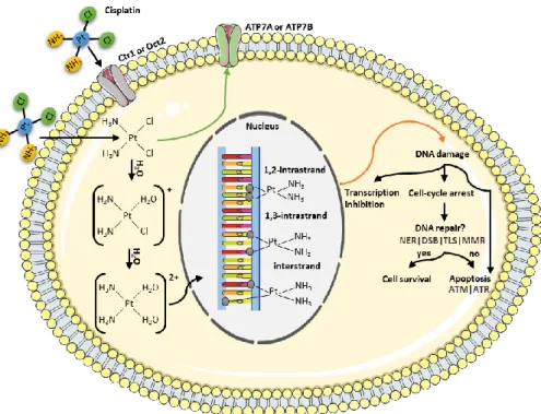
Figure 1. Mechanism of action of cisplatin. Cisplatin enters in the cell through passive diffusion or by the active copper transporter 1 (Ctr1) or the organic cation transporter 2 (Oct2). This drug can also be exported through the copper-transporting adenosine triphosphatase (ATPase) 1 (ATP7A) and the copper-transporting ATPase 2 (ATP7B). To react with deoxyribonucleic acid (DNA), the neutral cisplatin is activated by a series of spontaneous aquation reactions, forming [Pt(NH3)2Cl(OH2)]+ and [Pt(NH3)2(OH2)2]2+. Thus,
the platinum binds to guanine and adenine residues, forming cisplatin-DNA adducts and inducing intrastrand and interstrand crosslinks. All of the cisplatin-DNA adducts distort the DNA structure and can be recognized by several repair pathways, including the nucleotide excision repair (NER) pathway, the double-strand break (DSB) repair, the translesion synthesis (TLS) and the proteins involved in the mismatch repair (MMR) system. If the degree of damage is limited, the several pathways activated culminate in DNA repair. Contrarily, if DNA repair fails or is overwhelmed by the extension of DNA lesions, cells undergo death (frequently apoptosis). In the process of apoptosis, two kinases that function as sensors of DNA damage are activated, named ataxia telangiectasia mutated (ATM) and ATM and RAD3-related protein (ATR). Figure produced using the Servier Medical Art. .... 6
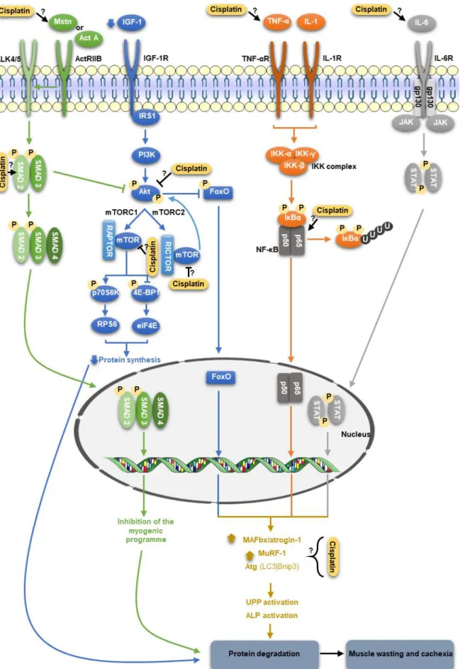
Figure 2. Signalling pathways modulated by cisplatin in skeletal muscle. In the myostatin (Mstn) pathway, this molecule binds to the activin A type IIB receptor (ActRIIB), resulting in its assembly with the activin A type IB receptor, ALK4 or ALK5. Consequently, SMAD2 and SMAD3 are activated by phosphorylation, which leads to the assembly with SMAD4. This heterodimer translocates to the nucleus and activate transcription of target genes. Thus, myostatin inhibits the myogenic programme, inducing muscle wasting. In addition, myostatin inhibits the insulin-like growth factor 1 (IGF-1)/phosphatidylinositol-3-kinase (PI3K)/Akt (also known as PKB – protein (IGF-1)/phosphatidylinositol-3-kinase B)/mammalian target of rapamycin (mTOR) pathway, and so, the levels of muscle atrophy F-box protein (MAFbx/atrogin-1), muscle-specific RING-finger 1 (MuRF1) and autophagy-related genes (Atg) increase, resulting in a decrease in protein synthesis and an increase in protein degradation. In the IGF-1/PI3K/Akt/mTOR pathway, IGF-1 activates the insulin receptor substrate 1 (IRS1) and then Akt blocks the repression of mTOR, which in turn, regulates the skeletal muscle mass by mTOR complex 1 and 2 (mTORC1 and mTORC2). In the mTORC1, which requires the regulatory-associated protein of mTOR (RAPTOR), occurs the phosphorylation and activation of p70 S6 kinase (p70S6K) and inhibition of eukaryotic translation initiation factor
iv 4E binding protein 1 (4E-BP1), which have as downstream targets the ribosomal protein 6 (RPS6) and the eukaryotic translation initiator (eIF4E), respectively, leading to protein synthesis. Regarding mTORC2 which binds to rapamycin insensitive companion of mTOR (RICTOR), this molecule phosphorylates Akt, resulting in maximum Akt activation. In addition, Akt phosphorylates and supresses the forkhead box O (FoxO) family of transcription factors. However, during catabolic diseases, this signalling pathway is silenced and, consequently, protein synthesis decreases and protein degradation increases. The prototypical nuclear factor kappa light-chain-enhancer of activated B cells (NF-κB) pathway can be activated by tumour necrosis factor alpha (TNF-α) and interleukin 1 (IL-1). These molecules lead to the activation of the IkB kinase (IKK) complex, which in turn, phosphorylates the NF-κB-bound inhibitors of NF-κB (IκBαs), which are target for polyubiquitination and degradation. The released NF-κB free dimers (p50/p65) translocate to the nucleus and induce the expression of MAFbx/atrogin-1 and MuRF1. Regarding interleukin 6 (IL-6), this cytokine can signalling through the Janus kinase/signal transducers and activators of transcription (JAK/STAT) pathway, resulting in the suppression of protein synthesis. Legend: Act A: activin A | ALP: autophagy-lysosome pathway | Bnip3: B cell lymphoma 2/adenovirus E1B 19-kDa protein-interacting protein 3 | gp130: glycoprotein 130 | IGF-1R: IGF-1 receptor | IL-1R: IL-1 receptor | IL-6R: IL-6 receptor | LC3: microtubule-associated protein 1 light chain 3 | TNF-αR: TNF-α receptor | UPP: ubiquitin-proteasome pathway. Figure produced using the Servier Medical Art. ... 12
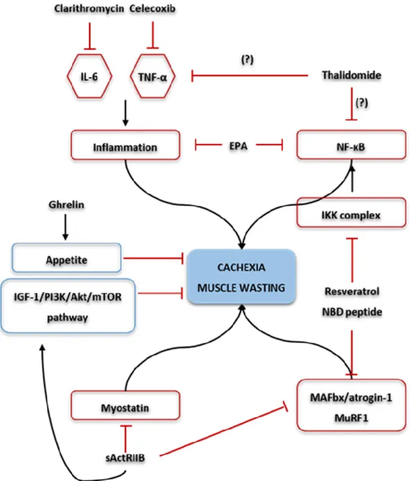
Figure 3. Possible therapeutic approaches for cachexia and muscle wasting. Ghrelin administration increases appetite and muscle mass. Clarithromycin decreases serum levels of interleukin 6 (IL-6), improving the cachectic status. Regarding thalidomide, this agent attenuates weight loss, possible (?) through downregulation of pro-inflammatory cytokines, such as tumour necrosis factor alpha (TNF-α) and /or through the inhibition of prototypical nuclear factor kappa light-chain-enhancer of activated B cells (NF-κB). In addition, the use of celecoxib demonstrated a significant decrease in serum levels of TNF-α and increase in lean body mass. The use of an activin A type IIB receptor (ActRIIB) decoy receptor (sActRIIB), results in the inhibition of the myostatin pathway, and also, prevents the activation of muscle atrophy F-box protein (MAFbx/atrogin-1) and muscle-specific RING-finger 1 (MuRF1). In addition, activate the insulin-like growth factor 1 (IGF-1)/phosphatidylinositol-3-kinase (PI3K)/Akt (also known as PKB – protein kinase B)/mammalian target of rapamycin (mTOR) pathway, thus preserving muscle mass. Resveratrol and the nemo-binding domain (NBD) peptide are inhibitors of the NF-κB
v pathway, and so, decrease the expression of MuRF1 and MAFbx/atrogin-1. Finally, the eicosapentaenoic acid (EPA), decreases pro-inflammatory cytokines release and inhibit NF-κB. ...20
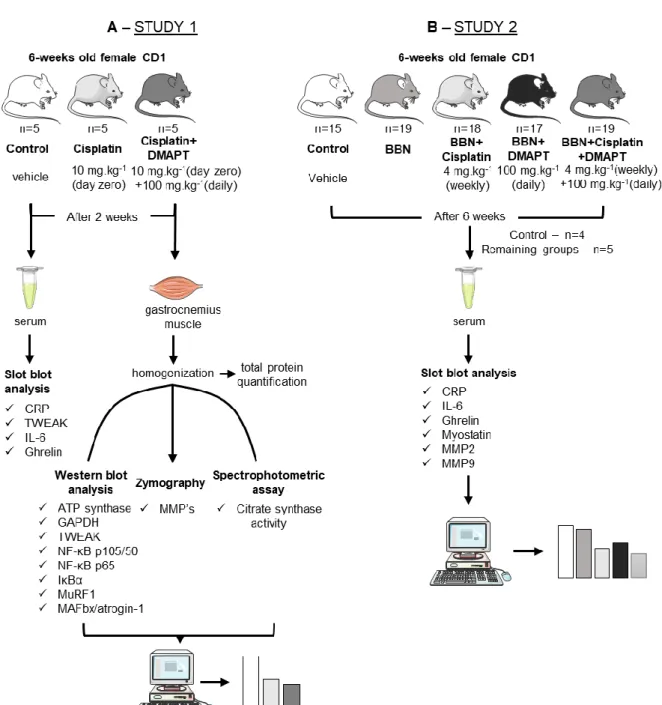
Figure 4. Schematic figure representing the experimental procedures performed in the (A) study 1 and (B) study 2. In study 1 the animals were divided in three groups – control (n=5), cisplatin (n=5) and cisplatin + dimethylaminoparthenolide (DMAPT) (n=5) –, whereas the study 2 was constituted by five experimental groups – control (n=15), N-butyl-N-(4-hydroxybutyl)-nitrosamine (BBN) (n=19), BBN + cisplatin (n=18), BBN + DMAPT (n=17), BBN + cisplatin + DMAPT (n=19). In both studies serum analysis was done through slot blot assays, while western blot analysis, zymography and spectrophotometric assay were only conducted in study 1 in order to analyse the gastrocnemius muscle. The proteins analysed are below the corresponding assays. Legend: ATP: adenosine triphosphate | CRP: C reactive protein | GAPDH: glyceraldehyde 3-phosphate dehydrogenase | IκBα: inhibitors of NF-κB | IL-6: interleukin-6 | MAFbx/atrogin-1: muscle atrophy F-box protein | MMP: metalloproteinase | MuRF1: muscle-specific RING-finger 1 | NF-κB: prototypical nuclear factor kappa light-chain-enhancer of activated B cells | TWEAK: tumour necrosis factor-related weak inducer of apoptosis. Figure produced using the Servier Medical Art..32
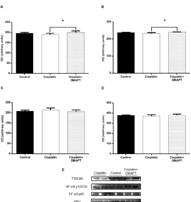
Figure 5. Effect of cisplatin or cisplatin plus DMAPT administration on (A) CRP, (B) TWEAK, (C) IL-6 and (D) ghrelin serum levels. Representative immunoblots are presented above the corresponding graphs. Values are expressed as mean ± standard deviation. (* p<0.05) .40
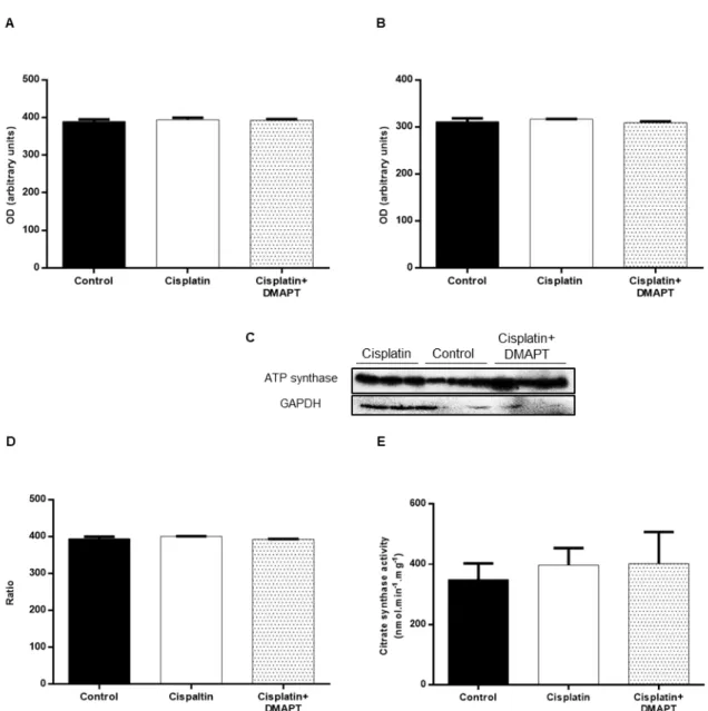
Figure 6. Effect of cisplatin or cisplatin plus DMAPT administration on the muscle expressions of (A) ATP synthase and (B) GAPDH. Representative immunoblots are presented in (C). It is also presented the (D) ATP synthase/GAPDH ratio. In (E) the citrate synthase activity can be observed. The values are expressed as mean ± standard deviation. ...41
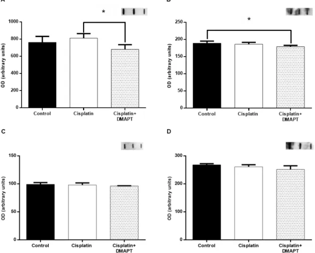
Figure 7. Effect of cisplatin or cisplatin plus DMAPT administration on the muscle expressions of (A) TWEAK, (B) NF-κB p105/50, (C) NF-κB p65 and (D) IκBα. Representative immunoblots are presented in (E). The values are expressed as mean ± standard deviation. (* p<0.05) ...42
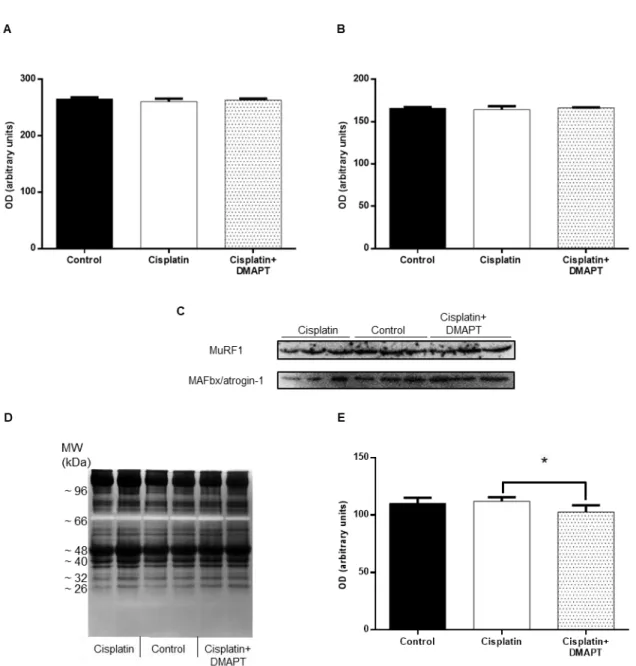
vi Figure 8. Effect of cisplatin or cisplatin plus DMAPT administration on the muscle expression of (A) MuRF1 and (B) MAFbx/atrogin-1. Representative immunoblots are presented in (C). (D) Representative zymography gel, evidencing one band with proteolytic activity. In (E) is presented the semi-quantitative analysis of the proteolytic activity for each group. The values are expressed as mean ± stan-dard deviation. (* p<0.05) ... 43
Figure 9. Effect of cisplatin or DMAPT or cisplatin plus DMAPT administration on the (A) grip strength or (B) grip strength-to-body weight ratio of BBN animals. The values are expressed as mean ± standard deviation. ... 45
Figure 10. Effect of cisplatin or DMAPT or cisplatin plus DMAPT administration on the serum levels of (A) CRP, (B) IL-6, (C) ghrelin, (D) myostatin, (E) MMP2 or (F) MMP9. Representative immunoblots are presented above the corresponding graphs. The values are expressed as mean ± standard deviation. (* p<0.05) ... 46
TABLE INDEX
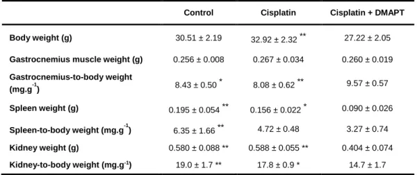
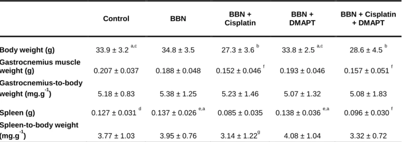
Table 1. The effect of cisplatin or cisplatin plus DMAPT on body weight, gastrocnemius muscle weight, spleen weight and kidney weight and on the ratios gastrocnemius-to-body weight, spleen-to-body weight and kidney-to-body weight. Values are expressed as mean ± standard deviation. ... 39
Table 2. The effect of cisplatin or DMAPT or the combination of cisplatin plus DMAPT on body weight, gastrocnemius muscle weight, spleen weight and the ratios gastrocnemius-to-body weight and spleen-to-gastrocnemius-to-body weight. Values are expressed as mean ± standard deviation. ... 44
Table 3. Characterization of the experimental groups used in study 2 concerning the percentage of invasive and muscle invasive lesions. ... 45
vii ABBREVIATIONS
4E-BP1 Eukaryotic translation initiation factor 4E binding protein Act A Activin A
ActRIIB Activin A type IIB receptor ALK4 Activin A type IB receptor ALK5 Activin A type IB receptor ALP Autophagy-lysosome pathway
AMPK 5´ adenosine monophosphate-activated protein kinase ANOVA Analysis of variance
Atg Autophagy-related genes ATM Ataxia telangiectasia mutated
ATP7A Copper-transporting adenosine triphosphatase 1 ATP7B Copper-transporting adenosine triphosphatase 2 ATP Adenosine triphosphate
ATPase Adenosine triphosphatase
ATR Ataxia telangiectasia mutated and RAD3-related protein BBN N-butyl-N-(4-hydroxybutyl)-nitrosamine
BMI Body mass index
Bnip3 B cell lymphoma 2/adenovirus E1B 19-kDa protein-interacting protein 3 BSA Bovine serum albumin
CC Cancer cachexia
CDDP cis-diammine-dichloro-platinum CHK1 Checkpoint kinase 1
viii COX-2 Cyclooxygenase 2
CRP C reactive protein Ctr1 Copper transporter 1
DMAPT Dimethylaminoparthenolide DNA Deoxyribonucleic acid DSB Double-strand break
DTNB 5,5'-dithiobis-(2-nitrobenzoic acid) E1 Ubiquitin-activating enzymes
E2s Ubiquitin-carrier or conjugating proteins E3s Ubiquitin-protein ligases
ECL Enhanced chemiluminescence eIF4E Eukaryotic translation initiator EPA Eicosapentaenoic acid
ERK Extracellular signal-regulated kinases FDA Food and Drug Administration
Fn14 Fibroblast growth factor-inducible 14 FoxO Forkhead box O
GAPDH Glyceraldehyde 3-phosphate dehydrogenase G-CSF Granulocyte colony-stimulating factor
GDF Growth differentiation factor
GH Growth hormone
GHS Growth hormone secretagogue
GHS-R1a Growth hormone secretagogue receptor gp130 Glycoprotein 130
ix IFN-γ Interferon gamma
IGF Insulin-like growth factor
IGF-1R Insulin-like growth factor 1 receptor
IκBs Inhibitors of nuclear factor kappa light-chain-enhancer of activated B cells IKK Inhibitors of nuclear factor kappa light-chain-enhancer of activated B cells
kinase
IL-1β Interleukin 1 beta IL-1R Interleukin 1 receptor IL-6 Interleukin 6
IL-6R Interleukin 6 receptor I.P. Intraperitoneally
IRS1 Insulin receptor substrate 1
JAK Janus kinase
JNKs c-JUN N-terminal kinases
LC3 Microtubule-associated protein light chain 3 LIF Leukemia inhibitor factor
MAFbx Muscle atrophy F-box protein MAPK Mitogen-activated protein kinase MMP Metalloproteinase
MMR Mismatch repair
MST1 Mammalian sterile 20-like protein kinase 1 Mstn Myostatin
mTOR Mammalian target of rapamycin
mTORC1 Mammalian target of rapamycin complex 1 mTORC2 Mammalian target of rapamycin complex 2
x MuRF1 Muscle-specific RING-finger 1
NBD Nemo-binding domain NER Nucleotide excision repair
NF-κB Nuclear factor kappa light-chain-enhancer of activated B cells Oct2 Organic cation transporter 2
p70S6K p70 S6 kinase
PGC-1α Peroxisome proliferator-activated receptor gamma coactivator 1-alpha PI3K Phosphatidylinositol-3-kinase
PKB Protein kinase B
RAPTOR Regulatory-associated protein of mammalian target of rapamycin RICTOR Rapamycin-insensitive companion of mammalian target of rapamycin RNA Ribonucleic acid
RPS6 Ribosomal protein 6
sActRIIB Soluble activin A type IIB receptor SAPK Stress-activated protein kinases
SDS-PAGE Sodium dodecyl sulphate-polyacrylamide gel electrophoresis SIRT1 Sirtuin 1
SIRT6 Sirtuin 6
SMD Skeletal muscle differentiation
STAT Signal transducers and activators of transcription TGF-β Transforming growth factor beta
TLS Translesion synthesis
TNF-α Tumour necrosis factor alpha
TNF-αR Tumour necrosis factor alpha receptor
xi UPP Ubiquitin-proteasome pathway
3 Cancer is the second leading cause of death worldwide and its occurrence is expected to increase because of the growth and aging of the population. [1,2] In 2012, about 14.1 million new cancer cases and 8.2 million deaths were estimated to have occurred. [2] In 2015, the number of deaths increased to over 8.7 million. [1] This complex disease is characterized by an uncontrolled growth of abnormal cells that have become capable to invade adjacent tissues and colonize other organs. [3] A common and debilitating complication of cancer is a metabolic syndrome termed cachexia. [4] The etiology of this syndrome is not well established and, therefore, no standardized assessment procedure exists nor therapies to prevent or treat cancer cachexia (CC). [5–7] This condition is characterized by the progressive wasting of fat tissue and skeletal muscle, despite adequate nutrition, thus negatively affecting the prognosis and quality of life of cancer patients. [4,8] Indeed, one of the main features of cachexia is the involuntary loss of skeletal muscle mass, known as muscle wasting, which contributes to the functional muscle impairment observed in cancer patients and to the difficulties related with the clinical delivery of chemotherapy. [9,10] Cancer treatments, such as chemotherapy, may also contribute to the development of cachexia. [11] In fact, chemotherapy negatively alters patient’s nutritional state, and also, decreases the body weight. [12,13] Some cytotoxic drugs may generate their own cachexia-like side-effects, through the increased activity of various mediators involved in the molecular pathways underlying muscle wasting. [12–14] Nevertheless, few studies have been dedicated to the comprehension of the association between cancer treatments and the development of cachexia, which is responsible for more than 20% of all cancer-related deaths. [4,11,12,15] Therefore, the preservation of body weight through the prevention and treatment of skeletal muscle wasting may increase survival chances of cancer patients and improve their quality of life. [16]
1. THE IMPACT OF CHEMOTHERAPY IN CANCER TREATMENT
In the middle of the 20th century, chemotherapy emerged. [17] One of the first events
that lead to the development and use of chemotherapeutic agents was performed by Sidney Farber, the father of the modern era of chemotherapy, in 1948. [18,19] He reported that folic acid antagonists could induce temporary remission in child patients with acute leukemia. [20] Since then, chemotherapy has become one of the principal treatment approaches in cancer treatment, influencing the treatment and survival of these patients. [17,21] In fact, chemotherapy is effective in the treatment of several malignancies, including choriocarcinoma, Hodgkin’s and non-Hodgkin’s lymphoma and germ cell, small cell lung, ovarian and testicular cancer. [17,22] This therapeutic approach usually involves the
4 administration of anticancer drugs in a standardized treatment regimen, which is specific for the type of cancer, with the aim of inducing tumour cell death. [13,23]
Chemotherapeutic drugs can be classified into alkylating agents (e.g. cisplatin and busulfan), antitumour antibiotics (e.g. mitoxantrone and bleomycin), antimetabolites (e.g. aminopterin and cytarabine), mitotic inhibitors (e.g. vinblastine and paclitaxel), topoisomerase inhibitors (e.g. etoposide and topotecan), corticosteroids, hormones and antagonists (e.g. tamoxifen and bicalutamide) and biologically targeted agents including tyrosine kinase inhibitors, antibiotics and other pathway inhibitors. [21] The agents that are frequently used induce cell death through apoptosis, mitotic catastrophe and premature senescence. The cytotoxic agents damage the deoxyribonucleic acid (DNA) or other critical molecules, which activate cellular effector systems, including the apoptotic machinery in cancer cells. However, cytotoxic chemotherapy is associated with many deleterious side-effects, such as myelosuppression and gastrointestinal and pulmonary toxicity. [15]
Conventional chemotherapy is frequently related with the development of drug resistance and systemic toxicity, which limits its therapeutic effectiveness. [24] This resistance to chemotherapeutics can be divided into intrinsic or acquired. [25] In intrinsic resistance, resistance-mediating factors pre-exist in the bulk of the tumour cells, making the treatment ineffective. Contrarily, in acquired resistance, the tumour is sensitive in the beginning of the treatment, but resistance is developed in the course of the therapy. This acquired resistance can be caused by mutations that appear during the treatment and by numerous other adaptive responses, including activation of alternative compensatory pathways and increased expression of the therapeutic target. Thus, alternative approaches are being development in order to decrease or abolish these therapy-related complications. [24] The improvements made so far include the use of chemotherapy in combination with other treatment modalities, such as immunotherapy, angiogenesis inhibition therapy, differentiation therapy and radiotherapy. These multiple treatment protocols have the aim to eliminate the complications related with cancer cell resistance to a specific drug or class of drugs and to decrease toxicity linked to high-dose chemotherapy. The drugs presently used for cancer treatment are not tumour-specific, and therefore, are frequently related with severe side-effects, such as immunosuppression and cardiotoxicity. Additionally, it is possible that chemotherapy may directly promote cachexia; however, the mechanisms involved are less well described. [11,15] Among the chemotherapeutic drugs related to cachexia, attention has been given to cisplatin; nevertheless, few studies have been addressed to the comprehension of the mechanisms responsible for cisplatin-induced muscle wasting. [13,15] Thus, this will be the agent studied in the present work.
5 1.1. The chemotherapeutic effect of cisplatin
Cisplatin (cis-diammine-dichloro-platinumII or cis-[PtCl
2(NH3)2] or CDDP), an organic
platinum-based chemotherapeutic agent, is one of the most potent and effective broad-spectrum anticancer drugs used. [13,23,26,27] It contains a platinum core with two amine nonleaving groups and two chloride leaving groups. [19] This anticancer agent was synthetized for the first time in 1845 and its structure deduced in 1893 by Alfred Werner. However, it was only in the 1960s that cisplatin was discovered as an anticancer drug and in 1978 was approved by the Food and Drug Administration (FDA) for the treatment of testicular and bladder cancer. [23,28] Nowadays, cisplatin integrates the standard of care for the treatment of several human tumours including head and neck, ovary and bladder cancer. [29] As a matter of fact, treatment with cisplatin has good efficacy in the therapy of these types of cancer, and also, of cervical cancer and can cure over 90% of all testicular cancer cases. [19] Consequently, these types of cancer received better prognosis, becoming less life threatening. [23] Cisplatin, which is administered intravenously, frequently results in an initial therapeutic success related to partial responses or disease stabilization. [28] Nevertheless, the use, efficacy and therapeutic profile of this anticancer agent are limited due to side-effects in normal tissues. [23,30] Such reversible and irreversible side-effects include nephrotoxicity, neurotoxicity, ototoxicity, hypomagnesemia, gastrointestinal toxicity, myelosuppression, hair loss, nausea and emesis. [15,27,30,31] The substantial neurotoxicity expresses as peripheral neuropathy, cachexia and anorexia. [32] In fact, it is usually observed in cancer patients treated with cisplatin a significant body weight loss, mainly due to muscle atrophy. [33] These complications frequently intensify the pain, anorexia and cachexia induced by the tumour itself. [32] Cisplatin-related toxicities are dose-dependent, so overdose may cause significant morbidity and/or mortality. [23] The incidence of cisplatin overdose is not known and, until now, general guidelines to manage a cisplatin overdose or a specific antidote do not exist. [23]
Cisplatin is a DNA-damaging agent, being generally accepted that genomic DNA is the primary target of this drug. [30] Since the discovery of the clinical benefits of cisplatin, studies have been made in order to understand how this chemotherapeutic agent works. [26,34–37] There are some gaps in the knowledge of the mechanisms underlying cisplatin-induced DNA damage. [38] In mammals, the uptake of cisplatin occurs through passive diffusion or involving the active copper transporter 1 (Ctr1) or the organic cation transporter 2 (Oct2) (Figure 1). [39,40] This drug can also be exported through copper-transporting adenosine triphosphatase (ATPase) 1 (ATP7A) and copper- transporting ATPase 2 (ATP7B), and can also be inactivated by reduced glutathione (GSH) or metallothionein. [40]
6 It is important to notice that the intracellular concentration of cisplatin determines the extent of the DNA lesions and, consequently, the efficacy of apoptosis machinery activation. To react with DNA, the neutral cisplatin needs to be activated through a series of spontaneous aquation reactions involving the successive replacement of cis-chloro ligands of the drug with water molecules. [26] Therefore, once inside the cell, cisplatin undergoes aquation to form [Pt(NH3)2Cl(OH2)]+ and [Pt(NH3)2(OH2)2]2+, a process that is facilitated by the low
cellular concentrations of chloride ions (approximately 2-10 mM when compared with approximately 100 mM in the extracellular milieu). [28,31] Moreover, the aquated form is a positively charged molecule more reactive towards the cellular targets, in particular with endogenous nucleophiles, including GSH, methionine, metallothioneins, proteins (via their sulfhydryl groups on cysteines) and nitrogen donor atoms on nucleic acids. [28,30,38,39]
Figure 1. Mechanism of action of cisplatin. Cisplatin enters in the cell through passive diffusion or by the
active copper transporter 1 (Ctr1) or the organic cation transporter 2 (Oct2). This drug can also be exported through the copper-transporting adenosine triphosphatase (ATPase) 1 (ATP7A) and the copper-transporting ATPase 2 (ATP7B). To react with deoxyribonucleic acid (DNA), the neutral cisplatin is activated by a series of spontaneous aquation reactions, forming [Pt(NH3)2Cl(OH2)]+ and [Pt(NH3)2(OH2)2]2+. Thus, the platinum binds to guanine and adenine residues, forming cisplatin-DNA adducts and inducing intrastrand and interstrand crosslinks. All of the cisplatin-DNA adducts distort the DNA structure and can be recognized by several repair pathways, including the nucleotide excision repair (NER) pathway, the double-strand break (DSB) repair, the translesion synthesis (TLS) and the proteins involved in the mismatch repair (MMR) system. If the degree of damage is limited, the several pathways activated culminate in DNA repair. Contrarily, if DNA repair fails or is overwhelmed by the extension of DNA lesions, cells undergo death (frequently apoptosis). In the process of apoptosis, two kinases that function as sensors of DNA damage are activated, named ataxia telangiectasia mutat
7 Due to the aquation of the chloride groups, the platinum binds to guanine and adenine (to a smaller extent) residues, forming cisplatin-DNA adducts and inducing intrastrand and interstrand crosslinks. [19,30,31] More specifically, the platinum atom of cisplatin forms covalent bonds with the N7 reactive centre on purine bases, resulting from this interaction between cisplatin and DNA: 1,2-intrastrand crosslinks involving adjacent guanine bases (GpG, 47-50%), 1,2-intrastrand crosslinks involving adjacent adenine and guanine bases (ApG, 23-28%), 1,3-intrastrand crosslinks involving nonadjacent guanines (cis-GNG), interstrand adducts (8-10%) and monofunctional binding to guanine (2-3%). [31,38,39] It has been indicated that the most important cisplatin-induced DNA lesions are the 1,2-intrastrand ApG and GpG, suggesting that these intrastrand adducts are responsible for most, if not all, cisplatin cytotoxicity. [26] All of the cisplatin-DNA adducts bend and unwind the DNA helix (i.e. distorting its structure), thus interfering with DNA replication and/or transcription, which result in DNA damage followed by cell-cycle arrest and cell death. [30,31,41] The Pt-DNA biofunctional adducts influence the cell life through two principal mechanisms – interaction with cellular proteins and inhibition of ribonucleic acid (RNA) transcription. [38] Among the various theories proposed to explain the mechanisms responsible for transcription inhibition, one of them is based on the blocking of the polymerase. This is due to the Pt-DNA crosslink, a barrier to translocation, whereby the bases in the platinated dinucleotide cannot rotate correctly in order to enter into the enzyme active site. [38]
The distortions in DNA can be recognized by numerous repair pathways. [28] It is thought that the nucleotide excision repair (NER) represents the principal mechanism for the removal of cisplatin adducts, since it is responsible for the repair of the intrastrand crosslinks. [28,40] With regard to the interstrand crosslinks, a complex mechanism for their repair is required, including the components of NER, double-strand break (DSB) repair and translesion synthesis (TLS). [40] Additionally, proteins involved in the mismatch repair (MMR) system also contribute to the recognition and resolution of cisplatin lesions. [28] If the degree of damage is limited, the cisplatin adducts activate several pathways that culminate in DNA repair. [26,28,40] One of these pathways involves the activation of cell cycle checkpoints, which temporally results in S-phase arrest, followed by inhibition of the cyclin A or B kinase, which in turn induces a durable G2/M arrest. [28] This process has cytoprotective effects, being re-established the DNA integrity through repair mechanisms and prevention of potentially abortive or abnormal mitoses. Contrarily, if DNA repair fails or
8 is overwhelmed by the extension of DNA lesions, cells undergo death (frequently apoptosis). [26,28,40]
When the cisplatin-induced DNA lesions lead to apoptosis, two kinases that function as sensors of DNA damage are activated, named ataxia telangiectasia mutated (ATM) and ATM and RAD3-related protein (ATR). [26,28] ATM is principally activated by cisplatin-induced DSBs, owing to continuous replication blockage, which causes collapse of replication forks, whereas ATR is activated due to stalled DNA replication forks. [40] Cisplatin preferentially activates ATR kinase, which phosphorylates tumour suppression protein p53 at serine-15 in order to initiate the activation of p53. [26] In addition, ATR activates the checkpoint kinase 1 (CHK1), which is the principal substrate and downstream effector of ATR. [26,28] CHK1 phosphorylates p53 on serine 20, stabilizing it. [28] Activated p53 signals to lethal functions through nuclear and cytoplasmic mechanisms that ultimately result in mitochondrial outer membrane permeabilization or increased signalling via death receptors followed by cell death. This process of apoptosis is initiated with the translocation of the cisplatin-induced Bax from the cytosol to the mitochondria. [26] In this organelle, a cascade of events occurs, including the release of apoptogenic factors (such as cytochrome c), which triggers the caspase-9/caspase-3 pathway, resulting in apoptosis. In addition, ATR has been linked to the activation of numerous specific branches of the mitogen-activated protein kinase (MAPK) cascade, including those mediated by extracellular signal-regulated kinases (ERK), c-JUN N-terminal kinases (JNKs or stress-activated protein kinases - SAPK) and p38 kinases, which phosphorylates p53 in various positions. [26,28] These MAPK members are involved in the regulation of cell proliferation, cell differentiation, cell survival and apoptosis. [26] The contribution of these pathways to the cytotoxic effects of cisplatin continues to be clarified. [28] Interestingly, ATM seems to be involved in cisplatin-induced cell cycle arrest but not cell death. Nonetheless, CHK2, which is the main downstream target of ATM, has been implicated in cisplatin-induced lethal signals in an ATM-independent manner. [28]
Cisplatin-based chemotherapy is related with high morbidity due to complications like malnutrition, cachexia, chemotherapy resistance and psychosocial distress, which adversely affects both quality of life and survival of cancer patients. [21,42,43] Cisplatin is regularly associated with marked side-effects, such as diarrhea, nausea, anorexia and fatigue. Cisplatin is a highly emetogenic chemotherapeutic agent, thus resulting in a decrease in appetite and, consequently, weight loss. [32] Regarding cachexia, cancer treatments may contribute to the development of this syndrome; however, its causative role is not completely understood, with few studies devoted to the comprehension of the
9 association between cancer treatment and the development of cachexia. [11,15] As a matter of fact, cancer patients with muscle wasting are more predisposed to develop severe drug-associated toxicity and present a poorer prognosis. [11] In opposition, patients with preserved muscle mass are normally more resistant to chemotherapy side-effects and may even tolerate higher doses of chemotherapy. [29] In fact, muscle fatigue is one of the most common side-effects of cisplatin during and after treatment. [13] Therefore, to increase the chances of a successful treatment and improve quality of life, the development of therapeutic strategies to counteract or attenuate these cancer-related complications is of enormous importance in the clinical practice. [13]
2. CANCER CACHEXIA: PATHOPHYSIOLOGIC MECHANISMS
A devastating complication of cancer is cachexia, a complex metabolic syndrome that means “bad condition”, from the Greek kakos (i.e. bad) and hexis (i.e. condition or appearance). [4,44] The common features among the several definitions of cachexia are weight loss, mostly because of the loss of skeletal muscle and body fat, and inflammation. [45] This life-threatening condition is related to numerous diseases, being very important in the setting of cancer since it affects more than 50% of all cancer patients and has a prevalence of about 86% in patients with advanced disease. [4,33,46] Cachexia is more prevalent in patients with solid tumours such as lung, pancreatic, colorectal or gastric cancer. [5,10] The development of cachexia is a strong predictor of morbidity and mortality, being responsible for 25-30% of all cancer-related deaths. Both weight loss and rate of weight loss correlate positively with mortality. [4] This wasting condition also decreases the efficacy and increases the toxicity of chemotherapy. [5,47] Presently, the moment when this condition appears in the clinical history of cancer patients is, generally, unpredictable. [48]
Pathophysiologically, CC is characterized by a negative protein balance (i.e. increased proteolysis and decreased protein synthesis) and energy imbalance (i.e. increased energy expenditure rate). [10,44] In fact, these profound alterations may be irreversible at the moment of evident weight loss. [48] Clinically, this syndrome is characterized by a weight loss of at least 5% in 6 months, in the presence of underlying illness and can be categorized as mild, moderate or severe corresponding to a weight loss of 5%, 10% or 15% of total body weight, respectively. [4] CC is associated with anorexia, early satiety, asthenia, fatigue, muscle weakness, skeletal muscle atrophy, low lean body mass, abnormal biochemistry (including increased inflammation, anaemia and low serum albumin) and impaired metabolic functions, like insulin resistance. [4,5,33,45,49]
Several mechanisms have been suggested to participate in the pathogenesis of CC, and therefore, it is considered a multifactorial and multi-organ syndrome, involving
10 metabolic pathways harboured in several organs (such as skeletal muscle and adipose tissue – both brown and white adipose tissues –, as well as brain, liver, gut and heart). [8,45] Although the etiology of this condition has not been established, several cellular and molecular mechanisms have been suggested, for instance, systemic inflammation, oxidative stress, metabolic disturbances and nutritional abnormalities. [7,8] By the fact that not all types of cancer promote CC, it is suggested that specific cancer cell-generated humoral factors are essential to the development of this wasting syndrome. [5] Despite the clinical studies performed so far and focused on the mechanisms of cachexia and targets for therapeutic interventions, no standardized assessment or treatments to prevent or treat cancer cachexia are currently available. [5,6,49] Thus, efforts continue to be made to better comprehend the molecular basis of CC envisioning the development of strategies to counteract the high morbidity and mortality associated with this syndrome. [50]
2.1. Cancer-induced muscle wasting
Skeletal muscle contributes to more than 40% of body weight – being the principal protein reservoir in the body – and seems to be the main wasted tissue in CC. [45,51] In fact, cachectic patients mainly demonstrate lean mass depletion particularly in the appendicular body compartment. [52] Since this area is predominantly composed of muscle mass, it seems that a specific loss of skeletal muscle occurs. Thereby, a key characteristic of cachexia is the involuntary loss of skeletal muscle mass, known as muscle wasting, which leads, along with other causes, to the functional impairment of skeletal muscle observed in CC and to the complications associated with the clinical delivery of chemotherapy. [9,10] Contrary to what happens in muscle atrophy caused by starvation or physical inactivity, muscle wasting in cachectic patients cannot be inverted by nutritional supplementation. [6] The loss of skeletal muscle mass seems to be closely related with the tumour size, stage and the type of anticancer drug used. [33] Several molecular mechanisms seem to be responsible for the cancer-induced loss of muscle mass, being modulated by the interaction between tumour and host factors. [5,33] Nevertheless, CC is not caused only by the cancer per se, being that the antineoplastic treatments, such as chemotherapy, may also contribute to the development and exacerbation of this syndrome. [53]
Cancer therapeutics are related with the development of systemic inflammation, intensification of the already-reduced energy and swallowing problems and anorexia due to nausea, which results in a negative effect on the nutritional intake of the patient. [12,53,54] Chemotherapy can induce anorexia, nausea, vomiting, mucositis, taste change or lethargy, thus interfering with the maintenance of the patient’s nutritional state. [12] These symptoms are associated with the nature and course of the chemotherapeutic drugs used and some
11 cytotoxic drugs may generate their own cachexia-like side-effects. In fact, administration of the chemotherapeutic agents cisplatin or folfiri decreases body weight, including the skeletal muscle mass. [11,13] In addition, several studies reveal that cisplatin administration increases the activity of various mediators involved in the molecular pathways related to the muscle wasting process. [13,14] However, the association between CC and the effects of chemotherapy remains to be fully understood, being that different chemotherapy agents induce cachexia through different mechanisms, and so, future research is needed in order to understand their transduction pathways. [12,54]
Among the molecular pathways involved in the regulation of muscle mass are the ubiquitin-proteasome pathway (UPP), the autophagy-lysosome pathway (ALP), the insulin-like growth factor (IGF)-1/phosphatidylinositol-3-kinase (PI3K)/Akt (also known as PKB – protein kinase B)/mammalian target of rapamycin (mTOR) pathway, the myostatin pathway and the MAPK pathway (Figure 2). [5,10,13,29] The UPP seems to be responsible for the degradation of myofibrillar and specific regulatory proteins involved in muscle protein expression; the ALP targets the degradation of mitochondria and other cellular components; the overexpression of IGF-1/PI3K/Akt/mTOR pathway leads to muscle hypertrophy; and the activation of the myostatin pathway leads to atrophy. [5,13] Moreover, CC-related muscle wasting is accompanied by systemic inflammation. [5,13]
Systemic inflammation is an important host-related alteration that promotes muscle wasting, through the overrepresentation of pro-inflammatory cytokines and down-expression of anti-inflammatory cytokines. [52,55,56] In cancer, inflammation is a double-edged sword because, apart from the natural function of the immune system in regulating tumour growth, cancer cells induce the immune system to produce particular cytokines that stimulate tumour growth, progression and survival. [46] In fact, pro-inflammatory cytokines secreted both by immune cells or tumours can directly stimulate pathways that promote skeletal muscle protein turnover. [45] In addition, pro-inflammatory cytokines increase the metabolic rate due to the activation of thermogenesis, inhibit skeletal myocyte differentiation and decrease food intake. [48] Several pro-inflammatory mediators such as interleukin (IL) 1 beta (IL-1β), IL-6, interferon gamma (IFN-γ), transforming growth factor beta (TGF-β) and leukemia inhibitor factor (LIF) have been associated with the pathogenesis of muscle wasting, but emphasis has been given to the pro-inflammatory cytokine tumour necrosis factor alpha (TNF-α). [9] These molecules, principally TNF-α, are powerful activators of the prototypical nuclear factor kappa light-chain-enhancer of activated B cells (NF-κB) transcription factor (p65/p50). [55] In contrast with the upregulation of the pro-inflammatory cytokines, a decrease in the expression of anti-inflammatory cytokines simultaneously
12
Figure 2. Signalling pathways modulated by cisplatin in skeletal muscle. In the myostatin (Mstn) pathway,
this molecule binds to the activin A type IIB receptor (ActRIIB), resulting in its assembly with activin A type IB rec
13 occurs, including IL-4, IL-10 and IL-12. [46] Furthermore, cisplatin treatment induces the expression of the pro-inflammatory cytokines TNF-α and IL-6 and diminishes the expression of the anti-inflammatory cytokine IL-10. [57] Despite the recognized importance of inflammation in the pathogenesis of CC, anti-inflammatory drugs had no success in the reversion of CC. [58] So, other biological processes might also be equally important in the pathogenesis of CC. [58]
2.1.1. Ubiquitin-proteasome pathway
The UPP is the principal pathway involved in the intracellular protein degradation of skeletal muscle fibres. [33] This pathway is composed by ubiquitin-activating enzymes (E1), ubiquitin-carrier or conjugating proteins (E2s) and ubiquitin-protein ligases (E3s), and involves two successive steps. [33,51] Firstly, occurs the polyubiquitination, that is, the covalent attachment of ubiquitin chains to the targeted protein substrate. This process
receptor, ALK4 or ALK5. Consequently, SMAD2 and SMAD3 are activated by phosphorylation, which leads to the assembly with SMAD4. This heterodimer translocates to the nucleus and activate transcription of target genes. Thus, myostatin inhibits the myogenic programme, inducing muscle wasting. In addition, myostatin inhibits the insulin-like growth factor 1 (IGF-1)/phosphatidylinositol-3-kinase (PI3K)/Akt (also known as PKB – protein kinase B)/mammalian target of rapamycin (mTOR) pathway, and so, the levels of muscle atrophy F-box protein (MAFbx/atrogin-1), muscle-specific RING-finger 1 (MuRF1) and autophagy-related genes (Atg) increase, resulting in a decrease in protein synthesis and an increase in protein degradation. In the IGF-1/PI3K/Akt/mTOR pathway, IGF-1 activates the insulin receptor substrate 1 (IRS1) and then Akt blocks the repression of mTOR, which in turn, regulates the skeletal muscle mass by mTOR complex 1 and 2 (mTORC1 and mTORC2). In the mTORC1, which requires the regulatory-associated protein of mTOR (RAPTOR), occurs the phosphorylation and activation of p70 S6 kinase (p70S6K) and inhibition of eukaryotic translation initiation factor 4E binding protein 1 (4E-BP1), which have as downstream targets the ribosomal protein 6 (RPS6) and the eukaryotic translation initiator (eIF4E), respectively, leading to protein synthesis. Regarding mTORC2, which binds to rapamycin insensitive companion of mTOR (RICTOR), this molecule phosphorylates Akt, resulting in maximum Akt activation. In addition, Akt phosphorylates and supresses the forkhead box O (FoxO) family of transcription factors. However, during catabolic diseases, this signalling pathway is silenced and, consequently, protein synthesis decreases and protein degradation increases. The prototypical nuclear factor kappa light-chain-enhancer of activated B cells (NF-κB) pathway can be activated by tumour necrosis factor alpha (TNF-α) and interleukin 1 (IL-1). These molecules lead to the activation of the IkB kinase (IKK) complex, which in turn, phosphorylates the NF-κB-bound inhibitors of NF-κB (IκBαs), which are target for polyubiquitination and degradation. The released NF-κB free dimers (p50/p65) translocate to the nucleus and induce the expression of MAFbx/atrogin-1 and MuRF1. Regarding interleukin 6 (IL-6), this cytokine can signalling through the Janus kinase/signal transducers and activators of transcription (JAK/STAT) pathway, resulting in the suppression of protein synthesis. Legend: Act A: activin A | ALP: autophagy-lysosome pathway | Bnip3: B cell lymphoma 2/adenovirus E1B 19-kDa protein-interacting protein 3 | gp130: glycoprotein 130 | IGF-1R: IGF-1 receptor | IL-1R: IL-1 receptor | IL-6R: IL-6 receptor | LC3: microtubule-associated protein 1 light chain 3 | TNF-αR: TNF-α receptor | UPP: ubiquitin-proteasome pathway. Figure produced using the Servier Medical Art.
14 implies the sequential action of the E1, E2 and E3. [51] Subsequently, this complex can be recognized by the 26S proteasome, which digests the substrate into peptides. [33,51] Two muscle-specific E3 ligases – muscle-specific RING-finger 1 (MuRF1) and muscle atrophy F-box protein (MAFbx - also known as atrogin-1) – appear to be overexpressed in various catabolic conditions. The transcription factor forkhead box O (FoxO) is responsible for the increased transcription of MuRF-1 and MAFbx/atrogin-1. [59] These molecules are responsible for the ubiquitination and degradation of muscle proteins by the proteasome. [33] In fact, the deletion of MAFbx/atrogin-1 and MuRF1 protects the skeletal muscle in cases of experimental atrophy. [60] Contrarily, administration of cisplatin increases the expression levels of MAFbx/atrogin-1 and MuRF1. [13] The UPP is up-regulated by several cytokines, hormones (such as glucocorticoids) and myostatin, and inhibited by insulin or IGF-1. [60] It is thought that the elevated activity of this pathway in cachexia is induced by the activation of FoxO and NF-κB transcription factors, inducing the MuRF-1 and MAFbx/atrogin-1, and so, the proteasome activity. [61]
2.1.2. Autophagy-lysosome pathway
Autophagy may occur through macroautophagy, microautophagy, selective macroautophagy and microautophagy. [62] Macroautophagy (hereafter referred as autophagy) is used by the skeletal muscle to transport cytoplasm, organelles and proteins to the lysosome for degradation. [63] In this process occurs the sequestration of cytoplasmic components (e.g. long-lived or malfunctioning proteins and obsolete or damaged organelles) into double-membrane vesicles (i.e. autophagosomes), which subsequently fuse with lysosomes and their contents are digested by hydrolases and recycled. [62,64] The principal steps of this process – autophagy initiation, nucleation and lysosome fusion/degradation – are controlled by a family of evolutionary and conserved autophagy-related genes (Atg) proteins. [63] Autophagy occurs at physiological levels in all eukaryotic cells in order to remove misfolded proteins and damaged organelles and to prevent protein aggregates accumulation. However, the excessive and abnormal activation of this process during catabolic states exacerbates the process of muscle wasting, with the consequent impairment of myofiber homeostasis, leading to an excessive removal of cellular components needed for normal activities. [65,66] Indeed, abnormal autophagic protein degradation has been associated with several human diseases, including cancer. [67] Accordingly, components of the wasting process seem to mediate autophagy, including Akt, mTOR, pro-inflammatory cytokines and NF-κB activation. [62,63] In addition, FoxO3 has been implicated in the control of the autophagic degradation pathway in skeletal muscle, through the regulation of the expression of key Atg, including the microtubule-associated
15 protein 1 light chain 3 (LC3) and B cell lymphoma 2/adenovirus E1B 19-kDa protein-interacting protein 3 (Bnip3). [63]
The FoxO family is constituted by three members, being of notice FoxO1 and FoxO3 that are probably activated in all types of atrophy and can induce the expression of UPP and Atg, including LC3 and Bnip3, which are important markers of autophagosome formation. [12,16,68] Actually, activation of FoxO3 alone is sufficient to activate proteolysis through UPP and autophagy and, consequently, to cause significant atrophy. [16] Both FoxO1 and FoxO3 activity is strongly controlled by various post-translational modifications. On the one hand, FoxO3 is stimulated by phosphorylation by mammalian sterile 20-like protein kinase 1 (MST1) or 5´-adenosina monophosphate-activated protein kinase (AMPK). On the other hand, FoxO3 transcriptional activity is inhibited through phosphorylation by Akt, deacetylation by sirtuin 1 (SIRT1), ubiquitination or binding to JUNB or peroxisome proliferator-activated receptor gamma coactivator 1-alpha (PGC-1α). [16]
2.1.3. IGF-1/PI3K/Akt/mTOR pathway
IGF-1 signalling is possibly the best-characterized pathway related to muscle hypertrophy. [45] In most cells, this pathway controls cell division. [16] However, in the non-dividing muscle fibres, the IGF-1/PI3K/Akt/mTOR pathway promotes protein synthesis and inhibits protein degradation. Thus, IGF-1 activates insulin receptor substrate 1 (IRS1)-PI3K-Akt signalling, and then (IRS1)-PI3K-Akt blocks the repression of mTOR, which in turn, regulates skeletal muscle mass by two different complexes, termed mTOR complex 1 and 2 (mTORC1 and mTORC2). [45,69] In the mTORC1, which requires an adaptor protein named regulatory-associated protein of mTOR (RAPTOR), occurs the phosphorylation and activation of p70 S6 kinase (p70S6K) and inhibition of eukaryotic translation initiation factor 4E binding protein 1 (4E-BP1), which have as downstream targets the ribosomal protein 6 (RPS6) and the eukaryotic translation initiator (eIF4E), respectively, leading to protein synthesis. [69– 71] Regarding mTORC2, which binds to rapamycin-insensitive companion of mTOR (RICTOR), this complex phosphorylates Akt at serine 473 through a mechanism of positive feedback, thus allowing maximum Akt activation. [71] However, mTORC2 inhibition induces autophagy mainly through FoxO3. [69,72] Furthermore, Akt phosphorylates and supresses the FoxO family of transcription factors, preventing their translocation to the nucleus and the expression of the two ubiquitin ligases, MAFbx/atrogin-1 and MuRF1, and autophagy genes. [56] Nevertheless, during catabolic diseases, this signalling pathway is silenced, and consequently, protein synthesis decreases whereas FoxO-mediated proteolysis (through FoxO-mediated expression of the atrogene programme) and fibre atrophy increases. The inhibition of IGF-1/PI3K/Akt/mTOR pathway can occur due to increased levels of myostatin
16 or its homologue activin A. Additionally, administration of cisplatin also decreases phosphorylation of the proteins Akt and mTOR. [13,57]
2.1.4. Myostatin pathway
Myostatin (also known as growth differentiation factor (GDF) 8), an autocrine/paracrine cytokine that negatively regulates skeletal muscle mass and growth, is a TGF-β-family ligand that is predominantly expressed and released by skeletal muscle. [16,45,73,74] Cardiac muscle and adipose tissue exhibit low levels of this molecule. This ligand binds to the high-affinity activin A type IIB receptor (ActRIIB) on muscle membranes, resulting in its assembly with the activin A type IB receptor, ALK4 or ALK5. [16,73,75] Consequently, the two transcription factors SMAD2 and SMAD3 are activated by phosphorylation, which leads to the assembly of SMAD2/3 with SMAD4. [75] This heterodimer is able to translocate to the nucleus and activate transcription of target genes. Thus, myostatin inhibits the myogenic programme, the process responsible for the generation of myoblasts in order to originate skeletal muscle tissue, and so myoblast proliferation decreases. [45,56] This means that satellite cells, the stem cells responsible for muscle fibres proliferation and differentiation, are inhibited. [16] In addition, protein synthesis decreases, due to the inhibition of the IGF-1/PI3K/Akt/mTOR pathway by myostatin, and therefore, the mRNA levels of MAFbx/atrogin-1 and MuRF1 increase and degradation-related gene expression is activated. [44,45,56] This disinhibition of FoxO also leads to the upregulation of autophagy genes. [70] So, protein degradation increases and protein synthesis decreases. [45]
Though the physiological and pathophysiological mechanisms that regulate myostatin secretion in several conditions are mostly unknown, it is known that FoxO1, NF-κB, SMAD2, SMAD3 and SMAD4 can increase myostatin expression. [16] The released myostatin acts in an autocrine and paracrine manner, facilitating its catabolic effects. Other factors associated with TGF family members, such as activin A and GDF11, can stimulate SMAD2 and SMAD3 production through their binding to ActRIIB, thus acting like myostatin and limiting normal muscle growth and inducing muscle wasting. [16] Furthermore, administration of cisplatin increases the levels of phosphorylated SMAD2. [13]
2.1.5. NF-κB pathway
NF-κB is a transcription factor present in several cell types, such as in the mature skeletal muscle and, through the regulation of the expression of cyclin D1, inhibits vertebrate skeletal muscle differentiation (SMD). [76] Additionally, NF-κB is an activator of anti-apoptotic genes. [77] The mammalian NF-κB family is composed by five proteins –
17 RelA (p65), RelB, c-Rel, NF-κB1 (p50 and p105) and NF-κB2 (p52 and p100) - and has a crucial role in controlling innate and adaptive immunity. [78,79] These five proteins regulate the transcription of target genes by associating with each other, creating homo- and heterodimeric complexes, being that the p50 and p65 proteins form the principal prototypical heterodimer present in almost all the cell types. [78,80] Transactivation of the NF-κB can be started by several stimuli associated with various biological processes, for instance, inflammation. [55] In addition, activation of this pathway also accelerates inflammation. [12] Under physiological conditions, the activity of NF-κB is regulated; nevertheless, in cachexia this molecule is constitutively activated. [74]
In skeletal muscle, the potential targets of this factor are anti-apoptotic genes. [77] In non-stimulated cells, NF-κB proteins are present in the cytoplasm, forming a complex with inhibitor proteins collectively known as inhibitors of NF-κB (IκBs). [81] The majority of agents that activate NF-κB use a common pathway where, in a general way, occurs the phosphorylation of IκBs, followed by ubiquitination, and successively, degradation of IκBs by the proteasome. [79,81] Thus, cell stimulation leads to the activation of the IkB kinase (IKK) complex. [77] This complex is constituted by two catalytic subunits, IKK-α and IKK-β, and a regulatory subunit, IKK-γ/NEMO. Activated IKK phosphorylates the two NH2-terminal
serines (serine 32 and serine 36) in NF-κB-bound IκBαs, which are targeted for polyubiquitination and degradation. [9,77,81] The released NF-κB free dimers (p50/p65) translocate to the nucleus and bind to their cognate DNA-binding sites in order to regulate the transcriptional activation of hundred target genes, including cytokines, chemokines, stress response proteins and numerous enzymes, such as those related to protein degradation by the UPP. [77,79–81] This classic NF-κB signalling pathway is only one of two major pathways that activate NF-κB transcription factors. [77] The second pathway or alternative pathway, includes the specific activation of p52:RelB heterodimers, and it is required for the generation of secondary lymphoid organs and for B-cell maturation and survival. [77] Thus, it is not directly involved in innate immunity and inflammation, but might be involved in mammary carcinogenesis. Additionally, NF-κB can be activated by another pathway (that has a small role in the physiological NF-κB activation) independent of IKK that might contribute to skin carcinogenesis, given that it is activated by ultraviolet radiation. This pathway is based on the activation of casein kinase 2 (CK2), where IκBα degradation is induced by the phosphorylation of carboxy-terminal sites. [77]
It has been demonstrated that NF-κB is a potent inducer of myostatin, but the specific mechanisms by which NF-κB promotes atrophy remain undefined. [16] Nonetheless, increased activity of NF-κB may lead to muscle wasting possibly through three
18 potential mechanisms: i) NF-κB may improve the expression of various proteins involved in the UPP, which in turn, are responsible for the degradation of specific muscle proteins; ii) NF-κB may increase the expression of inflammatory molecules that directly or indirectly promote muscle wasting; iii) NF-κB may affect the myogenic differentiation process that may be necessary for regeneration of atrophied skeletal muscles. [80] Thus, muscle-specific activation of NF-κB induced severe skeletal muscle atrophy similarly to the observed in CC. [9] Contrarily, selective blockade of NF-κB in muscle reduces the muscle wasting, and also, prolongs the survival in a mouse model (Lewis lung carcinoma) of CC. Additionally, NF-κB is involved in the deregulated activation and expansion of the satellite cell pool, which aggravates the wasting process. [46] This happens because satellite cells are incapable of complete differentiation. In addition, administration of cisplatin in healthy mice results in the activation of NF-κB (i.e. phosphorylation of its p65 subunit) and increases 4-fold the NF-κB DNA binding activity in myotubes. [14] Besides that, the upstream signals that control NF-κB function in muscle wasting are unknown. [16]
In cancer, as in other catabolic conditions, various pro-inflammatory cytokines such as TNF-α and IL-1 might induce the expression of NF-κB and, consequently, the upregulation of the expression of MURF1 and MAFbx/atrogin-1. [45] This pro-inflammatory response results particularly from the formation of p50/p65 dimers. [12] Recently, it was described an inflammatory cytokine named TNF-related weak inducer of apoptosis (TWEAK) that is strongly associated with muscle wasting. [16,54] This small pleiotropic cytokine is a member of the TNF-α superfamily and has several biological functions, such as induction of apoptosis and stimulation of pro-inflammatory cytokines. [54] This molecule causes NF-κB activation through binding to the surface fibroblast growth factor-inducible 14 (Fn14) receptor. [16] Moreover, TWEAK upregulates the expression of MAFbx/atrogin-1 and MuRF1 through the p38 MAPK and the Janus kinase/signal transducers and activators of transcription (JAK/STAT) pathway. Likewise, IL-6, which is secreted principally from tumour and immune cells, can signal through the JAK/STAT pathway, resulting in suppression of protein synthesis in muscle cells, since prolonged activation of this signalling is associated with muscle wasting observed in CC. [70,82] Until now, the efforts made to block a particular cytokine in order to prevent or slowdown the process of muscle wasting have been unsuccessful. [16,54] However, small-molecule inhibitors of IKK-β and suppressors of NF-κB activation have been described, but their specificity and potency are still uncertain. [16]