CHARACTERIZATION OF DIFFERENT BREAST CANCER STEM
CELL PHENOTYPES IN PROLIFERATIVE, PRE-MALIGNANT AND
NEOPLASTIC LESIONS OF THE BREAST: ASSOCIATIONS WITH
BREAST CANCER BEHAVIOR AND PROGRESSION
Tese de Candidatura ao grau de Doutor em Patologia e Genética Molecular submetida ao Instituto de Ciências Biomédicas Abel Salazar da Universidade do Porto.
Orientador – Professor Doutor Carlos Alberto da Silva Lopes
Categoria – Professor Catedrático Jubilado
Afiliação – Departamento de Patologia e Imunologia Molecular, Instituto de Ciências Biomédicas Abel Salazar, Universidade do Porto
Co-Orientador – Professor Doutor Jorge Sérgio Reis-Filho Categoria – Professor Convidado
Afiliação – Departamento de Patologia, Memorial Sloan-Kettering Cancer Center, Nova Iorque, E.U.A.
Financial support:
PhD fellowship: SFRH/BD/74307/2010 Fundação para a Ciência e Tecnologia (FCT)
Ao abrigo do disposto do nº 2, alínea a) do artigo 31º do Decreto-Lei n.º 230/2009 declara-se que o autor desta dissertação contribuiu activamente na conceptualização, execução, interpretação e escrita dos seguintes manuscritos aceites, submetidos e em preparação:
A. Da Cruz Paula, O. Marques, A. Rosa, F. Faria, A. Rêma, C. Lopes, Co-expression of stem cell markers ALDH1 and CD44 in non-malignant and neoplastic lesions of the breast, Anticancer Res., 3 (2014) 1427-1434
A. Da Cruz Paula, O. Marques, R. Sampaio, A. Rosa, J. Garcia, A. Rêma, F. Faria, P. Silva, R. Vizcaíno, C. Lopes, Characterization of CD44+ ALDH1+ Ki-67 -cells in non-malignant and neoplastic lesions of the breast, Anticancer Res., 36 (2016) 4629-4638
A. Da Cruz Paula, C. Leitão, O. Marques, A. Rosa, A. Santos, A. Rêma, F. Faria, A. Rocha, J. Luís Costa, M. Lima, C. Lopes, Characterization of CD44+/CD24-/Ck+/CD45- cells in non-malignant and neoplastic lesions of the breast through flow-cytometry and massive parallel sequencing (Submitted for publication)
Foi preciso ter percorrido um longo caminho para ter conseguido finalizar esta Tese. Durante esse percurso, tive o privilégio de ter a ajuda de inúmeras pessoas que me ensinaram muito a nível profissional e que me deram muita força a nível pessoal. A todas as pessoas que estiveram incansavelmente ao meu lado durante os últimos anos, o meu maior agradecimento.
Ao Professor Doutor Carlos Lopes, orientador deste trabalho, por me ter acompanhado e guiado durante os últimos oito anos. Muito obrigado pelas oportunidades dadas para o meu desenvolvimento profissional, pelos conselhos científicos partilhados e também pela motivação incutida e direcionada para a autoaprendizagem. Independentemente daquilo que o futuro me reserva, o Professor será sempre uma referência para mim, a todos os níveis.
Ao Professor Doutor Jorge Reis-Filho, co-orientador desta tese, pela transmissão dos seus conhecimentos científicos que em muito contribuíram para a minha aprendizagem enquanto aluno de doutoramento. Foi um enorme privilégio ter sido seu aluno no Memorial Sloan-Kettering Cancer Center e ter feito parte da sua equipa de investigação no seu laboratório. Será sempre um prazer ouvir as suas palestras e ideias que tanto alargaram a minha visão científica. Por tudo isto, o meu maior agradecimento.
À Doutora Margarida Lima pela disponibilidade e por me ter dado todas as condições para a realização da técnica de citometria de fluxo. Muito obrigado por ter sempre acreditado em mim e por me ter acompanhado e ajudado quando mais necessitei para conseguir levar esta tese até ao fim. Terei sempre uma enorme admiração pelo seu excelente trabalho, rigor e dedicação.
À Professora Doutora Fátima Gärtner pela constante disponibilidade demonstrada ao longo deste percurso e por ter sempre facilitado a realização das experiências científicas inerentes a este projecto.
To Doctor Britta Weigelt, I want to thank you for your kindness and support during the development of highly demanding scientific projects in the Laboratory of
knowledge and sense of responsibility.
À Doutora Catarina Leitão pela constante disponibilidade e dedicação para com este trabalho. Muito obrigado por teres sempre acreditado em mim e no projecto desenvolvido bem como por todos os teus conhecimentos científicos transmitidos. Foram muitas as adversidades que surgiram durante o desenvolvimento deste trabalho, mas graças ao esforço e à dedicação tudo se consegue. Por todo o teu esforço e dedicação, o meu maior agradecimento.
To Doctor Luciano Martelotto, thank you so much for having been my "Master" during the time we worked together, it was definitely a great pleasure for me. Thank you for everything you taught me, for the strength you gave me in the bad times and for the patience you had with me. More than a great professional, you are also a great friend.
To Doctor Salvatore Piscuoglio I wish to thank you for having helped integrate me into the research team and for everything you taught me. Thank you also for your companionship.
To all others elements of the research team from the Laboratory of Professor Jorge Reis-Filho I want to thank you all for the good work that we did together, for your support and transmission of knowledge.
À Professora Doutora Fátima Carneiro por amavelmente ter permitido a utilização do microscópio de microdissecção a laser e também pelas suas valiosas críticas referentes à análise da tripla-imunohistoquímica desenvolvida neste trabalho. À Doutora Paula Silva pela ajuda e dedicação durante as horas intermináveis de trabalho. Muito obrigado por todos os conselhos dados durante o meu longo percurso que me ajudaram a tornar-me num aluno e numa pessoa mais responsável.
Ao Doutor José Luís Costa pelos conselhos científicos fornecidos que permitiram aumentar o meu campo de conhecimento na área da sequenciação e pela disponibilidade demonstrada relativamente ao desenvolvimento do projecto.
obtenção de amostras valiosas e primordiais para todas as investigações que foram realizadas neste trabalho, assim como pela sua disponibilidade para as discussões científicas em torno da patologia.
Aos internos do Serviço de Anatomia Patológica do Hospital de Santo António, nomeadamente à Doutora Rita Sampaio, ao Doutor José Garcia, à Doutora Filipa Moreno e à Doutora Renata Dias pela amizade, acompanhamento e por todas as nossas enriquecedoras discussões científicas.
Aos patologistas e técnicos do Serviço de Anatomia Patológica do Hospital de Santo António pelas críticas construtivas e disponibilidade sempre demonstrada ao longo de todo este trabalho. Um profundo agradecimento também para as “Senhoras da Secretaria” do mesmo Serviço pela amabilidade e carinho evidenciado durante estes últimos anos. Foi um prazer ter feito parte desta casa. A todos os elementos do Laboratório de Citometria do Hospital de Santo António pela amabilidade, apoio prestado e pela transmissão de conhecimentos inerentes à citometria de fluxo. Um agradecimento especial à Helena Santos que me acompanhou de um modo exemplar.
À Alexandra Rêma e Fátima Carvalho do Laboratório de Patologia Veterinária do Instituto de Ciências Biomédicas Abel Salazar por todo o esforço, dedicação e ajuda durante o desenvolvimento deste projecto e também por todos os conhecimentos imunológicos e histopatológicos transmitidos.
À Ana Rosa, pela incansável ajuda e pelo profissionalismo demonstrado na realização de importantes técnicas laboratoriais, contribuindo e muito para o desenvolvimento e finalização da Tese. Muito obrigado também pela amizade e apoio constante.
Sendo impossível agradecer individualmente a todos os meus amigos por serem numerosos, agradeço a todos aqueles que estiveram sempre ao meu lado durante esta caminhada. Um agradecimento especial à Oriana, não apenas por ser a grande amiga que é, mas também por me ter acompanhado ao longo do meu percurso académico e me ter apoiado constantemente. Também ao Rui
relativos a esta tese. Muito obrigado aos que me deram força para nunca desistir acreditando sempre em mim, tenho uma sorte tremenda em ter-vos a todos até porque muito daquilo que sou hoje é graças a vocês. À vila de Ponte de Lima por me ter proporcionado momentos de descontração e lazer, sobretudo em alturas de maior nervosismo e angústia. A todos vós, amigos, o meu maior agradecimento.
Por fim e o mais importante, aos meus pais e às minhas irmãs que me apoiaram incansavelmente durante toda a minha vida e que de tudo fizeram para me tornarem na pessoa que hoje sou. Depois deste árduo caminho percorrido espero que possam ficar orgulhosos de mim! Por todo o carinho demonstrado e pela força transmitida, o meu enorme agradecimento.
“If you understand failure you won't be afraid of it anymore. You only fail when you decide to not try again, so it's entirely in your control. Once you understand failure, it's impossible to fail”
Diretivas Legais ...v
Table of Contents ... .xiv
Figures Index ... .xvii
Tables Index.. ... ..xx
Abbreviations ... ..xxiii
Abstract ... .xxvii
Resumo ... xxx
Chapter 1. General Introduction ...1
1. Breast Anatomy ... 3
2. Breast Cancer ... 7
2.1. Epidemiology ... 7
2.2. Risk Factors ... 8
2.3. Prognostic Markers and Therapeutic Strategies in Breast Cancer ... 10
2.4. Molecular Subtyping of Breast Cancer ... 15
3. Breast Tumorigenesis ... 18
3.1. Benign Lesions ... 18
3.2. In situ Breast Carcinoma ... 19
3.3. Invasive Breast Carcinoma ... 20
4. Breast Cancer Progression ... 23
4.1. Theories of Breast Cancer Progression ... 23
4.2. Transition from DCIS to IBC ... 26
4.3. Breast Cancer Progression and Next-Generation Sequencing ... 29
5. Theories of Cancer Evolution ... 31
5.1. Clonal Evolution Model ... 32
5.2. Cancer Stem Cell Model ... 33
5.3. Co-existence of CSC and Clonal Evolution Models ... 34
6. Cancer Stem Cells ... 36
6.1. Origin of CSCs ... 36
6.2. CSC Niche ... 37
6.3. The Role of Quiescence in CSCs ... 38
6.4. CSCs and Intra-tumor Heterogeneity ... 39
7. Breast Cancer Stem Cells ... 41
7.1. CD44+/CD24-/low Phenotype ... 42
7.2. ALDH1 ... 44
7.3. CD44+/ALDH1+/high Phenotype ... 47
7.4. Prominin-1 (CD133) ... 48
7.5. Integrins ... 50
7.6. BCSCs and Next-Generation Sequencing ... 51
Chapter 3. Co-expression of Stem Cell Markers ALDH1 and CD44 in
Non-malignant and Neoplastic Lesions of the Breast ... 89
Chapter 4. Characterization of CD44+ ALDH1+ Ki-67- Cells in Non-malignant and Neoplastic Lesions of the Breast ... 99
Chapter 5. Characterization of CD44+/CD24-/Ck+/CD45- Cells in Non-malignant and Neoplastic Lesions of the Breast through Flow Cytometry and Massive Parallel Sequencing ... 111
Chapter 6. Discussion and Conclusions ... 137
1. General Discussion ... 139
References ... 146
Chapter 1 Figures
Figure 1. Schematic diagram of the breast...4
Figure 2. A. Breast cancer estimated age-standardized incidence and mortality rates in
the World per 100000. B. Incidence and mortality rates of the most common types of cancer for both sexes in Portugal–estimates presented for the year 2012 ... 8
Figure 3. Historical model of breast cancer development and evolution based on
morphological features and epidemiological studies. A. Classic model of breast cancer progression of the ductal type; B. Alternative model of breast ductal cancer progression;
C. Model of lobular neoplasia. ... 23 Figure 4. Low- and high-grade multistep model of breast cancer progression based on
morphological, immunophenotypical and molecular features. Solid arrows represent links between morphological entities demonstrated by morphological and/or molecular data. Dotted arrows represent hypothetical links still needed to be demonstrated. ... 25
Figure 5. Hypothetical models of progression from in situ to invasive breast cancer. A.
Progression from DCIS to IDC as a convergent phenotype. B. Progression from DCIS to IDC as an evolutionary bottleneck. Abbreviations: DCIS, ductal carcinoma in situ; IBC, invasive breast carcinoma ... 28
Figure 6. Theoretical synthesis of the clonal evolution and CSC concepts. Top to bottom:
clonal evolution model. 1. A first oncogenic mutation (lightning arrow) occurs in a stem cell (or, alternatively, in a progenitor or even a differentiated cell) of a normal epithelium, leading to the formation of a genetically homogeneous benign lesion. 2. A second mutation occurs in one of the cells in the benign lesion leading to the growth of a malignant clone with invasive potential within the primary tumor. 3. A third mutation occurs in a cell within the malignant subclone resulting in the entry into the blood vessel for distant metastasis. 4. A final mutation give rise to a tumor completely taken over by cells that behave as CSCs. Left to right: at each stage of this clonal evolution process, tumors and subclones within tumors contain cells that behave as CSCs. Since the final hit (4) causes all cells to behave as CSCs, the CSC concept became meaningless at this stage. ... 35
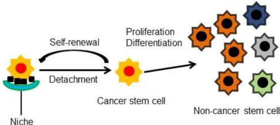
detachment of CSCs from their niche leads to their asymmetric or symmetric division. ... 38
Chapter 3 Figures
Figure 1. Representatives images of ALDH1 staining in a) non-malignant (x20), b) DCIS
(x40) and c) IDC samples (x20) and CD44 staining in d) non-malignant (x20), e) DCIS (x40) and f) IDC samples (x40) ... 94
Chapter 4 Figures
Figure 1. Identification of hyaluronan receptor(CD44)+aldehyde dehydrogenase-1(ALDH1)+Ki-67− breast cells. Positive controls for CD44 (A), ALDH1 (B) and Ki-67 (C) (×20). D: Illustrates a breast tissue sample without ALDH1 expression; black arrow points to one CD44+ALDH1−Ki-67+ breast cell (×40). E: A breast tissue sample without CD44 expression; black arrow points to one CD44−ALDH1+KI-67+ breast cell (×40). F: A breast tissue sample without CD44 and ALDH1 expression (×40). G, H: Breast tissue samples containing CD44+ALDH1+Ki-67− breast cells (black arrows); white arrow in (G) points to one CD44+ALDH1+Ki-67+ breast cell (×40)... 103
Figure 2. Representative images of the triple immunohistochemistry for hyaluronan
receptor (CD44), aldehyde dehydrogenase-1 (ALDH1) and Ki-67 in breast tissue sections. Normal (A) and non-malignant (Fibroadenoma) (B) breast tissue section; black arrows points to CD44+ALDH1+Ki-67− breast cells (×40).C Overview of a ductal carcinoma in situ (C) and an invasive ductal carcinoma (E) breast tissue (×20). Insets (D and F) show the same tissues at higher magnification (×40). D: Examples of CD44+ALDH1+Ki-67− tumor cells (black arrows); the white arrow demonstrates an example of one cell without ALDH1 expression. F: Exhibits examples of CD44+ALDH1+Ki-67− invasive tumor cells (black arrows); the white arrow demonstrates an example of one CD44+ALDH1+Ki-67+ cell….104
Figure 3. Pools of CD44+ALDH1+Ki-67− breast cells (dotted black circular boxes) in ductal carcinoma in situ (A) and invasive ductal carcinoma (B) breast tissues respectively (×40). ... 105
Figure 4. Univariate analysis of overall survival according to hyaluronan
Figure 1. Gating strategy for the identification of CD44+/CD24-/Ck+/CD45- breast cells: (A), all cells were gated on a forward scatter (FSC) versus side scatter (SSC) dot plot to exclude dead cells and debris (B), epithelial mammary cells were identified and hematopoietic cells were eliminated from the analysis by gating cells on cytokeratin-FITC versus CD45-PercP dot plots (C) and CD44+/CD24-/Ck+/CD45- breast cells were identified by gating cells on CD44-APC versus CD24-PE. Negative control: cells labeled only with the IgG1-FITC isotype control. FMO CD44: Fluorescence-minus-one control lacking the CD44-APC antibody. FMO CD24: Fluorescence-minus-one control lacking the anti-CD24-PE antibody. Sample test: cells labeled with anti-C45-PerCp, anti-Cytokeratin-FITC, anti-CD44-APC and anti-CD24-PE antibodies. Q1: CD44+/CD24-/Ck+/CD45- cells; Q2: CD44+/CD24+/Ck+/CD45- cells; Q3: CD44-/CD24-/Ck+/CD45- cells; Q4: CD44 -/CD24+/Ck+/CD45- cells ... 119
Figure 2. Identification of CD44+/CD24-/Ck+/CD45- breast cells. (A) fibroadenoma; (B) ductal carcinoma in situ (DCIS) and (C) Invasive ductal carcinoma (IDC). Q1: CD44+/CD24-/Ck+/CD45- cells; Q2: CD44+/CD24+/Ck+/CD45- cells; Q3: CD44-/CD24 -/Ck+/CD45- cells; Q4: CD44-/CD24+/Ck+/CD45- cells. ... 123
Figure 3. Representative images of the immunohistochemistry for E-cadherin and
Vimentin in breast tissue sections. (A) and (C) Negative and positive expression of E-Cadherin, respectively. (B) and (D) Negative and positive expression of vimentin, respectively. ... 125
Chapter 6 Figures
Figure 1. Clonal co-existence. Most tumors have their origin in a single mutated cell and
the cells in the resultant clone will have the same founder mutation (black asterisk). During progression various selection pressures (nutritional or immune status, oxygenation, therapy) could result in the emergence of new clones through genetic and epigenetic alteration. ... 143
Chapter 1 Tables
Table 1. Characteristics of molecular subtypes of IBCs. ... 16
Table 2. Tenets of the cancer stem cell and clonal evolution models. ... 32
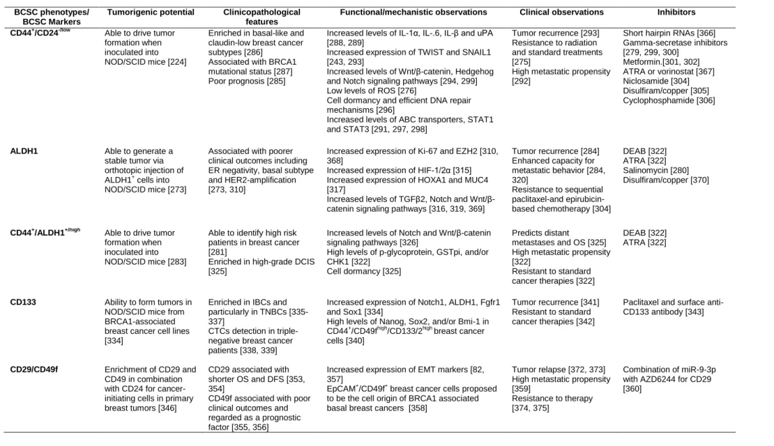
Table 3. Characteristics of the different assessed BCSC phenotypes and BCSC markers. ... 53
Chapter 3 Tables Table I. Clinical characteristics of samples. ... 92
Table II. Invasive ductal carcinoma patients’ hormone receptor status. ... 92
Table III. ALDH1 and CD44 positivity in breast tissues samples. ... 93
Table IV. ALDH1 and CD44 expression in breast tissue samples. ... 93
Table V. ALDH11 and CD44 expression according to patients’ hormone receptor status ... 95
Table VI. ALDH1 and CD44 combined score mean rank expression ... 95
Chapter 4 Tables Table I. Prevalence of hyaluronan receptor (CD44)+ aldehyde dehydrogenase-1 (ALDH1)+ Ki-67– cells in breast tissue samples ... 106
Table II. Mean percentage of hyaluronan receptor (CD44)+ aldehyde dehydrogenase-1 (ALDH1)+ Ki-67– breast cells in breast tissue samples ... 107
Table III. Mean percentage of hyaluronan receptor (CD44)+ aldehyde dehydrogenase-1 (ALDH1)+ Ki-67– breast cells according to ductal carcinoma in situ nuclear grade ... 107
Table IV. Mean percentage of hyaluronan receptor (CD44)+ aldehyde dehydrogenase-1 (ALDH1)+ Ki-67– breast cells according to invasive ductal carcinoma clinicopathological markers of clinical progression and outcome ... 108
epidermal growth factor receptor (HER)-2 status. ... 108
Chapter 5 Tables
Table 1. Mean fluorescence intensities of CD44, CD24 and Cytokeratin expression in
breast tissue samples ... 122
Table 2. Prevalence and mean percentages of CD44+/CD24-/Ck+/CD45- breast cells in breast tissue samples ... 123
Table 3. Mean percentages of CD44+/CD24-/Ck+/CD45- breast cells according to IDC clinicopathological markers of clinical progression. ... 124
Table 4. Mean percentages of CD44+/CD24-/Ck+/CD45- breast cells according to patient’s hormone receptor status and to E-Cadherin and vimentin expression ... 125
Table 5. Characteristics of the Targeted Sequencing applied in the DNA samples of
CD44+/CD24-/Ck+/CD45- breast cells sorted from 6 mastectomies samples ... 126
Table 6. Description of mutations identified in CD44+/CD24-/Ck+/CD45- breast cells of each mastectomy sample ... 127
ABC – ATP-binding cassette AD – Adenosis
ADH – Atypical Ductal Hyperplasia
AKT – v-Akt Murine Thymoma Viral Oncogene Homolog 1 and 2 ALDH1 – ALdehyde DeHydrogenase 1
ALH – Atypical Lobular Hyperplasia APC – Adenomatous Poliposis Coli APC – AlloPhyCocyanin
ASCO – American Society of Clinical Oncology ASR – Age Standardized Rate
ATRA – All-Trans Retinoic Acid BCSC – Breast Cancer Stem Cell BRCA – BReast CAncer genes 1 and 2 BSA – Bovine Serum Albumin
BSC – Breast Stem Cell
CAF – Cancer Associated Fibroblast CCND1 – CyCliN D1
CDKN2A – Cyclin-Dependent Kinase Inhibitor 2A CHK1 – Checkpoint Homolog 1
CHPv2 – Ion Torrent Cancer Hotspot Panel v2 CGH – Comparative Genomic Hybridization CK – Cytokeratin
CMF – Cyclophosphamide, Methotrexate, 5-Fluorouracil CSF1 – Colony Stimulating Factor 1
CSFR1 – Colony Stimulating Factor Receptor 1 CSC – Cancer Stem Cell
CXCL7 – C-X-C chemokine Ligand 7 DAB - DiAminoBenzidine
DCIS – Ductal Carcinoma In Situ DEAB – DiEthylAminoBenzaldehyde DHP – Ductal HyperPlasia
DFS – Disease Free Survival ECM – ExtraCellular Matrix e.g – exampli gratia
EGFR – Epidermal Growth Factor Receptor EMT – Epithelial-Mesenchymal Transition ER – Estrogen Receptor
EU28 – European Union Member States EZH2 – Enhancer of Zeste Homolog 2
FACS – Fluorescence-Activating Cell Sorting FAD – Fibroadenoma
FEA – Flat Epithelial Atypia
FFPE – Formalin-Fixed Paraffin-Embedded FGFR1 – Fibroblast Growth Factor Receptor 1 FITC – Fluorescein IsoThioCyanate
FMO – Fluorescence-Minus-One FSC – Forward SCatter
GSTpi – Glutathione-S-Transferase pi H&E – Hematoxylin and Eosin
HER2 – Epidermal Growth factor receptor 2 HIF-1 and 2 – Hipoxia Inducible Factor 1 and 2 HOXA1 – HOmeoboX A1
HR – Hormone Receptors
HRAS – Harvey Rat SArcoma viral oncogene homolog IBC – Invasive Breast Cancer
IDC – Invasive Ductal Carcinoma
IDC-NOS – Invasive Ductal Carcinoma Not Otherwise Specified ILC – Invasive Lobular Carcinoma
IHC - ImmunoHistoChemistry INDEL – INsertion and DELetion iPSC – induced Pluripotent Stem Cell IL – Interleukin
LCIS – Lobular Carcinoma In Situ LOH – Loss Of Heterozigosity
KRAS – Kirsten RAt Sarcoma viral oncogene homolog mAb – monoclonal Antibody
MAP2K4 – Mitogen-Activated Protein Kinase kinase 4
MAP3K1 – Mitogen-Activated Protein Kinase kinase kinase 1 MET – Mesenchymal-Epithelial Transition
MGA – MicroGlandular Adenosis MNP – Multi-Nucleotide Polymorphism MPS – Massive Parallel Sequencing mTOR – mammalian Target of Rapamycin MUC4 – MUCin 4
NF1 – Neurofibromin 1
NGS – Next Generation Sequencing
NOD/SCID – Non-Obese Diabetic/Severe Combined ImmunoDeficiency NOTCH1 – NOTCH homolog 1
NST – No Special Type
OCT – Optimum Cutting Temperature OS – Overall Survival
PBS – Phosphate Buffered Saline PE – PhycEerythrin
Pgp – P-glycoprotein
PR – Progesterone Receptor
PTEN – Phosphatase and TENsin homolog RB1 – Retinoblastoma 1
RET – RET proto-oncogene ROS – Reactive Oxygen Species SC – Stem Cell
SD – Standard Deviation
SEM – Standard Error of the Mean
SERM – Selective Estrogen Receptors Modulator SKT11 – Serine/Threonine Kinase 11
SMARCB1 - SWI/SNF Related, Matrix Associated, Actin Dependent Regulator Of Chromatin, Subfamily B, Member 1
SNAIL1 – SNAIL Family Zinc Finger 1 SNP – Single-Nucleotide Polymorphism SNV – Single Nucleotide Variant
SSC – Side SCatter
STAT1 – Signal Transducer and Activator of Transcription 1 STAT3 – Signal Transducer and Activator of Transcription 3 TDLU – Terminal Duct Lobular Unit
TGFβ2 – Transforming Growth Factor Beta 2 TMA – Tissue Microarray
TP53 – Tumor Supressor p53
UDH – Usual epithelial Ductal Hyperplasia UN – Unknown
uPA – urokinase-type Plasminogen Activator WHO – World Health Organization
The Cancer stem cell (CSC) model became an attractive concept to explain several poorly understood clinical phenomena due to its inherent theoretical properties. Such properties are based on the molecular features of normal stem cells (SCs). Thus, CSCs are believed to have the ability to renew themselves and to last a lifetime and to be resistant to electromagnetic and chemical insults. This resistance ability allows them to stagnate for long periods of time and consequently, to colonize other parts of the body. With this notion, a search for specific surface and intracellular biomarkers has been ongoing in recent years for the identification, isolation and characterization of CSCs in several cancers, like in breast cancer. In fact, breast cancer stem cells (BCSCs) were initially defined by the presence and absence of the cell-surface proteins CD44 and CD24, respectively. The CD44+/CD24-/low phenotype has been demonstrated to have tumor initiating properties and has been associated with stem cell-like characteristics, enhanced invasive properties, radiation resistance and with distinct genetic profiles suggesting an association with adverse prognosis. However, and due to the high levels of heterogeneity associated with this disease, some breast tumors were shown not to harbor any CD44+/CD24-/low breast cell. As a consequence, additional SC markers like ALDH1 have been reported.
Hence, our first goal in this study was to compare by immunohistochemistry (IHC), two of the most reliable SC markers ALDH1 and CD44 and to correlate their expression in different breast lesions. Moreover, we combined these markers with Ki-67 to evaluate quiescence and to identify, assess its distribution and estimate the mean percentages of CD44+/ALDH1+/Ki-67- cells in non-malignant and malignant lesions.
CD44 and ALDH1 expression was commonly observed in distinct breast lesions and a higher combined expression of these markers was noticed in ductal carcinomas in situ (DCIS) when compared with invasive ductal carcinomas (IDCs). Such result was subsequently strengthened by the enrichment of CD44+/ALDH1+/Ki-67- tumor cells observed in DCIS. Besides that, our results also demonstrated that this phenotype may favor distant metastasis being able of predicting overall survival (OS).
the most studied phenotype in breast cancer. The latter was recently associated with epithelial-mesenchymal transition (EMT) markers and was demonstrated to have several signaling pathways dysregulated. Thus, massive parallel sequencing (MPS) can be regarded as an interesting approach to deepen the molecular characterization of CD44+/CD24-/low cells since it allows the analysis of hundreds of genes in just one population of cells. In fact, standardized Next-Generation Sequencing (NGS) kits are currently available providing reliable sequencing results in routine cancer diagnostics, like the Ion Torrent Cancer Hotspot Panel v2 (CHPv2). Such assay includes 50 genes known to be involved in the pathogenesis of many human cancers.
In this way, our second goal was to characterize CD44+/CD24 -/Cytokeratin(Ck)+/CD45- cells through flow cytometry (FCM) in a cohort containing non-malignant and malignant breast lesions. The CHPv2 assay was used for the identification of somatic mutations in the DNA extracted from isolated CD44+/CD24-/Ck+/CD45- cells. The expression of E-Cadherin and vimentin was also analyzed in the malignant lesions.
The percentage of CD44+/CD24-/Ck+/CD45- cells increased significantly from non-malignant to malignant lesions and was negatively correlated with tumor size. A significant association with vimentin was also observed. From the MPS analysis, the non-malignant lesion harbored only a single-nucleotide polymorphism (SNP). Mutations in the tumor suppressor p53 (TP53), NOTCH homolog 1 (NOTCH1), Phosphatase and tensin homolog (PTEN) and v-akt murine thymoma viral oncogene homolog 1 (AKT1) genes were found in isolated CD44+/CD24 -/Ck+/CD45- cells from DCIS. Additional mutations in the colony-stimulating factor 1 receptor (CSF1R), ret proto-oncogene (RET) and TP53 genes were also identified in IDCs.
In conclusion, CD44+/ALDH1+/Ki-67- tumor cells may have a higher tumorigenic effect in breast cancer than CD44+/CD24-/low tumor cells. Due to its role, ALDH1 can be determinant for the behavior of BCSCs, for their ability to resist to chemotherapeutic agents and their dissemination to other parts of the body, which can be aided by the role of CD44. Additionally, quiescence seems to be crucial for tumor progression, resistance to chemotherapeutic agents and metastatic spread of BCSCs. Further studies to infer about the tumorigenic and
The characterization of CD44+/CD24-/Ck+/CD45- cells supports the existence of a tumor initiation capability from these cells which can be strengthened by the acquisition of an EMT state. All of the mutated genes that were found in this study can play important roles for the development and transformation of CD44+/CD24-/Ck+/CD45- breast cells into a malignant phenotype, for stemness maintenance and activation of the EMT state. Functional analyses are now required to determine the tumorigenic effect of each mutation. Subsequent applications of NGS technologies are also demanded to better understand the malignant progression of breast stem cells (BSCs) and to design an effective mutational profile of these cells.
O modelo das células estaminais cancerosas tornou-se num conceito atractivo para explicar vários fenómenos clínicos pobremente compreendidos devido às suas propriedades teóricas inerentes. Tais propriedades baseiam-se nas características moleculares das células estaminais normais. Deste modo, acredita-se que as células estaminais cancerosas são capazes de se renovarem e de durarem uma vida inteira e de serem resistentes aos insultos químicos e electromagnéticos. Esta capacidade de resistência permite-lhes estagnar por longos períodos de tempo e consequentemente, de colonizarem outras partes do corpo. Com esta noção, tem sido feito nos últimos anos uma procura por biomarcadores de superfície e intracelulares específicos para a identificação, isolamento e caracterização das células estaminais cancerosas em vários tipos de cancros, como no cancro da mama. De facto, as células estaminais cancerosas da mama foram inicialmente definidas pela presença e ausência das proteínas de superfície celular CD44 e CD24, respectivamente. O fenótipo CD44+/CD24-/baixo foi demonstrado ter propriedades de iniciação tumoral e tem sido associado com características típicas das células estaminais, propriedades invasoras aumentadas, resitência à radiação e com distintos perfis genéticos que sugerem uma associação com um prognóstico adverso. No entanto e devido aos elevados níveis de heterogeneidade associados com esta doença, alguns tumores da mama foram demonstrados não conter nenhuma célula CD44+/CD24-/baixo. Consequentemente, outros marcadores de células estaminais foram reportados como o ALDH1.
Deste modo, o nosso primeiro objectivo neste estudo foi comparar, por imunohistoquímica, dois dos marcadores de células estaminais mais fiáveis, o ALDH1 e o CD44 e correlacionar as suas expressões em diferentes lesões da mama. Mais ainda, combinámos estes marcadores com o Ki-67 para avaliar a quiescência e para identificar, determinar as suas distribuições e estimar as percentagens das células CD44+/ALDH1+/Ki-67- em lesões benignas e malignas.
A expressão do CD44 e do ALDH1 foi comummente observada em diferentes lesões da mama e uma elevada expressão combinada destes marcadores foi observada em carcinomas in situ quando comparada com carcinomas invasores. Este resultado foi subsequentemente reforçado pelo
este fenótipo pode favorecer metástases à distância sendo capaz de prever a sobrevida global.
Apesar destes resultados, o fenótipo CD44+/CD24-/baixo continua a ser de longe o fenótipo mais estudado no cancro da mama. Este último foi recentemente associado com marcadores de transição epitélio-mesenquimal e foi demonstrado ter várias vias de sinalização desreguladas. Assim sendo, a sequenciação massiva em paralelo pode ser vista como uma abordagem interessante para aprofundar a caracterização molecular deste fenótipo já que permite a análise de centenas de genes apenas numa população celular. De facto, encontram-se disponíveis kits de sequenciação de última geração fornecendo resultados de sequenciação fidedignos em rotinas de diagnóstico do cancro, como o Ion Torrent Cancer Hotspot Panel v2. Este teste inclui 50 genes conhecidos por estarem envolvidos na patogénese de vários cancros humanos.
Desta forma, o nosso segundo objectivo foi caracterizar células CD44+/CD24-/Ck+/CD45- por citometria de fluxo num grupo contendo lesões benignas e malignas da mama. O teste Ion Torrent Cancer Hotspot Panel v2 foi utilizado para a identificação de mutações somáticas no DNA extraído das células CD44+/CD24-/Ck+/CD45- isoladas. A expressão da E-Caderina e da vimentina foi também analisada nas lesões malignas.
A percentagem das células CD44+/CD24-/Ck+/CD45- aumentou significativamente a partir das lesões benignas para as malignas e foi negativamente correlacionada com o tamanho tumoral. Uma associação significativa com a vimentina foi também observada. A partir da análise da sequenciação massiva em paralelo, a lesão benigna conteve apenas um polimorfismo de nucleótido único. Foram encontradas mutações nos genes TP53, NOTCH1, PTEN e AKT1 nas células CD44+/CD24-/Ck+/CD45- isoladas dos carcinomas in situ. Mutações adicionais nos genes CSFR1, RET e TP53 foram também identificadas nos carcinomas invasores.
Em conclusão, as células tumorais CD44+/ALDH1+/Ki-67- podem ter um maior efeito tumorigénico no cancro da mama do que as células tumorais CD44+/CD24-/baixo. Devido à sua função, o ALDH1 pode ser determinante para o comportamento das células estaminais cancerosas da mama, para a capacidade
outras partes do corpo, a qual pode ser auxiliada pela função do CD44. Mais ainda, o estado de quiescência parece ser crucial para a progressão tumoral, para a resistência aos agentes quimioterapêuticos e pela propagação metastática das células estaminais cancerosas da mama. Estudos posteriores têm ainda de serem descritos para inferir sobre a capacidade tumorigénica e metastástica das células tumorais CD44+/ALDH1+/alto combinadas com o seu estado de quiescência.
A caracterização das células CD44+/CD24-/Ck+/CD45- suporta a existência de uma capacidade de iniciação tumoral por parte destas células a qual pode ser fortalecida pela aquisição de um estado de transição epitélio-mesenquimal. Todos os genes mutados que foram detectados neste estudo podem desempenhar funções cruciais para o desenvolvimento e transformação das células de mama CD44+/CD24-/Ck+/CD45- para um fenótipo maligno, para a manutenção da estaminalidade e activação do estado de transição epitélio-mesenquimal. Análises funcionais são agora necessárias para determinar os efeitos tumorais de cada mutação. Aplicações subsequentes das tecnologias de sequenciação de última geração são também necessárias para melhor compreender a progressão maligna das células estaminais da mama e para traçar um perfil mutacional efectivo destas células.
Chapter 1
1. Breast Anatomy
The mammary system is very different from other organ systems. From birth through puberty, pregnancy, and lactation, the breast is affected by several dramatic changes in size, shape and function [1].
The adult breast lies on the anterior chest wall between the second and sixth ribs, and from the sternal edge medially to the mid-axillary line laterally. Breast tissue also projects into the axilla as the axillary tail of Spence [2].
Anatomically, the breast lies in a space within the superficial fascia, although microscopic extensions of glandular parenchyma occasionally traverse these boundaries. Superiorly this layer is continuous with the cervical fascia and inferiorly with the superficial abdominal fascia of Cooper. Extensions of fibrous strands from the dermis into the breast form the suspensory ligaments of Cooper, which attach the skin and nipple to the breast [3].
An interesting work regarding the anatomy of the nipple suggested the existence of more than 20 lobes that are defined by the major lactiferous ducts that open on the nipple [4]. A lobe resembles to a tree, whose trunk, branches and leaves are hollow. These arborizing networks transport milk from the lobules to the nipple and are called the terminal portions of the duct system. One lobule is formed by multiple blunt-ending ducts in a cluster like the fingers of a glove. These fingers form the glandular acini of the lobule being surrounded by specialized connective tissue, histologically different from the stromal connective tissue existent in the rest of the breast. The lobule is then formed together by the glandular acini and specialized connective tissue. A terminal duct and its lobule are collectively called the terminal duct lobular unit (TDLU) which are found as immediate branches of the major ducts [1, 2] (Figure 1).
The lobular acini are invested by a loose, fibrovascular intralobular stroma with different numbers of lymphocytes, plasma cells, macrophages, and mast cells. This specialized intralobular stroma is sharply delimited from the surrounding denser, more highly colagenized, paucicellular interlobular stroma and stromal adipose tissue [5].
The number of acini per lobule and the size of mammary lobules have been found to be extremely variable. During the menstrual cycle, the morphological changes displayed by the lobules are seen in both epithelial and stromal
components [6-9]. Although the high variability of such changes that exist among lobules in the same breast or even among immediately adjacent lobules, a dominant morphologic pattern is typically current in each phase [5].
The nipple areola complex is a circular area of skin that exhibits increased pigmentation and contains numerous sensory nerve endings. The nipple is placed centrally and is prominent above the surrounding areola. Near the periphery of the areola are elevations (tubercles of Montgomery) formed by the openings of modified sebaceous gland, whose secretion protect the nipple during breastfeeding [5, 10]. A keratinizing, stratified squamous epithelium covers both the nipple and the areola and extends for a short distance into the terminal portions of the lactiferous ducts. During lactation, epithelial cells in both the terminal duct and lobule endure secretory changes. Thus, the terminal ducts are responsible for both secretion and transport of the secretions to the extra-lobular portion of the ductal system [11].
Figure 1. Schematic diagram of the breast. Adapted from [1].
Cells that form the duct epithelium are of two types: columnar cells, lining the lumen having cytoplasm endowed with abundant organelles involved in secretion and expressing a variety of low-molecular weight CKs including CKs 7, 8, 18 and 19 [12-16], and myoepithelial cells being distributed in a discontinuous manner in the epithelium [17]. Myoepithelial cells lay between the epithelial layer
and the basal lamina forming a network of slender processes investing the overlying epithelial cells. These cells range in appearance from barely visible, flattened cells with compressed nuclei to prominent epithelioid cells with abundant clear cytoplasm. In some cases, the myoepithelial cells have a myoid appearance featuring a spindle cell shape and dense, eosinophilic cytoplasm, reminiscent of smooth muscle cells. Immunohistochemical stains for several markers are used to discriminate these cells, including S-100 protein, actins, calponin, smooth muscle myosin heavy chain, p63, and CD10, among others [18-21]. A third cell type in normal breast tissue was also recently proposed. These cells were seen to be dispersed irregularly throughout the ductal lobular system expressing the basal cytokeratin CK5. Due to their ability to differentiate into both glandular epithelial and myoepithelial cells, such cells are believed to be progenitor cells [15].
The epithelial-stromal junction comprises an epithelial-mesenchymal layer within the duct, the basal lamina, and a surrounding zone of delimiting fibroblasts and capillaries. Elastic tissue fibers are variably present around normal ducts but in the premenopausal breast, these fibers tend to be less pronounced. Besides elastic fibers, the normal periductal stroma contains a sparse scattering of lymphocytes, plasma cells, mast cells, and histiocytes. Ochrocytes are periductal histiocytes with cytoplasmic accumulation of lipofuscin pigment. Such pigmented cells tend to be more frequent in the post-menopausal breast and are associated with inflammatory or proliferative conditions [3].
Considering that the mature breast is subjected to deep changes associated with the menstrual cycle, pregnancy, lactation and menopause, the normal microscopic anatomy of the lobules is not constant. Furthermore, and regardless of the physiologic conditions, there are variations in the functional state of individual lobules suggesting that these individual lobules or lobules in regions of the breast may have intrinsic differences in response to hormonal stimuli [22]. Due to this intrinsic dynamic ability of breast cells to be continuously influenced and remodeled, it is believed that they can be susceptible to carcinogenesis. In fact, loss of heterozygosity (LOH) is one of the genetic alterations that have been detected in histologically normal-appearing lobular epithelium [23], but other genetic changes have also been found in epithelium and myoepithelial cells. Although the frequency of these alterations has not been established yet,
increasing existing data suggest that they are detected more often in histologically normal lobules from patients with carcinoma than in breasts without carcinoma [3].
2. Breast Cancer
2.1. Epidemiology
Breast cancer is a global health problem and one of the major causes of female morbidity and mortality [24-26]. Its incidence, prevalence and the economic burden it imposes on national health services make it a major public health including both the developed and developing countries [27].
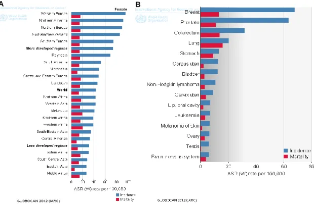
This disease affects women aged less than 45 years being more prevalent in the 45-65 years age group. Currently, breast cancer continues to be the leading cause of female death from cancer worldwide. In 2012, ~1.67 million new breast cancer diagnoses were made throughout the world and 522000 women died of breast cancer. In the same year, the incidence in Europe Union Member States was ~362000 new cases with 92000 deaths, accounting for a mean incidence rate of 66.5 and a mean mortality rate of 16.0 per 100000 women (world age-standardized rate, ASR-W) [28]. The differences in the incidence, mortality and survival rates are due to different risk factors, availability of organized screening programs and access to effective treatments [24]. Although the mortality rate is higher in less developed countries [29], the incidence of breast cancer in Western and Northern Europe is between the highest in the world [28]. For this reason, breast cancer prevention continues to be a major public health goal (Figure 2A).
Portugal applied its first region-based screening program in 1990 but nationwide screening was only attained in 2005. The Health Ministry aims at 60% coverage by the end of 2016 [30] implementing a program that offers digital mammography to women aged 45-69 years. Nonetheless, ~6000 new breast cancer cases are diagnosed annually and ~1500 women die due to this disease (Figure 2B). Indeed, the incidence rate of 67.6 cases per 100000 people is currently higher when compared with the mean established for European Union countries. Even if the mortality rates (13.1 cases per 100000 people) are still lower than the European ratio, early diagnosis of abnormalities is extremely important to better understand the risk of breast cancer progression [28].
Figure 2. A. Breast cancer estimated age-standardized incidence and mortality rates in the World
per 100000. B. Incidence and mortality rates of the most common types of cancer for both sexes in Portugal – estimates presented for the year 2012. Adapted from [28].
2.2. Risk Factors
In the past several years, significant improvements have occurred in our understanding of the causes and prevention of breast cancer. Factors like age, family history of breast cancer and experiences of reproductive life have long been known to be associated with breast cancer risk [31]. Age is by far the strongest risk factor for breast cancer in women. The incidence of this disease increases steeply with the greatest rate increment seen in postmenopausal women, where the risk doubles with each decade of life up to age 80. Race and ethnicity also constitute a marked risk factor, being highest in white women, following by black and Hispanic women [32]. Such dissimilarities may be explained by multifactorial inherited factors, genetic variations in the biology of the tumors or even cultural differences [33, 34]. Reproductive events, such as menarche, pregnancies and live births, lactation and menopause, all mark substantial changes that can influence breast cancer risk. Incidence may be affected by the effects of physical alterations due to reproduction and long-term modifications in hormonal exposures [32]. Increasing age at menarche is associated with decreasing breast cancer
A B
incidence; the risk of breast cancer decreases 5% for each year increment in age at menarche [35]. Concomitantly, increasing the reproductive span with a late age at menopause increases the risk of developing breast cancer due to a greater lifetime exposure to circulating hormones [36]. Parity, especially at an early age is associated with a decreased risk of breast cancer. Women whose first birth occurred at age 35 years or older have a higher risk of developing the disease when compared to women under 18 at the time of first pregnancy [35]. In parous women, lactation further decreases the risk. Nevertheless, the overall reduction in risk varies considerably within the population studied [37].
Recently, new risk factors have emerged including low physical activity, obesity, alcohol intake, and exogenous hormone use. In fact, some of these new factors seem to be related to perturbations in circulating estrogens, which are believed to be the major cause of breast cancer [31]. Regarding the exogenous hormones, studies concerning the use of oral contraceptives and hormonal therapy for the menopause are still inconclusive. However, the last is associated with increased risk of breast cancer, especially when comparing its use during short and long periods of time [38, 39].
Women with a family history of breast cancer, particularly in a first-degree relative, have approximately double the risk of developing the disease compared to women without such a history [40]. Studies of high risk families provided evidence of an autosomal dominant inheritance of breast cancer [41, 42]. Gene linkage studies and cloning [43, 44] identified two genes, BRCA1 and BRCA2 that appear to be associated with the majority of inherited breast cancers accounting for 2-5% of all breast cancers [45]. Lifetime risk of diseases associated with BRCA1 and BRCA2 mutations ranges from 20 to 80% [46], since that these genes are tumor suppressor genes with several important cell functions like transcription, regulation of cell cycle checkpoints, genomic stability and DNA repair [47-49].
Benign breast lesions can also influence risk. Overall, women with benign breast lesions without hyperplasia have a 1.5-fold increased risk of breast cancer compared to women without any benign breast lesions. The risk between women with hyperplasia varies by whether or not atypia is present: women having atypical hyperplasia and hyperplasia without atypia have a 2.6-fold and a 1.8-fold increased risk, respectively. Women harboring fibroadenomas have an independent increased risk for breast cancer [50]. Benign breast disease may be
sensitive to the same risk factor as for invasive cancer. Hence, benign lesions should be considered as a first step in breast cancer progression [31].
2.3. Prognostic Markers and Therapeutic Strategies in Breast Cancer Breast cancer progression evolves through a series of intermediate processes, beginning with ductal hyperproliferation, followed by subsequent evolution to carcinoma in situ, invasive carcinoma, and finally into metastatic disease [51]. Considering the marked heterogeneity of breast cancer, the identification of markers that can predict tumor behavior is particularly important. In fact, these markers can be seen as a useful tool for the clinical management of cancer patients, assisting in diagnostic procedures, staging, evaluation of therapeutic response, detection of recurrence, distant metastasis and prognosis [52].
Currently, the standard for assessing the prognosis of patients with newly diagnosed stage I-III breast cancer is to use an integrated prognostic model that includes information about tumor size, tumor grade, tumor proliferation index, lymph node involvement, distant metastasis, estrogen- and progesterone-receptors (ER and PR) and epidermal growth factor receptor type 2 (HER2) status. Such combination and integration converge into a single risk prediction score for prognostic stratification [53-55].
One of the most important prognostic factors is the gross measurement of tumor size. Several studies have shown that survival time decreases gradually with increasing tumor size. Patients with stage I tumors smaller than 1 cm have a 20-year disease-free survival (DFS) rate of 88%, whereas patients with tumors 1-2 cm have a 20-year DFS rate of only 68%. The incidence of lymph node metastases is 10% for patients with tumors having 1 cm or less, compared with 75-80% for patients with tumors larger than 10 cm [56].
Another important prognostic factor for breast cancer is the presence or absence of axillary lymph node metastases and the number of axillary lymph nodes with metastatic tumor. Overall survival time, recurrence and time to recurrence, metastases and treatment failure, all significantly correlate with the number of positive axillary lymph nodes [57-59]. The 10-year DFS rate is around
70-80% when axillary lymph nodes are negative for metastatic tumor. With each positive node, the OS rate gradually decreases [60].
In patients with invasive breast cancer (IBC), histologic grading has been constantly shown to predict DFS and OS and is recommended for all invasive carcinomas to provide an estimation of differentiation. A histologic grading system endorsed by World Health Organization (WHO) and based on criteria established by Bloom and Richardson and Eslton and Ellis evaluates three parameters: tubule formation, degree of nuclear pleomorphism and number of mitotic figures identified on histologic sections. Elston and Ellis found that patients with grade 1 tumors had a significantly better OS than those with grade 2 or grade 3 tumors [61].
In respect to breast cancer treatment, all cancer therapy can be divided into two basic types: local and systemic therapy. Local therapy refers to those treatments that control disease in the specific area of the tumor. Surgery to remove the breast mass is one of the first treatment methods but the extent of surgery depends on the features of a specific tumor. Even if a mastectomy may be mandatory for some patients, in several cases, only a small portion of the breast at the tumor site is removed. Surgery may be followed by radiotherapy in order to kill any cancer cells that may remain after surgery. In fact, clinical studies have shown that for the majority of breast cancers, removal of the breast lump followed by radiotherapy is as effective as surgery to remove the whole breast [62].
After the surgery and before radiation begins, it is important to discuss the role of systemic adjuvant therapy that includes endocrine therapy, chemotherapy, and/or targeted therapy. After examining the risks versus the benefits of the therapy, the use of such therapy is recommended or not. The benefit of adjuvant or endocrine therapy is in proportion to the risk of breast cancer recurrence; however, and despite the existence of prognostic factors, no parameter is completely predictive of recurrence [63, 64]. The majority of the clinicians agree that many women with node-negative breast cancer should receive adjuvant chemotherapy, especially those with larger tumors. Women with tumors smaller than 1 cm, a low-grade malignancy, positive ER/PRs, negative HER2 status and a low proliferative rate have the lowest risk of recurrence. Conversely, women with tumors larger than 2 cm, a high-grade malignancy, negative ER/PRs, positive HER2 status, triple negative receptor status (ER, PR and HER2 negative) and a high rate of proliferation are at highest risk for tumor recurrence [63]. Currently,
ER, PR and HER2 scores have been used along with the Ki-67 proliferation marker as predictive factors for identifying a high-risk phenotype and also for the selection of the most efficient therapies [65].
- Ki-67
Ki-67 is a nuclear protein expressed in all phases of the cell cycle other than the G0 phase and is used to assess proliferation in breast cancer. A high percentage of Ki-67-positive cells are associated with poor prognosis and the optimal cutoff between high and low risk of developing distant metastasis has been suggested to be 14% of positive cells [66].
The analysis of the value of Ki-67 as a predictive and prognostic tool is useful for neoadjuvant setting. Some studies examining complete pathological response have identified a high Ki-67 proliferation rate as a predictive factor for a higher rate of complete pathological response. It was found however, that patients in whom progression occurred had a higher proliferation rate than those who responded to neoadjuvant chemotherapy. These results suggest a nonlinear effect of Ki-67 on treatment response and probably on prognosis as well [67, 68]. Nonetheless, proliferation as assessed by the percentage of Ki-67 positive cells combined with ER, PR and HER2 scores has a strong prognostic power especially in patients with ER-positive breast cancers.
- Hormonal Receptors
The clinical significance of ER and PR status in IBC has been well recognized for decades and the measurement of ER and PR levels became a standard practice when evaluating patients with primary breast cancer [69-74].
ERs and PRs belong to a super family of nuclear hormone receptors that function as transcription factors when they are bound to their respective ligands [75, 76]. These receptor proteins are generally divided into six functional regions. The ligand-binding now called ERα contains an amino-terminal, hormone-independent activation function (AF-1) domain, a centrally located DNA-binding domain followed by a hinge region, the hormone-binding domain with its integral hormone-dependent region (AF-2) and a carboxyl-terminal F domain. Binding of hormone to ERα triggers the activation of the receptor leading to the
disassociation of receptor coregulatory repressor proteins and histone deacetylase complexes. Conversely, the recruitment of several coactivator protein complexes with histone acetylase activity acts in coordination to induce the transcription of estrogen-responsive genes. ERβ contains a similar structure, encoding however a smaller AF-1 domain as well as the PR-A and PR-B isoforms which also differ predominantly in their amino-terminal regions [77]. The detailed structure-function studies of AF-1 and AF-2 regions were fundamental insights into a molecular basis for hormone receptors due to the demonstration of different activities contained within these two regions [78]. It was shown that in a cell where AF-2 domain is dominant, antiestrogens such as tamoxifen act as pure antagonists. However, in a cell where AF-1 domain is dominant, tamoxifen behave as a partial agonist stimulating ERα transcriptional activity and proliferation [79, 80]. This modulation is the reason why different antiestrogens like tamoxifen or raloxifene are also termed selective ER modulators or SERMs: they can function as antagonists in the breast, but also as ERα agonists in other tissues, like the bone and cardiovascular systems [78].
Although the majority of studies suggest that like ERα, ERβ protein expression is also associated with a better outcome in untreated patients with breast cancer, the prognostic value of ERβ still need to be depicted. When tamoxifen binds to ERα, the estrogen-stimulated growth of tumor cells is inhibited leading to a significant reduction of cancer recurrences and to an increment in survival in patients with ERα positive IBCs of all stages [81]. More recently, tamoxifen has also been shown to reduce subsequent breast cancer in patients with ERα positive DCIS [82] and in patients who are cancer-free but at high risk for developing breast cancer [83]. ERα loss as a significant mechanism of acquired hormonal resistance is still an important question to be solved because even if ERα is reduced in tamoxifen-resistant tumors, the development of hormonal resistance is more frequently associated with the maintenance of ERα at the time of progression [84]. For this reason, the clinical use of other endocrine agents with distinct mechanisms of action, such as the steroidal antagonist faslodex has emerged. Currently, about two-thirds of tamoxifen-resistant breast cancer patients respond to second-line therapy with faslodex, exhibiting no ERα agonist activity in cells [85]. Additionally, a third generation of anastrozole and letrozole was shown to be effective in postmenopausal tamoxifen-resistant patients [86]. Thus,
tamoxifen cannot be assumed as a global hormonal resistance antagonist, suggesting a multiple mechanism of hormonal resistance and involving a combined pool of SERMs.
- HER2
The HER2 gene (also known as c-ErbB2) encodes a 185-kDa transmembrane tyrosine kinase receptor [87] belonging to the epidermal growth factor receptor (EGFR) family. Such family also includes HER1, HER3, and HER4. These receptors are sensitive to signals that stimulate cell growth being expressed in a variety of tissues of epithelial, mesenchymal, and neuronal origin [87]. The homo- and heterodimerization with one another allows the auto-phosphorylation of tyrosine molecules, and consequently, the propagation of intracellular signaling through the mitogen-activated protein kinases (HER1/HER2 heterodimers) and phosphoinositide 3-kinase (PI3K) pathways (HER2/HER3 heterodimers). HER2 is unique due to its ability to dimerize with any of the other three receptors and besides that, it does not require ligand binding for activation [88].
This oncogene is amplified in 20 to 30% of breast cancers and since that its overexpression is associated with an aggressive phenotype of tumor cells, resistance to antihormonal and cytotoxic therapies and poor OS, it is considered as a marker of poor prognosis [52]. Currently, the humanized monoclonal antibody trastuzumab targets the extracellular domains of HER2 being thus indicated for the treatment of HER2-positive breast cancers. Several studies demonstrated the high efficacy of trastuzumab through its significant inhibitory effect on tumor growth and chemotherapy sensitizer [89]. Such anti-tumor effect is believed to be conferred by the inhibition of receptor-receptor interaction, receptor decreasing by endocytosis, blockade of extracellular domain cleavage of the receptor and activation of antibody-dependent cellular cytotoxicity [90, 91]. Besides trastuzumab, other therapeutic strategies have been designed to target HER2 protein such as lapatinib, a tyrosine kinase inhibitor which showed enhanced efficacy after failure of trastuzumab therapy [92].
2.4. Molecular Subtyping of Breast Cancer
Although heterogeneity in breast cancer has long been recognized, recent microarray-based gene expression profiling analysis [93, 94] brought this concept to the vanguard of breast cancer research.
Studies published by Perou et al and Sorlie et al have demonstrated that breast carcinomas can be classified according to the similarity between their transcriptomic profiles. For instance, the authors developed an “intrinsic gene set” (i.e genetic differences between samples from different patients and samples from the same patient) and through hierarchical clustering analysis, tumors were classified into four main groups: (A) luminal, comprising ER-positive tumors and expressing genes belonging to the ER pathway with profiles corresponding to “normal luminal epithelial cells”; (B) basal-like cancers, which are hormone receptor negative/low tumors expressing genes that are usually expressed by basal/myoepithelial cells; (C) HER2 tumors with HER2 overexpression and genes belonging to the HER2 amplicon, and (D) normal breast-like group, which clusters together with normal breast samples and fibroadenomas [95, 96].
However, the molecular taxonomy of breast cancer is constantly being modified since that in each publication different intrinsic gene lists are demonstrated. As a consequence, slightly different subtypes have emerged like an interferon-rich group [97], the molecular apocrine subtype [98, 99] and the claudin-low subtype [100] (table 1).
Table 1. Characteristics of molecular subtypes of IBCs. Adapted from [101].
Molecular subtype ER PR HER2 Basal
markers
Proliferation Histology
Luminal A +++ +++ - - Low
Low grade ductal, cribiform, tubular, classic lobular
Luminal B + ± ± ± High Ductal , micropapillary
HER2-enriched +/- +/- + ± High High-grade ductal
Basal-like - - - + High High-grade ductal,
metaplastic, medullary, adenoid-cystic
Molecular apocrine
- - ± ± High Apocrine, ductal,
lobular
Claudin-low - - - ± High High-grade ductal,
metaplastic +++ High expression, + moderate expression, ± variable expression, - no expression
ER-positive expression markedly defines the luminal molecular subtype, which is also characterized by the relatively high expression of several genes expressed by breast luminal cells like CK8/18, GATA3 and estrogen-regulated genes. Luminal tumors are divided into luminal A and luminal B tumors due to their different ERα expression levels, proliferation rates assessed by Ki-67 and clinical outcomes [102]. However, patients with luminal tumors have better outcomes and a broader range of treatment options than patients with other types of breast cancer [103].
The overexpression of the HER2 protein on the cell membranes is due to the genomic amplification of the 17q22.24 region, which constitute the HER2-enriched molecular subtype. HER2-enriched tumors can be ER-positive and/or PR-positive and have a more aggressive phenotype than HER2 normal tumors. Still, overexpression of HER2 makes the majority of these tumors highly responsive to HER2 inhibitors such as trastuzumab [104]. Intrinsically, ER-positive, HER2-enriched tumors have a poorer response to systemic therapy than ER-negative, HER2-enriched tumors [103].
Basal-like breast cancers have the worst clinical outcome when compared with all others breast cancer groups. They are characterized by an aggressive phenotype with high histologic grade, high proliferation levels pushing borders of
invasion and large areas of tumor necrosis, representing 15-20% of breast cancers [105]. Such tumors express high levels of basal-markers like CKs 5, 14 and 17 but do not express ERα, PR or HER2. Consequently, anti-hormonal approaches for these tumors are unfeasible and also, the poor genetic knowledge regarding the transformation and progression of this tumor subtype makes it difficult to target with current therapies [106].
Accounting for 8-14% of breast cancers, the molecular apocrine subgroup was shown to be distinct from luminal and basal-like breast cancers. It is characterized by increase androgen signaling and is frequently associated with apocrine histological features. This molecular phenotype is thought to be associated with poor long-term survival and has specific surrogate immunohistochemical markers, including androgen receptor- and gamma-glutamyltransferase 1 [103, 107].
Claudin-low tumors account for approximately 5% of IBCs and are characterized by low or absent expression of EMT markers, presence of immune response genes and cancer stem cell-like features [108]. Clinically, the majority of claudin-low tumors carry poor prognosis and are ER-negative, PR-negative and HER2-negative (triple-negative) tumors that have a higher frequency displaying metaplastic and medullary differentiation [109].
It should be noted however, that this molecular taxonomy, as referred above, remains a working model in progress and not a definitive classification system, as additional molecular subtypes have been and may be identified in the future.
![Figure 1. Schematic diagram of the breast. Adapted from [1].](https://thumb-eu.123doks.com/thumbv2/123dok_br/15145386.1012286/36.892.219.720.552.927/figure-schematic-diagram-breast-adapted.webp)
![Table 1. Characteristics of molecular subtypes of IBCs. Adapted from [101].](https://thumb-eu.123doks.com/thumbv2/123dok_br/15145386.1012286/48.892.131.791.141.519/table-characteristics-molecular-subtypes-ibcs-adapted.webp)


![Table 2. Tenets of the clonal evolution and cancer stem cell models. Adapted from [218]](https://thumb-eu.123doks.com/thumbv2/123dok_br/15145386.1012286/64.892.110.785.149.685/table-tenets-clonal-evolution-cancer-stem-models-adapted.webp)