Universidade de Lisboa
Faculdade de Ciências
Departamento de Biologia Animal
Natural Genetic Variation of Male Sperm Size in the
Nematode Caenorhabditis elegans
Nuno Filipe da Silva Soares
Dissertação
Mestrado em Biologia Evolutiva e do Desenvolvimento
2012
Universidade de Lisboa
Faculdade de Ciências
Departamento de Biologia Animal
Natural Genetic Variation of Male Sperm Size in the Nematode
Caenorhabditis elegans
Nuno Filipe da Silva Soares
Mestrado em Biologia Evolutiva e do Desenvolvimento
Orientador externo:
Christian Braendle, PhD, Institut de Biologie Valrose, CNRS & University of Nice
Orientador interno:
Élio Sucena, PhD, Faculdade de Ciências da Universidade de Lisboa
I
Acknowledgments
First,I would like to express my sincere gratitude to Christian Braendle for accepting to be my supervisor and for its guidance during my project. Also a special thanks to all the team in Nice, in particular to Nicolas Callemeyn, for the help with R and GWAS, and to Anne Vielle, for her patiente and help. To Erik Andersen I kindly thank for the strains provided for my project and for the help with GWAS.
I also would like to thank the Erasmus Placement program for the opportunity it gave me of doing my project abroad.
My sincere gratitude to Nicole for helping me with the life in Nice when I needed.
Ao Professor Élio quero agradecer pelos conselhos e pela disponibilidade enquanto meu orientador interno.
Um sincero e sentido obrigado a todos os meus amigos que fizeram-me rir no momento certo, deram-me alento quando precisava e ajudaram-me a ultrapassar este ano. Um obrigado aos momentos fantásticos que partilhei e às coisas que me ensinaram. É um obrigado para amigos da FCUL, para amigos de histórias antigas, para amigos de Nice. Não nomeio ninguém, mas acho que todos a quem agradeço sabem quem são e como foram importantes para mim. Esta tese é dedicada a todos vocês.
Um muito especial obrigado à minha família que sempre me apoiou e que tornou esta tese possível. Deram-me a força necessária para continuar e nunca me deixaram sentir sozinho, mesmo estando longe. Um obrigado especial para minha mãe pelo apoio, para o meu pai pelas conversas e para o meu irmão pela companhia. Esta tese também é dedicada a vocês.
Por último, um agradecimento especial à Anna, pela sua companhia, pela sua força e pela sua paciência. Um grande obrigado pelas suas surpresas, pelos jantares e pelos livros. Também lhe dedico esta tese, pois ela também a tornou possível.
Obrigado a todos/ thank you all, Nuno
II
Abbreviations
CSA Cross-Sectional Area
DIC Differential Interference Contrast FB-MO Fibrous Body-Membranous Organelle GWAS Genome-Wide Association Study MSP Major Sperm Protein
NGM Nematode Growth Medium QTL Quantitative Trait Loci
III
Abstract
Sperm competition is a major evolutionary force driving diversity in sperm morphology. In Caenorhabditis elegans, an androdioecious nematode, sperm competition may occur between sperm of different males competing for fertilization of a hermaphrodite or between hermaphrodite self-sperm and male sperm. Upon outcrossing, male sperm consistently outcompetes hermaphrodite sperm, and the main factor increasing sperm competitiveness is sperm size, which is always larger in males. Previously observed variation in male sperm size in C. elegans thus potentially reflect adaptive responses to sperm competition. However, the extent of natural genetic variation in male sperm size has not been quantitatively studied and its underlying proximate causes remain elusive.
Here we quantitatively assessed natural genetic variation of male sperm size in C. elegans using a world-wide set of 97 wild isolates, covering a maximum of reported genetic variation. The aim of the project was to initiate the genetic characterization of male sperm size using Genome-Wide Association Study (GWAS) and build the basis for future Quantitative Trait Loci (QTL) mapping.
This work provides the most extensive analysis of natural genetic variation in C. elegans male sperm size up to date. We report significant heritable variation in male sperm size; however, we did not detect any significant candidate genomic regions using GWAS. Sperm cell area ranging from approximately 16 µm2 to 27 µm2; with most isolates showing an intermediate sperm size of approximately 22 µm2. The analysis detected two isolates as clear outliers: JU561 and LSJ1, which show a much smaller sperm size compared to the rest of isolates. We discuss the significance of these results and outline future approaches. In addition, we report pilot experiments focusing on the effects of male age and mating on sperm size.
Keywords: Sperm Evolution; Sperm Competition; Sperm Size; Natural Genetic Variation;
IV
Resumo
A reprodução sexual é um tipo de reprodução comum que envolve a fusão de dois gâmetas normalmente referidos como oócitos, os femininos, e espermatozóides, os masculinos. Os espermatozóides são, por norma, células móveis com a única função de encontrar e fertilizar os maiores e imóveis oócitos femininos para originar o zigoto. No entanto, os espermatozóides apresentam uma grande diversidade morfológica em diversos taxa. Esta diversidade morfológica é atribuída principalmente a três fatores: adaptação a novos ambientes; interações macho-fêmea e macho-macho; fenómenos de especiação. Devido à dificuldade de acesso a fêmeas e/ou à quantidade limitante de oócitos disponíveis para fertilização, é usual existir competição entre machos. Em casos de poliandria, ou em sistemas semelhantes, onde a mesma fêmea acasala com mais do que um macho, a competição masculina desce para um nível de competição espermática assumindo-se como um fator de seleção pós-copulatória, tendo um impacto no fitness masculino.
A competição espermática assume um papel preponderante na evolução da morfologia dos espermatozóides. Estes ao competirem dentro do trato reprodutor feminino podem ter várias características a serem selecionadas para um melhor desempenho. O aumento do número/densidade de espermatozóides para aumentar as hipóteses de fertilização, conhecido como “fair raffle”, é uma das características mais selecionadas. Contudo, características morfológicas, como o tamanho e a mobilidade, podem melhorar a qualidade dos espermatozóides e aumentar a capacidade destes de fertilização face a outros competidores. A competição espermática, apesar de ser um fenómeno associado a machos, pode-se encontrar também em espécies hermafroditas ou androdióicas, sempre que promiscuidade dos recetores permita o encontro de esperma de diferentes dadores.
O modelo utilizado neste projecto foi o Caenorhabditis elegans, um nemátode bastante utilizado em ciências biológicas como organismo modelo. Foi o primeiro animal com o genoma sequenciado e possui diversas técnicas e ferramentas moleculares disponíveis e características favoráveis à sua manutenção e manipulação. C. elegans possui um sistema sexual androdióico sendo as suas populações constituídas por hermafroditas suficientes (XX) e machos (XO) e o seu principal método de reprodução consiste na auto-fecundação. Os machos representam uma percentagem mínima da população, cerca de 1%, e surgem espontaneamente devido a erros de disjunção dos cromossomas sexuais ou resultam do cruzamento entre machos e hermafroditas (50% da descendência do macho). Os hermafroditas produzem espermatozóides num período limitado durante a última fase larvar e quando se
V tornam adultos mudam para oogénese irreversivelmente. A competição espermática em C. elegans existe assim maioritariamente entre hermafroditas, quando fecundados, e machos.
Em C. elegans, tanto os espermatozóides masculinos como os hermafroditas são semelhantes: ambos quando imaturos são esféricos; quando ativados desenvolvem características amebóides deslocando-se através dum pseudópode; ambos fertilizam os oócitos quando estes atravessam a espermateca a caminho do útero. Contudo, os espermatozóides masculinos são maiores e mais competitivos que os dos hermafroditas. Este maior tamanho dos espermatozóides confere uma maior capacidade de mobilidade e aderência bem como capacidade de deslocar espermatozóides mais pequenos. Durante a passagem dos oócitos pela espermateca os espermatozoides são arrastados com estes para o útero, necessitando de voltar para a espermateca para a fertilização. É neste retorno que espermatozóides maiores terão vantagem sobre os mais pequenos. Estudos anteriores indicam que o facto de espermatozóides maiores serem mais competentes que os de menor tamanho é uma característica partilhada no género Caenorhabditis. Espécies gonocorísticas, onde o risco de competição espermática é maior, também possuem espermatozóides maiores que espécies androdióicas. Porém, este maior tamanho dos espermatózoides aparenta também estar associado a um maior custo para os indivíduos, fazendo com que sejam produzidos mais lentamente. Um elevado nível de variação do tamanho dos espermatozóides masculinos foi também observado, tanto entre estirpes de C.elegans, assim como entre outras espécies do género Caenorhabditis. Adicionalmente, demonstrou-se que em condições de evolução experimental, onde a competição espermática era maior, machos com espermatozóides maiores foram selecionados. Este facto prova a importância do tamanho dos espermatozóides em C. elegans no fitness masculino assim como prova que é uma característica onde a variação genética tem relevância.
Atualmente desconhece-se como é regulado o tamanho de espermatozóides em Caenorhabditis, tanto a nível genético como de desenvolvimento. São conhecidos três mutantes, spe-10, spe-17 e spe-39, que diminuem o tamanho dos espermatozóides, contudo os espermatozóides mutantes não são competentes e não ilustram uma variação natural. Evidências recentes indicam que o reduzido tamanho dos espermatózoides hermafroditas deve-se a constrangimentos induzidos pelo soma feminino. Nematodes fêmeas artificialmente induzidos a produzir espermatozóides (como um hermafrodita artificial) apresentam uma predisposição para criá-los mais pequenos que os dos machos. O tamanho dos espermatozóides provavelmente é influenciado por diversos fatores, como a cooperação entre o intestino e a gónada somática, onde o sexo do nematode é relevante. Especula-se que a
VI diferença de tamanho entre espermatózoides masculinos/hermafroditas surja como a combinação da pressão selectiva sobre ambos os tipos de espermatozóides e dos constrangimentos de desenvolvimento existentes no hermafrodita.
O trabalho aqui apresentado teve como objectivo compreender o papel da variação genética natural em C. elegans na variação do tamanho dos espermatozóides masculinos e identificar os fatores genéticos por detrás desta.
Começou-se por explorar a possibilidade de usar um de dois conjuntos de Recombinant Inbred Lines (RIL) para uma análise Quantitative Trait Loci (QTL). Os resultados obtidos revelaram que as diferenças no tamanho dos espermatozóides masculinos das estirpes parentais não era suficiente para prosseguir com a análise QTL e o projecto foi descartado.
A abordagem passou a centrar-se na caracterização da variação natural do tamanho de espermatozóides masculinos em C. elegans e a na realização de um Genome-Wide Association Study (GWAS) para a mesma característica. Para isso mediu-se os espermatozóides masculinos de uma coleção de 97 estirpes com origem natural de C. elegans, com base num estudo recente em que a sua caracterização genética foi feita. Os nossos resultados revelaram uma variação significativa entre estirpes, tal como era esperado, e documentou pela primeira vez o tamanho dos espermatozóides masculinos para várias estirpes. Duas estirpes, LSJ1 e JU561, distinguiram-se das outras pelo pequeno tamanho de espermatozóides. Esta descoberta é particularmente interessante devido ao facto de LSJ1 diferir substancialmente no tamanho dos espermatozóides da estirpe N2, também medida na caracterização. LSJ1 deriva de N2 e ambas são extremamente próximas geneticamente, contudo LSJ1 foi mantida em meio líquido durante várias décadas. Em meio líquido o cruzamento entre machos e hermafroditas é inexistente, criando um cenário onde apenas os espermatozóides hermafroditas são relevantes na reprodução. LSJ1 aparenta representar um caso de evolução extremamente rápido em resposta a uma pressão artificial. É possível que o tamanho dos espermatozóides masculinos tenha sido afetado pela seleção sobre os espermatozóides hermafroditas, visto ambos partilharem genes responsáveis pela espermatogénese. Uma rápida acumulação de mutações afetando o tamanho dos espermatozóides masculintos também pode explicar esta observação.
O mapeamento através do GWAS não resultou em nenhuma região genómica candidata responsável pela variação do tamanho dos espermatozóides, apontando esta característica como poligénica com uma provável complexa arquitectura genética. Este resultado pode dever-se também ao relaxamento da pressão seletiva sobre os
VII espermatozóides masculinos devido ao reduzido número de machos presentes nas populações naturais, permitindo a acumulação de polimorfismos.
Adicionalmente, foi realizado um estudo piloto, abordando o efeito do envelhecimento dos indivíduos e do cruzamento (esgotamento da reserva de espermatozóides) no tamanho dos espermatozóides masculinos. Analisaram-se em três alturas diferentes, duas estirpes com diferentes tamanhos de espermatozóides masculinos, N2 e AB1, submetidas a dois tratamentos: um em que os machos estavam em isolamento e outro onde estavam na presença de hermafroditas, com os quais se podiam cruzar. Os resultados aqui apresentados indicam que o tamanho dos espermatozóides varia com a idade dos machos, tendo os machos mais jovens espermatozóides mais pequenos. Um estudo anterior observou a mesma tendência num outro nemátode, suportando os resultados do nosso estudo.
Concluindo, o trabalho aqui descrito contribui para uma melhor compreensão da evolução espermática de C. elegans. Complementa o conhecimento sobre espermatozóides masculinos já existente, documentando o tamanho destes para várias estirpes de origem natural de C. elegans e relaciona-o com a variação genética natural. A nossa caracterização permitiu-nos escolher as melhores estirpes para a futura criação de um conjunto de RILs, com o objectivo de realizar uma análise QTL sobre variação do tamanho de espermatozóides masculinos. Adicionalmente, o trabalho aqui apresentado comprova que fatores, como o envelhecimento, são relevantes para tamanho dos espermatozóides e, consequentemente, para a competição espermática.
Palavras-Chave: Evolução Espermática; Competição Espermática; Tamanho de
VIII
Index
Acknowledgments ... I Abbreviations ... II Abstract ...III Resumo... IV Index ... VIII Index of figures ... X Index of tables ... X Index of supplementary tables ... XI Index of supplementary figures ... XIChapter I ... 1
1. Introduction ... 1
1.1. Sperm size and sperm competition ... 1
1.2. The model – Caenorhabditis elegans ... 2
1.3. Spermatogenesis and Spermiogenesis in C. elegans ... 3
1.4. Sperm size in C. elegans ... 5
1.5. Proximate mechanisms regulating sperm size ... 9
2. Aims ...10
Chapter II ...11
3. Materials and methods ...11
3.1. Cultures and maintenance of the nematodes ...11
3.2. Dissection and Scoring ...11
3.3. Male induction and Male cultures...12
3.4. Measurement and Calculus of Sperm Cross-sectional Area ...12
3.5. Natural genetic variation in C. elegans male sperm size ...13
3.6.1. Male sperm size in C. elegans isolates used for RIL construction ...13
3.6.2. Genome-wide association study ...14
3.7. Effects of age and mating on male sperm size...14
3.8. Statistics ...15
Chapter III ...16
4. Results ...16
IX
4.1.1. Male sperm size in C. elegans isolates used for RIL construction ...20
4.1.2. Genome-wide association study ...21
4.2. Effects of age and mating on male sperm size...22
Chapter IV ...24
5. Discussion ...24
5.1. Natural genetic variation in C. elegans male sperm size ...24
5.1.1. Male sperm size in C. elegans isolates used for RIL construction ...26
5.1.2. Genome-wide association study ...27
5.2. Effects of age and mating on male sperm size...27
6. Conclusions and Perspectives ...28
Supplementary Information ... i
Annex A ... i
X
Index of figures
Figure 1 – C. elegans sexual anatomy.. ... 3
Figure 2 – Spermatogenesis in C. elegans.. ... 4
Figure 3 – Sperm competition and fertilization in C. elegans.. ... 7
Figure 4 – Male sperm size variation among Caenorhabditis species.. ... 8
Figure 5 – Male and hermaphrodite sperm size variation among Caenorhabditis androdioecious species... 9
Figure 6 – Male C. elegans spermatids. ...11
Figure 7 – Measurement of male C. elegans spermatids.. ...12
Figure 8 – Wild-isolate strains geographical origin.. ...13
Figure 9 – C. elegans wild-isolates phylogeny and male sperm size. ...18
Figure 10 – Male sperm size distribution for JU393 individuals. ….………19
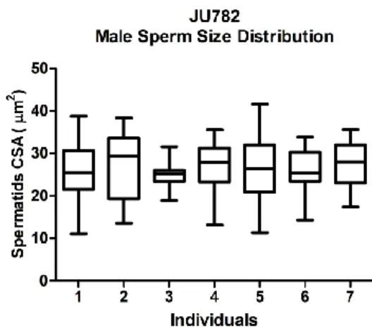
Figure 11 – Male sperm size distribution for JU782 individuals.. ...19
Figure 12 – Male sperm size distribution for JU1530 individuals.. ...19
Figure 13 – Male sperm size distribution for JU1213 individuals.. ...19
Figure 14 – Male sperm size distribution for JU561 individuals. ...19
Figure 15 – Male sperm size distribution for LSJ1 individuals.. ...19
Figure 16 – Male sperm size distribution for QTL preliminary study CB4856-N2.. ...20
Figure 17 – Male sperm size distribution for QTL preliminary study AB2-CB4856.. ...21
Figure 18 – C. elegans male sperm size mapping.. ...21
Figure 19 – AB1 Male Sperm Size.. ...23
Figure 20 – N2 Male Sperm Size.. ...23
Index of tables
Table 1 – Variation of C. elegans male sperm size.. ... 17Table 2 – QTL preliminary study CB4856-N2.. ... 20
Table 3 – QTL preliminary study AB2-CB4856.. ... 21
Table 4 – AB1 male sperm size. ... 22
XI
Index of supplementary tables
Supplementary Table 1 – C. elegans wild-isolate information. ... ii
Statiscal Table 1 – C. elegans natural male sperm size variation (95 isolates) ... iii
Statiscal Table 2 – RIL set CB4856-N2. ... iii
Statiscal Table 3 – RIL set AB2-CB4856. ... iii
Statiscal Table 4 – Ageing and mating effect on AB1 ... iii
Statiscal Table 5 – Ageing and mating effect on N2 ... iv
Index of supplementary figures
Supplementary Figure 1 – GWAS diameter #1... iv1
Chapter I
1. Introduction
1.1. Sperm size and sperm competition
Sexual reproduction involves the fusion of male and female gametes, commonly referred to as sperm and oocytes. Usually, the male gamete is a free-moving cell that fertilizes the larger, immobile female gamete. Spermatozoa exhibit a huge morphological diversity in various taxa (Birkhead & Møller, 1998). For instance, spermatozoa vary from tadpole morphology, as in mammals, to multiflagellate cells in termites (Baccetti & Dallai, 1978), to amoeboid cells in nematodes (Lamunyon & Ward, 1994; Ward et al. 1981), and to cooperative cells with distinct phenotypes in beetles (Higginson et al. 2012). This diversity is thought to have arisen through adaptation to new environments, male-female and male-male interactions and species differentiation (LaMunyon & Ward, 2002). Given the general limitation of female oocyte availability (Bateman, 1948; Birkhead & Møller, 1998), mating and access to females and ultimately oocyte fertilization may lead to competition among males. Male competition for females is a very common phenomenon in sexual species and may be expressed in many ways, such as behaviors and/or morphological and/or physiological traits that will hinder the approach/mating of other males (Hartmann & Loher, 1999; Chasnov et al. 2007). In cases of polyandry, in which females mate with more than one male, male competition may be postcopulatory. At this level, sperm competition, when sperm of different males compete for the same oocytes in the female’s reproductive tract, along with cryptic female choice, have an important role on male fitness (Bateman, 1948; Birkhead & Møller, 1998; Birkhead & Pizzari, 2002).
Sperm competition is considered to be a major selective pressure driving the evolution of sperm traits and diversity (LaMunyon & Ward, 2002; Parker & Pizzari, 2010). One of the most common sperm traits to be enhanced by sperm competition is sperm number/density to increase the probability of fertilization, a phenomenon known as “fair raffle” (Parker & Pizzari, 2010). But sperm quality may be also influenced by its individual morphology or behaviour, e.g. its size and motility, and thus influence male fitness. For instance, larger sperm may move faster or displaces smaller sperm (Gomendio & Roldan, 2008; Immler et al. 2010; Snook, 2005). Sperm competition is not restricted to sexual species (male/female), since it may happen when the promiscuity of sperm receptors allows the encounter of sperm from different donors. Therefore, hermaphroditic species, including sequential and simultaneous hermaphrodites, and
2 also androdioecious species, where hermaphrodites and males can be found in the same population, are systems where sperm competition may occur (Anthes et al. 2006; LaMunyon & Ward, 1998, 1999; Murray & Cutter, 2011). The study and comparison of all these different systems of sexual reproduction provides valuable insights for the understanding of how sperm competition shapes sperm evolution and morphology.
1.2. The model – Caenorhabditis elegans
Caenorhabditis elegans is a free-living bactivorous nematode and a well-established model organism in biology (Brenner, 1974) whose genome was the first animal genome to be fully sequenced (C. elegans Sequencing Consortium, 1998). With all life stages being optically transparent, suitable for imaging and microscopy, easy maintenance and manipulation, C. elegans assumed a leading role as a research model in the life sciences. During postembryonic development (usually 3 to 3.5 days at 20 °C), this nematode passes through 4 larval stages, L1 to L4, before reaching adulthood.
Androdioecy evolved three times independently in the genus Caenorhabditis: in C. elegans, C. briggsae and C. sp 11. All other Caenorhabditis species are gonochoristic (Kiontke et al., 2011). A androdioecious species, such as C. elegans, reproduce through self-fertilizing hermaphrodites and facultative outcrossing with males (Fig.1). C. elegans male frequency observed in natural and laboratory populations seems to be very low (Barrière & Félix, 2005; Teotonio et al., 2006). Although rare, male C. elegans remain capable of outcrossing and mating with hermaphrodites even if they are not as efficient as males of gonochoristic species (Chasnov et al., 2007). Males, generated by non-disjunction of the X chromosomes during meiosis, are XO, while the hermaphrodites are XX. Alternatively, males result from cross-fertilization between males and hermaphrodites.
Hermaphrodites are self-fertile and produce a limited number of sperm during the last larval stage, L4, after which they switch permanently to oocyte production (Hubbard & Greenstein, 2005; Kimble & Crittenden, 2007). In standard conditions, the hermaphroditic sperm is used for self-fertilization with almost 100% of efficiency (Ward & Carrel, 1979). Alternatively to self-fertilization, the hermaphrodites may out-cross with males, but they cannot mate between themselves: essentially, Caenorhabditis hermaphrodites are females capable of producing a limited amount of sperm, but with no reproductive organs to transfer sperm. Thus, sperm competition occurs only between self-sperm and male sperm, as well as between sperm of different males. Since the majority of hermaphrodites do not mate with males, given their rarity,
3 selfing is the main mode of reproduction. Nevertheless, when hermaphrodites out-cross with males, production of oocytes is stimulated therefore increasing the number of progeny produced by hermaphrodites compared to selfing individuals (Ward & Carrel, 1979).
1.3. Spermatogenesis and Spermiogenesis in C. elegans
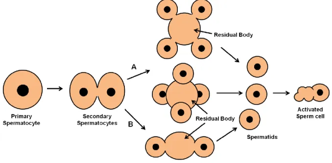
Sperm is generated after undifferentiated germ cells go through two processes: spermatogenesis and spermiogenesis. In C. elegans, spermatogenesis (Fig. 2) begins when the primary spermatocyte buds off from the rachis and enters meiosis I giving origin to two secondary spermatocytes with complete or partial cytokinesis. Each of these secondary spermatocytes goes through meiosis II forming two haploid spermatids that will bud off from an anucleate residual body. This last division is asymmetrical and the residual body retains great part of secondary spermatocyte constituents, like all ribosomes (there is no protein synthesis from this step on), the Golgi apparatus, the endoplasmic reticulum and cytoskeleton components actin and tubulin. On the other hand, the nucleus, several mitochondria and MOs (fibrous body-membranous organelle) migrate with the spermatids (Ward et al., 1981). FB-MOs have the function of correctly directing the asymmetrical division of proteins between spermatids and the residual body. The membranous organelle is formed from the Golgi apparatus and the fibrous body is formed in association with it, having MSP (Major Sperm Protein) as the principal component, in the primary spermatocyte (Roberts et al., 1986). In hermaphrodites, spherical spermatids are accumulated in the spermatheca whilst in males they are accumulated in the seminal vesicle and produced during the individual’s adult life (Ward & Carrel, 1979). In both sexes, a common regulatory genetic pathway controls spermatogenesis (L’Hernault, 2006).
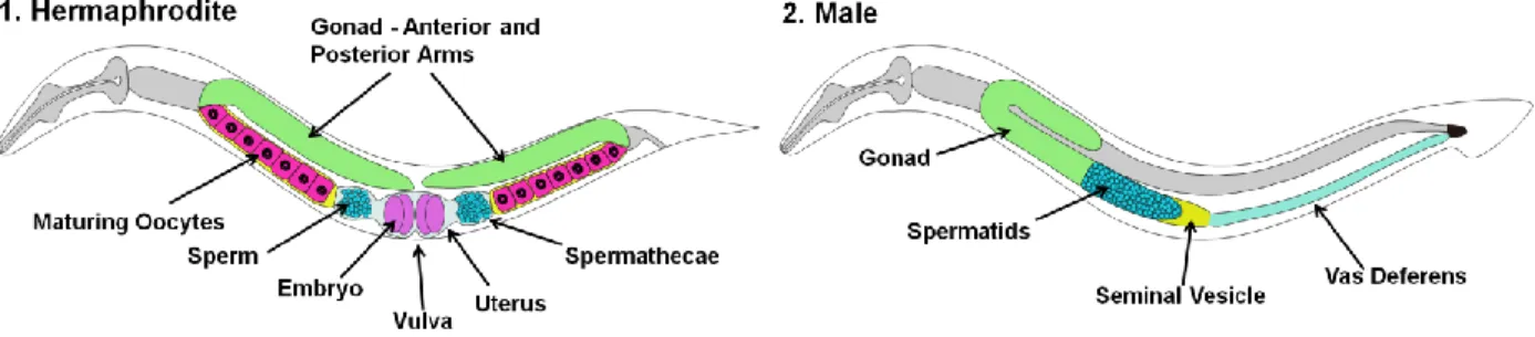
Figure 1 – C. elegans sexual anatomy. (1) Adult hermaphrodites possess two symmetric gonadal arms. In each
arm the oocytes maturate before reaching the spermatheca, where they will be fertilized by sperm (self or male sperm, depending on the situation). The new embryos pass to the uterus and will exit through the vulva. (2) Adult male possess a single gonad arm. Spermatids are constantly being produced and are accumulated in the seminal vesicle. When mating, spermatids will travel through the vas deferens and are transferred to the hermaphrodite.
4 Spermiogenesis in C. elegans is the process by which the round spermatid becomes an amoeboid motile sperm cell. This process is characterized by the fusion of the MOs with the plasma membrane, releasing its contents and maintaining a pore in the plasma membrane, while the FBs disassemble releasing the MSP fibers, which become free to create the pseudopod (Roberts et al., 1986; Ward et al., 1981). These MSP fibers will generate motile forces through assembling and disassembling in the pseudopod, allowing the sperm to crawl forward (Italiano et al., 2001; Stewart & Roberts, 2005; Ward & Klass, 1982). Although having most aspects of spermatogenesis and spermiogenesis in common, there is evidence suggesting that male and hermaphrodite sperm differ in the activation mode of spermiogenesis. Hermaphroditic spermatids become active with the beginning of ovulation while male spermatids become active only when they are transferred to hermaphrodites during copulation (Geldziler et al., 2005; Nance et al., 2000; Shakes & Ward, 1989; Smith & Stanfield, 2011; Stanfield & Villeneuve, 2006).
Figure 2 – Spermatogenesis in C. elegans. Primary spermatocyte undergoes meiosis I giving origin to two
secondary spermatocytes. The secondary spermatocytes may have an incomplete cytokinesis (A) or a complete one
(B). For the first case, secondary spermatocytes undergo meiosis II giving origin to a residual body with four budding
haploid spermatids. The four spermatids can bud off in parallel (on the top) or the secondary spermatocytes may twist and spermatids bud off forming a tetrahedron (on the center). For the complete cytokinesis the process is similar, each secondary spermatocyte originates a residual body with two haploid spermatids. In the end, free quiescent spermatids enter in spermiogenesis when activated and give origin to amoeboid sperm.
5
1.4. Sperm size in C. elegans
In C. elegans, male and hermaphroditic sperm have similar features: amoeboid morphology, spherical immature sperm is activated and develops a pseudopod (each sex having specific pathways as mentioned before), and both fertilize the oocytes in the spematheca (LaMunyon & Ward, 1994; Ward & Carrel, 1979). During fertilization, either type of sperm may be displaced out of the spermatheca into the uterus, and sperm have the capacity to “crawl” back into the spermatheca (Ward & Carrel, 1979). Male and hermaphrodite sperm show, however, one significant difference: male sperm is larger and more competitive (LaMunyon & Ward, 1998; Murray et al., 2011; Singson et al., 1999).
LaMunyon and Ward’s work greatly contributed to the understanding of sperm competition and evolution in C. elegans (LaMunyon & Ward, 1995; LaMunyon & Ward, 1994, 1998, 1999, 2002). In 1994, exploring artificial insemination in C. elegans, they demonstrated that male sperm had consistent precedence over hermaphrodite sperm. It had already been known from simple male-hermaphrodite matings that cross-progeny dominated over self-progeny. The artificial insemination experiment, however, established clearly that male sperm itself caused the superior competitiveness over hemaphrodite sperm.
In the following year they dismissed the activation mode, the interval between activation and competition and the presence of seminal fluid has possible functions of male sperm precedence. The experiments proved that male sperm is able to displace and outcompete hermaphrodite sperm and it would be a trait from the cell itself. When males mated with self-sterile hermaphrodites, paternity was proportionally shared placing the competitive difference between male and hermaphrodites (LaMunyon & Ward, 1995).
Further exploration of the competitive superiority of male sperm revealed sperm size as a major determinant of sperm competitiveness in C. elegans (LaMunyon & Ward, 1998). It was reported that male sperm is significantly larger than hermaphrodite sperm as well as the sperm found in the spermathecae compared to the uterine one. Two strains were analyzed and compared: N2 (standard strain and the one used in the previous studies) and AB1 (a wild isolate strain from Australia). AB1 revealed larger male sperm and outcompeted N2’s male sperm when competitiveness between the two strains was tested, further supporting the authors’ hypothesis. Sperm motility was also shown to increase with larger sperm size. Larger sperm showed in vitro a better capacity to crawl whereas the smaller hermaphrodite smaller sperm was nearly immobile suggesting that it is near the minimum size to allow motility. These advantages of larger sperm together with its better capacity of adhering to the spermatheca wall
6 explain how male sperm displaces the smaller hermaphrodite sperm gaining physical precedence into the spermatheca (Fig. 3). However, larger sperm have a slower production rate and is very likely more costly to produce than smaller sperm. Since C. elegans ancestors probably had large sperm, the authors suggested has an explanation for the reduced size of hermaphrodite sperm that selection for faster selfing reproduction selected smaller, less costly, hermaphrodite sperm.
Additional past research, extending to the analysis of multiple Caenorhabditis species, including C. elegans, and more distantly related nematode species, indicates that larger sperm is more competitive across many nematodes (LaMunyon & Ward, 1999). Sperm size variation was considerable between species as well as between individuals, but no clear phylogenetic pattern was observed. Hermaphrodite sperm was the smallest sperm while the largest sperm belonged to gonochoristic species where sperm competition among males is likely to be strong. Presumably, sperm competition may have represented an important selective force in shaping of sperm size diversity. Unpublished data from our lab on male sperm size variation in the Caenorhabditis genus is consistent with these initial observations (Fig.4, Fig.5).
An experimental evolution study to assess if C. elegans sperm size responded to an increased degree of sperm competition showed that higher sperm competition may indeed select for larger male sperm (LaMunyon & Ward, 2002). Lines resulting from the combination of four wild isolate strains (CB4855, DR1345, DR1350 and AB1) and N2 self-sterile phenotype were used in the experiment. After sixty generations, males experiencing higher sperm competition increased sperm volume by 20%.
The studies mentioned above focused mainly on the standard strain N2 that has been maintained in lab conditions for decades. A recent study analyzed a subset of 7 C. elegans wild isolates with distinct genetic backgrounds (AB1, CB4855, CB4856, DR1350, JU440, MY2 and PD4790) and which vary in mating and sperm traits (Murray et al., 2011). The authors confirmed the presence of ample heritable variation in C. elegans male sperm size. The comparison of the relative importance of mating and sperm traits for male fertilization success and male competition confirmed sperm size as a dominant factor, thus proving its relevance for male fitness.
The studies described above present solid evidence that sperm competition is a major selective force shaping sperm size in Caenorhabditis nematodes. It is considered that sperm size of both sexes result from the equilibrium between sperm competition and the opposing pressure of the higher cost to produce larger sperm. It was suggested that hermaphrodite smaller sperm was selected has a selfing trait to achieve an optimum on sperm production rate,
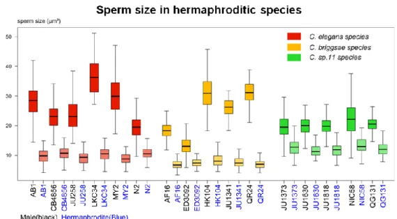
7 since males are rare, and to allow an advantageous rate of outcrossing in order to increase variability on the populations. Male sperm size would follow the reduction of hermaphrodite sperm size, whether by reduced competition or by indirect selection by the pleiotropy of common spermatogenesis genes for both sexes. Unpublished data from our lab on sperm size of the three androdioecius species shows substantial species and isolate differences (Fig 5). C. elegans and C. briggsae male sperm size is much more variable than hermaphrodite sperm and the difference between sperm types on C. sp 11 is reduced compared to the others. Nevertheless this relationship has never been deeply looked into and further studies are needed to fully understand it.
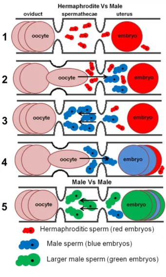
Figure 3 – Sperm competition and fertilization in C.
elegans. (1) Virgin hermaphrodite, already ovulating, with
activated sperm waiting in the spermatheca for passing oocytes from the oviduct to fertilize them. Some sperm was flushed out to the uterus from the spermathecae by previous crossing oocytes/embryos and crawls back to the spermathecae to fertilize new oocytes. (2-4) A male mates with the hermaphrodite and larger male sperm enters in competition with the hermaphroditic one. After a cross of an embryo to the uterus, sperm will crawl back to the spermathecae, but male sperm will recover faster and displace the smaller hermaphrodite’s sperm gaining precedence in the access to oocytes. With time, the continuous passage of oocytes through the spermathecae and embryos through the uterus flush out of the worm all the smaller hermaphrodite sperm and male sperm fertilizes all the oocytes until its exhaustion.
(5) Male vs. male sperm competition works in a similar
way to the previous hermaphrodite vs male sperm competition – the larger sperm will have an advantage and outcompete the smaller sperm.
8
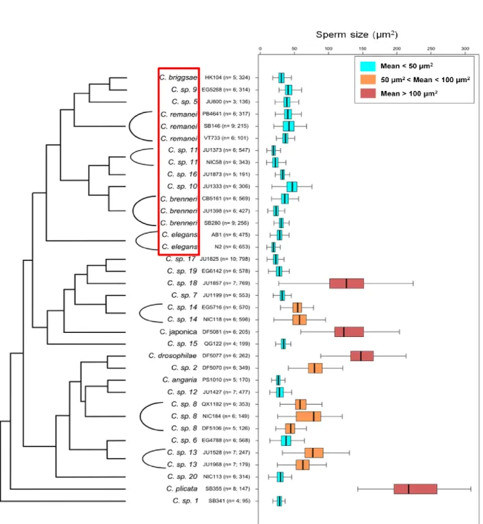
Figure 4 – Male sperm size variation among Caenorhabditis species. Among the extensive variation in male
sperm size, one notes the independent and repeated evolution of extremely large sperm size (mean>100 µm2) in the
Japonica/Drosophilae/Angaria groups of the phylogeny. The red box highlights the Elegans group where, both
gonochoristic and androdioecious species exhibit relatively small male sperm size (mean<50 µm2) (Callemeyn & Braendle, unpublished data).
9
1.5. Proximate mechanisms regulating sperm size
Virtually nothing is known about the genetic and developmental factors regulating sperm size or the potential genetic differences that could contribute to intra and inter-specific variation in Caenorhabditis sperm size. Only the mutants spe-10, spe-17 and spe-39 show abnormal sperm phenotypes in which sperm size is affected and sperm cells are smaller than wild-type (Nishimura & L’Hernault, 2010), but these obviously do not reflect natural genetic variation of sperm size. When spe-10, a four-pass trans-membrane protein with a DHHC-CRD zinc finger domain, is mutated the FBs (Fibrous-Body) do not interact in a normal way with MOs (Membranous-organelles) and do not form the FB-MOs complexes that migrate to the forming spermatids, leaving the FBs in the residual body (Gleason et al., 2006). The mutant spe-17 (a highly charged protein rich in serine and threonine) has spermatids with abnormalities in the FB-MO’s like spe-10, in this case FB-MOs segregate correctly, but ribosomes travel into budding spermatids associated to them (L’Hernault et al., 1993). The spe-39 protein has a wide somatic expression in C. elegans and also has orthologs for several organisms including humans. In C. elegans, the spe-39 mutant is often not capable of producing spermatids, and when produced spermatids are smaller and with small vesicles, interpreted as an earlier stage of MO biosynthesis (Zhu & Hernault, 2003).
A recent study showed that mutationally induced hermaphrodites in Caenorhabditis remanei females (a gonochoristic species), had smaller sperm compared to normal male sperm
Figure 5 – Male and hermaphrodite sperm size variation among Caenorhabditis androdioecious species. C.elegans in red, C. briggsae in yellow and C. sp 11 in green. On
the X axis, strains name on black for males and on blue for hermaphrodites (Callemeyn & Braendle, unpublished data).
10 size (Baldi et al., 2011). Therefore, there is an apparent predisposition to make sexually dimorphic sperm in Caenorhabditis. In the same study, using C. elegans sex-determination mutants, it was demonstrated that the X chromosome ratio does not affect sperm size. On the other hand, it was demonstrated that the somatic gonad and the intestine cooperate to nurture larger sperm in males. The combination of oogenesis and spermatogenesis also proved to influence sperm size, probably due to competition for resources of both types of gametes. The authors speculate that sperm size results from the cooperation between selection, as mentioned before, and a developmental bias caused by the use of spermatogenesis genes by hermaphrodites.
2. Aims
The study of natural genetic variation in C. elegans sperm size provides (a) an ideal system to study the evolution of a trait under sexual selection and (b) a potential entry point to identify genetic factors influencing sperm size. Initially the aim of this thesis was to understand the role of natural genetic variation in male sperm size and its proximate causes in C. elegans using a set of previously constructed F2 RIL (Recombinant Inbred Lines) to map male sperm size variation using a QTL (Quantitative Trait Loci) analysis (e.g. Palopoli et al., 2008). However, we did not detect substantial differences in sperm size between parental lines, and we therefore decided to score a world-wide panel of 97 C. elegans wild isolates (Andersen et al., 2012). As the isolates had been SNP-genotyped at the whole genome level, this allowed us to perform a GWAS (Genome-Wide Association Study). In addition, this survey of C. elegans male sperm size was used to determine the best possible candidate isolates to construct a new F2 RIL panel. The main results of this thesis consist of the survey of male sperm size variation in C. elegans wild isolates and its statistical analysis, including the GWAS. In addition to the main work we conducted a pilot experiment testing the effect of ageing and mating in males on male sperm size.
11
Chapter II
3. Materials and methods
3.1. Cultures and maintenance of the nematodes
All strains used were cultured and maintained on NGM (Nematode Growth Medium) in Petri dishes (diameter 55mm) at 20ºC. To feed the nematodes, a drop of Escherichia coli OP50 was added to each plate (Brenner, 1974). All cultures were maintained and regularly transferred in order to avoid starvation.
3.2. Dissection and scoring
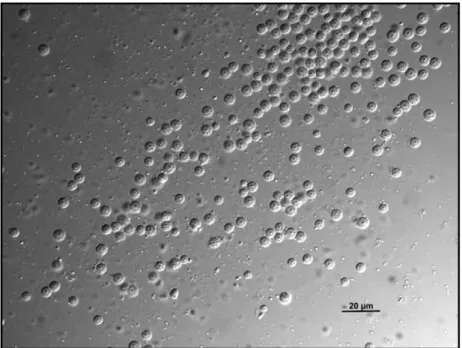
For the isolation of sperm, nematodes were dissected in a 8 µL drop of sperm medium (Nelson & Ward, 1980) with 30G needles on coated slides (coating solution and sperm medium composition in Annex A) before the sperm size measurement using a previously described method (LaMunyon & Ward, 1999). Using a DIC (Differential Interference Contrast) microscope, pictures of the spermatids were taken to measure the CSA (Cross-Sectional Area). Activated nematode sperm moves by means of pseudopods and has an irregular shape, but the spermatids prior to activation are spherical (Fig. 6) and have the same volume as the sperm, thus spermatids were used for all measurements (Roberts et al., 1986).
Figure 6 – Male C. elegans spermatids. DIC picture (60x) of round male
12 For the analysis of male sperm size of C. elegans isolates used on RIL construction, all data was collected using an Olympus BX 61 microscope with a Cool Snap HQ2 camera using a 60x objective lens. For the pilot experiment testing the effects of age and mating as well as for the male sperm characterization of 97 wild isolates, all data was collected using a Zeiss Axio Imager.A1 microscope with a Scion Corporation – Model CFW 1312c - camera using a 63x objective lens.
Before each dissection L4 males, last juvenile state before adulthood, were isolated in plates with 8-12 individuals in a standard plate for 20 to 30 hours in order to guarantee virginity. During dissection, the incision was made in order to cut the seminal vesicle to release the accumulated spermatids.
3.3. Male induction and male cultures
The occurrence of males in C. elegans is rare. In order to generate male populations for sperm size analysis, around five hermaphroditic L4 worms were isolated in a plate and then heat shocked for approximately 1h30 min, at 37ºC. This procedure is a common way of inducing non-disjunction errors of the X chromosome during gametogenesis so that males can be generated (Hodgkin & Doniach, 1997). The resulting males were crossed with L4 hermaphrodites in an approximate ratio of two males to a single hermaphrodite on new plates in order to have sufficient males for the experiments. These male cultures were maintained at least two generations before male dissection to ensure that males analyzed did not experience maternal effects resulting from the heat-shock treatment.
3.4. Measurement and calculus of sperm cross-sectional area
The collected photographs were analyzed in ImageJ® and for each spermatid two perpendicular diameters were measured (Fig.7) in order to calculate the CSA, approximating to an ellipse as follows:
A=πRARB where RA= DA/2 and RB = DB/2, with A being cell’s cross-sectional area; DA and DB the two diameters measured; RA and RB the radius correspondent to DA and DB, respectively.
Figure 7 – Measurement of male C. elegans spermatids. Red arrows represent
13
3.5. Natural genetic variation in C. elegans male sperm size
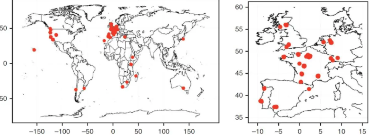
We quantified natural genetic variation in C. elegans male sperm size using a world-wide set of 97 wild isolates (Andersen et al., 2012), isolate origin and information in Annex A. These 97 isolates (reflecting all unique haplotypes) capture a maximum range of genetic variation within C. elegans after assessing a total of 200 wild isolates (Andersen et al., 2012) (Fig. 8). After thawing of frozen stocks, isolates were maintained at 15ºC and the worms transferred to new plates each 4-5 days. Given the large number of isolates to be assayed, male induction and subsequent sperm size quantification was carried out over a time period of six months. Throughout this period, isolates were selected at random for experiments performed at a given time point. In order to score the sperm size without bias, we attributed to each strain a code for stocks, cultures and male plates. We collected data for 94 of the 97 isolates plus data for the reference strain N2.
We let male plates pass through two generations either after male induction, as described previously, or after the strains came from the stocks in cases when some spontaneous males were found. For each strain, we measured 20 sperm of each of seven males (140 spermatids/isolate). Prior to dissection, L4 males were isolated on fresh plates, as described previously, for 24 hours.
3.6.1. Male sperm size in C. elegans isolates used for RIL construction
One of the sets of RILs had been derived from the two parental isolates, CB4856 and N2, generated by the Kruglyak lab (Seidel et al., 2008). The strain CB4856, a wild isolate from
Figure 8 – Wild-isolate strains geographical origin. Red dots mark the origin of the 97 C. elegans wild
14 Hawaii, is genetically divergent relative to the commonly used N2 lab strain. Two other strains were also included in this preliminary assay: QG1 (N2 genetic background with npr-1 allele of CB4856) and CX11400 (CB4856 genetic background with npr-1 allele of N2). The N2 npr-1 allele stems from a lab mutation not found in any other C. elegans wild isolate. Because the N2 npr-1 allele has profound effects on many traits (e.g. McGrath et al., 2009), these strains were included to control for any potential effects of npr-1 on male sperm size. For each strain, 10 individuals were dissected and for each individual 50 spermatids were measured making a total of 500 spermatids per strain. The strains used in this experiment were kindly provided by Erik Andersen (Kruglyak Lab, Princeton University).
The second set of RILs results from crosses between CB4856 and AB2 wild isolates, kindly provided by the Rockman lab, NYU University. We scored male sperm size for QX1199 (him-5 mutation in CB4856 background) and QG5 (him-5 mutation in AB2 background), respectively. The him-5 mutation populations have a higher ratio of males, since the mutation induces more spontaneous males by the non-disjunction of the sexual chromosomes. For QG5, 20 spermatids of each of seven males were measured (total 140 cells). For QX1199, 50 spermatids of each of seven males were measured (total 350 cells).
3.6.2. Genome-wide association study
We performed a GWAS for male sperm size using the SNP (Single Nucleotide Polymorphism) data set collected by Andersen et al. (2012). We used the software package Efficient Mixed-Model Association, EMMA, (Kang et al., 2008) for R® to do our association analysis. All SNP data used for the association has been described in the previously referred paper (Andersen et al., 2012). Briefly, the genomic data resulted from restriction-site-associated DNA sequencing analysis using EcoRI covering 8% of the C. elegans genome (100 Mb), and 6089 SNPs were used for the association mapping.
3.7. Effects of age and mating on male sperm size
We conducted a pilot experiment to test whether age and mating (sperm depletion) affect C. elegans male sperm size. We used N2 and AB1, two isolates that strongly differ in male sperm size (LaMunyon & Ward, 1998, 2002; Murray et al., 2011). To test for a mating (sperm depletion treatment) effect, we placed 15 L4 males together with 30 L4/young adult hermaphrodites on 55mm plates. To test for an effect of male age (isolation treatment), 15 L4
15 males were isolated on a standard plate. For each treatment and for each strain, three plate replicates were done at the same time. Per plate, 2 random males were picked and dissected to measure sperm size at three different time points: 24, 48 and 72 hours after the beginning of treatment. All males dissected had 30 spermatids measured making 180 cells (2 males, 3 replicates) for each timepoint in each treatment (with exception of AB1 ageing 72h that one of the replicates only had 1 male). An excess of males was used given their propensity to crawl out of the plates when isolated from hermaphrodites.
At 48h all worms, adult males and hermaphrodites, on sperm depletion treatment were picked to new standard plates to avoid overcrowding with the hermaphrodites’ progeny. There were also some contaminations, probably due to the daily manipulation on the same plate during treatment and picking for dissection, on the ageing treatment. All replicates from N2 and one replicate of AB1 at 48 hours of ageing treatment had minor contaminations outside the E. coli drop. All worms in contaminated plates were transferred to fresh plates in a tentative to avoid contamination. Nevertheless, at 72 hours of the ageing treatment, all replicates of both strains had, even if not extensive, contaminations. For AB1, only 5 males were scored for 72 hours of the ageing treatment, since in one replicate there was only one male.
For statistical analysis we performed a general linear model using the software STATISTICA with treatment and time considered as factors; replicates were nested in each time point and individuals were nested into the replicates.
3.8. Statistics
The mean sperm size for each strain in all experiments was calculated as the grand mean of the individual means. For each experiment, we did ANOVAs to compare sperm size, between strains. In all analyses, individuals were nested in strain to account for individual variation. Analyses were done with software STATISTICA®. Data were transformed where necessary to meet ANOVA requirements.
16
Chapter III
4. Results
4.1. C. elegans natural male sperm size variation
To characterize C. elegans natural genetic variation in male sperm size, we examined 95 wild isolates plus the reference strain N2. Three isolates (CB4851, JU397 and KR314) of the wild isolate set (Andersen et al., 2012) could not be measured due to problems with male induction.
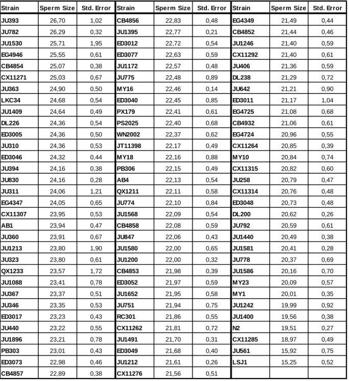
Mean male sperm size ranged from 15.25 µm2 of LSJ1 to 26.70 µm2 of JU363 (Table 1) with a global mean of all isolates of 22.25 µm2. Mean for each isolate was calculated as the grand mean of individual means. LSJ1 and JU561 appear as clear outliers with small sperm while JU1213, JU1530 and QX1233 show a high standard error indicating substantial inter-individual variation. Figure 9 depicts the phylogenetic relationship of C. elegans isolates (Andersen et al., 2012) with respective mean male sperm size. For brevity, given the number of strains, only the two strains with largest/smallest sperm size as well the two strains with the highest standard error are represented in detail, indicating their sperm size distribution among individuals (Fig.10, 11, 12, 13, 14 and 15).
The characterization of C. elegans male sperm size revealed considerable natural variation for this trait both at an inter- and intra-strain level. Our statistical analysis showed that, as expected, strains differed significantly for male sperm size (nested ANOVA F94,12635=26.6, p<0.001; Statistical Table 1 in Annex B).
17
Strain Sperm Size Std. Error Strain Sperm Size Std. Error Strain Sperm Size Std. Error JU393 26,70 1,02 CB4856 22,83 0,48 EG4349 21,49 0,44
JU782 26,29 0,32 JU1395 22,77 0,21 CB4852 21,44 0,46
JU1530 25,71 1,95 ED3012 22,72 0,54 JU1246 21,40 0,59
EG4946 25,55 0,61 ED3077 22,63 0,59 CX11292 21,40 0,61
CB4854 25,07 0,38 JU1172 22,57 0,48 JU406 21,36 0,59
CX11271 25,03 0,67 JU775 22,48 0,89 DL238 21,29 0,72
JU363 24,90 0,50 MY16 22,46 0,14 JU642 21,21 0,90
LKC34 24,68 0,54 ED3040 22,45 0,85 ED3011 21,17 1,04
JU1409 24,64 0,49 PX179 22,41 0,61 EG4725 21,08 0,68
DL226 24,36 0,54 PS2025 22,40 0,68 CB4932 21,06 0,61
ED3005 24,36 0,50 WN2002 22,37 0,62 EG4724 20,96 0,55
JU310 24,36 0,53 JT11398 22,17 0,49 CX11264 20,85 0,39
ED3046 24,32 0,44 MY18 22,16 0,88 MY10 20,84 0,74
JU394 24,16 0,38 PB306 22,15 0,49 CX11315 20,82 0,60
JU830 24,16 0,28 AB4 22,13 0,54 JU258 20,79 0,47
JU311 24,06 1,21 QX1211 22,11 0,58 CX11314 20,76 0,48
EG4347 24,05 0,65 JU774 22,10 0,84 ED3048 20,73 0,48
CX11307 23,95 0,53 JU1568 22,09 0,54 DL200 20,62 0,26
AB1 23,94 0,47 CB4858 22,08 0,59 JU792 20,59 0,61
JU360 23,91 0,67 JU847 22,06 0,43 JU1440 20,49 0,38
JU1213 23,80 1,90 JU1580 22,00 0,65 JU1581 20,41 0,28
JU323 23,80 0,61 JU1200 22,00 0,32 JU778 20,37 0,69
QX1233 23,57 1,72 CB4853 21,98 0,39 JU1586 20,16 0,70
JU1088 23,41 0,78 ED3052 21,97 0,59 MY23 20,09 0,57
JU367 23,37 0,51 JU1652 21,95 0,58 MY1 20,01 0,35
JU346 23,35 0,53 JU751 21,94 0,75 JU1242 19,99 0,92
ED3017 23,23 0,43 RC301 21,86 0,55 JU1400 19,56 0,38 JU440 23,22 0,55 CX11262 21,81 0,72 N2 19,51 0,27 JU1896 23,21 0,78 JU1491 21,70 0,31 CX11285 18,97 0,49 PB303 23,01 0,43 ED3049 21,68 0,40 JU561 15,92 0,75 ED3073 22,98 0,46 JU1212 21,61 0,26 LSJ1 15,25 0,52 CB4857 22,89 0,38 CX11276 21,56 0,51
Table 1 – Variation of C. elegans male sperm size. Mean and standard error for CSA of spermatids for each
wild isolate (µm2). For each strain, we measured 20 sperm of each of seven males (140 spermatids/isolate). Means represent grand means of the seven individual means.
18
Figure 9 – C. elegans wild-isolates phylogeny and male sperm size. Mean and standard error for CSA of
spermatids for measured isolates and neighbor-joining tree of wild-isolate strains of C. elegans, adapted from (Andersen et al., 2012).
19
Figure 10 – Male sperm size distribution for JU393 individuals. Boxplot of all measurements
for each individual (20 spermatids/male), whiskers 2.5-97.5 percentile.
Figure 11 – Male sperm size distribution for JU782 individuals. Boxplot of all measurements
for each individual (20 spermatids/male), whiskers 2.5-97.5 percentile.
Figure 12 – Male sperm size distribution for JU1530 individuals. Boxplot of all measurements
for each individual (20 spermatids/male), whiskers 2.5-97.5 percentile.
Figure 13 – Male sperm size distribution for JU1213 individuals. Boxplot of all measurements
for each individual (20 spermatids/male), whiskers 2.5-97.5 percentile.
Figure 14 – Male sperm size distribution for JU561 individuals. Boxplot of all measurements
for each individual (20 spermatids/male), whiskers 2.5-97.5 percentile
Figure 15 – Male sperm size distribution for LSJ1 individuals. Boxplot of all measurements for
each individual (20 spermatids/male), whiskers 2.5-97.5 percentile.
20
4.1.1. Male sperm size in C. elegans isolates used for RIL construction
Our preliminary studies to assess the viability of using one of the two RIL sets for a QTL analysis for male sperm size revealed significant differences between sperm size for the CB4856-N2 set of RILs and none for the AB2-CB4856 set. Mean for each strain was calculated as the grand mean of individual means.
The parental strains, CB4856 and N2, showed a difference of approximately 3 µm2 in their mean sperm size (Table 2), while CX11400’s (CB4856 genetic background with npr-1 allele of N2) mean is closer to N2 than CB4856. The smallest mean belongs to QG1 (N2 genetic background with npr-1 allele of CB4856), but this result might not be reliable due to contaminations (also, only 6 individuals were measured for this strain). The mean of CX11400 is similar to the N2 but sperm size range assumes a distribution closer to CB4856 (Fig. 16). The statistical analysis revealed small, yet significant differences in male sperm size between the strains tested for the CB4856-N2 set (nested ANOVA F3,1764=117.57, p<0.001; Statistical Table 2 in Annex B). The differences between individuals of the same strain were also significant (nested ANOVA F32,1764=20.09, p<0.001; Statistical Table 2 in Annex B).
For the alternative set of RILs, AB2-CB4856, the preliminary results revealed similar male sperm size (Table 3) and no significant differences of this trait between the parental strains (nested ANOVA F1,476=0.012, p=0.913; Statistical Table 3 in Annex B).. For both strains, QX1199 (him-5 mutation in CB4856 background) and QG5 (him-5 mutation in AB2 background), the sperm size range was similar (Fig. 17).
Strain Sperm Size (µm2) Std. Error
CB4856 23,60 1,14
CX11400 20,06 0,87
N2 20,90 0,40
QG1 18,41 0,71
Figure 10 – Male sperm size distribution for QTL preliminary study CB4856-N2. (On left) Boxplot of all
measurements for each strain, whiskers 2.5-97.5 percentile. For each strain, we measured 50 sperm of each of ten males (500 spermatids/strain).
Table 2 – QTL preliminary study CB4856-N2. Mean
and standard error for CSA of spermatids for each strain. Means represent grand means of the ten individual means.
21
4.1.2. Genome-wide association study
Following the characterization of male sperm size variation in C. elegans we proceeded to a GWAS for this trait. Our characterization provided us with the data necessary for this study with the strains used differing significantly in male sperm size. Spermatid CSA showed a heritability of 49.3% (+/- 15.2%) meaning that approximately half of male sperm size variation may be attributed to genetic differences. However, GWAS results did not detect any significant candidate genomic region for this trait (Fig. 18). Two GWAS were also realized using spermatids’ diameters with the same results (Annex B). The results point to a likely complex genetic architecture for male sperm size variation.
Strain Sperm Size (µm2) Std. Error
QG5 23,38 1,36
QX1199 23,49 1,31
Figure 11 – Male sperm size distribution for QTL preliminary study AB2-CB4856. (On left) Boxplot of all
measurements for each strain, whiskers 2.5-97.5 percentile. For QG5, we measured 20 sperm of each of seven males (140 spermatids/strain). For QX1199, we measured 50 sperm of each of seven males (500 spermatids/strain).
Table 3 – QTL preliminary study AB2-CB4856. Mean
and standard error for CSA of spermatids for each strain. Means represent grand means of the seven individual means.
Figure 12 – C. elegans male sperm size mapping. Result from GWAs for male sperm size in C. elegans. Autossomes I to V and sex chromosome X represented by boxes from I to V and box X,
respectively. Grey line at the top represent significance frontier, no genomic region was significant to male sperm size variation.
22
4.2. Effects of age and mating on male sperm size
In an additional experiment, we further explored the effect of age and mating on male sperm size. The overall analysis indicates that age has stronger influence than the applied treatments on male sperm size.
In AB1, age was a significant factor influencing male sperm size (GLM F2,1021=203.6, p<0.001; Statistical Table 4 in Annex B) with sperm size getting larger with age (Table 4). The other factors were not significant aside the difference between individuals (like in the other experiments).
In N2 both treatment (GLM F1,1059=68.43, p<0.001) and age (GLM F2,1059=26.52, p<0.001), as well as their interaction (GLM F2,1059=27.37, p<0.001) were significant for sperm size variation. Once more, the difference between individuals was significant (Statistical Table 5 in Annex B).
There is a clear increase of male sperm size between 24 hours to 48 hours, for both strains and treatments (Fig. 19 and 20). Also, at 72 hours for ageing (isolation) treatment the intra-individual variation was much larger comparing to other time points.
Treatment Time points Sperm Size (µm2) Std. Error
Sperm Depletion 24h 22,44 0,65 Sperm Depletion 48h 23,98 0,42 Sperm Depletion 72h 23,51 0,42 Isolation 24h 22,31 0,58 Isolation 48h 23,83 0,57 Isolation 72h 22,84 0,88
Table 4 – AB1 male sperm size. Mean and standard error for CSA of spermatids for each
treatment and time point. Means represent grand means of the six individual means for each time point.
Treatment Time points Sperm Size (µm2) Std. Error
Sperm Depletion 24h 21,67 0,27 Sperm Depletion 48h 23,74 0,17 Sperm Depletion 72h 24,81 0,43 Isolation 24h 21,05 0,46 Isolation 48h 22,51 0,36 Isolation 72h 21,18 0,79
Table 5 – N2 male sperm size. Mean and standard error for CSA of spermatids for each treatment
23
Figure 19 – AB1 Male Sperm Size. Mean (points)
and standard error (bars) for AB1 male sperm size. Mating (Sperm depletion) treatment on black and ageing (Isolation) treatment on blue. For each time point, we measured 30 sperm of each of six males (180 spermatids/time point).
Figure 20 – N2 Male Sperm Size. Mean (points)
and standard error (bars) for N2 male sperm size. Mating (Sperm depletion) treatment on black and ageing (Isolation) treatment on blue. For each time point, we measured 30 sperm of each of six males (180 spermatids/time point).
24
Chapter IV
5. Discussion
5.1. Natural genetic variation in C. elegans male sperm size
We provide a comprehensive view on natural genetic variation of male sperm size in C. elegans using a recently established wild isolate collection, covering the largest experimentally recorded amount of genetic diversity in this species. In agreement with previous studies (LaMunyon & Ward, 1999, 2002; Murray et al., 2011), our results demonstrate that C. elegans has considerable and significant genetic variation in male sperm size.
We observed that male sperm size in C. elegans is comprised mainly between 20 and 27 µm2. Our statistical analysis confirmed that male sperm size differed significantly between isolates, demonstrating natural genetic variation for the trait. However, C. elegans male sperm size variation does not seem to greatly vary with divergence among strains (Fig. 9): the four most divergent wild isolates from N2: CB4856 (22.83 µm2), DL238 (21.29 µm2), JU775 (22.48 µm2) and QX1211 (22.11 µm2) have similar sperm size, close to the global mean (22.15 µm2). This fact suggests three possible scenarios that might co-exist and were already mentioned by other authors (Baldi et al., 2011; Murray et al., 2011). A likely scenario is that male sperm size variation is a polygenic trait. Several genes may influence the phenotype with a small effect, instead of a single or a small number of genes with a stronger effect. A second scenario is the possible existence of relaxed selection for male sperm size. C. elegans is mainly a selfing organism and male sperm competition lost relevance with the appearance of hermaphroditism. As long as male sperm continue to be larger and able to outcompete hermaphrodite sperm, mutations with a small or neutral effect can be accumulated and male sperm size may decrease/degenerate. Lastly, it is possible that male sperm size evolution is driven by selection on hermaphrodite sperm size. As was mentioned previously, males and hermaphrodites, share the genetic pathway for spermatogenesis. Like this, pleiotropic genes could be selected for optimal hermaphrodite sperm characteristics, affecting male sperm characteristics as a byproduct of hermaphrodite selection. With the other scenarios this could contribute to mask phylogenetic patterns for male sperm size variation.
LSJ1 (15.25 µm2) and JU561 (15.92 µm2) appeared as clear outliers with the smaller sperm differing approximately 7 µm2 to the global mean. This result is quite interesting since closely related strains to both LSJ1 and JU561 have “normal” male sperm size. LSJ1, in