Substances on the Nematode
Caenorhabditis elegans
Yoko Honda1, Yasunori Fujita2, Hiroe Maruyama3, Yoko Araki3, Kenji Ichihara3, Akira Sato4, Toshio Kojima4,5, Masashi Tanaka1, Yoshinori Nozawa2,6, Masafumi Ito2, Shuji Honda1*
1Department of Genomics for Longevity and Health, Tokyo Metropolitan Institute of Gerontology, Sakaecho, Itabashiku, Tokyo, Japan,2Department of Longevity and Aging Research, Gifu International Institute of Biotechnology, Naka-fudogaoka, Kakamigahara, Gifu, Japan,3API Company Limited, Nagaragawa Research Center, Nagarayamasaki, Gifu, Japan,4Computational Systems Biology Research Group, Advanced Science Institute, RIKEN, Suehiro-cho, Tsurumi-ku, Yokohama, Kanagawa, Japan,5Hamamatsu University School of Medicine, Handayama, Higashi-ku, Hamamatsu, Shizuoka, Japan,6Department of Food and Health, Tokai Gakuin University, Naka-kirinocho, Kakamigahara, Gifu, Japan
Abstract
Background: One of the most important challenges in the study of aging is to discover compounds with longevity-promoting activities and to unravel their underlying mechanisms. Royal jelly (RJ) has been reported to possess diverse beneficial properties. Furthermore, protease-treated RJ (pRJ) has additional pharmacological activities. Exactly how RJ and pRJ exert these effects and which of their components are responsible for these effects are largely unknown. The evolutionarily conserved mechanisms that control longevity have been indicated. The purpose of the present study was to determine whether RJ and its related substances exert a lifespan-extending function in the nematode Caenorhabditis elegansand to gain insights into the active agents in RJ and their mechanism of action.
Principal Findings:We found that both RJ and pRJ extended the lifespan ofC. elegans. The lifespan-extending activity of pRJ was enhanced by Octadecyl-silica column chromatography (pRJ-Fraction 5). pRJ-Fr.5 increased the animals’ lifespan in part by acting through the FOXO transcription factor DAF-16, the activation of which is known to promote longevity inC. elegansby reducing insulin/IGF-1 signaling (IIS). pRJ-Fr.5 reduced the expression ofins-9,one of the insulin-like peptide genes. Moreover, pRJ-Fr.5 and reduced IIS shared some common features in terms of their effects on gene expression, such as the up-regulation ofdod-3and the down-regulation ofdod-19, dao-4andfkb-4.10-Hydroxy-2-decenoic acid (10-HDA), which was present at high concentrations in pRJ-Fr.5, increased lifespan independently of DAF-16 activity.
Conclusions/Significance: These results demonstrate that RJ and its related substances extend lifespan in C. elegans, suggesting that RJ may contain longevity-promoting factors. Further analysis and characterization of the lifespan-extending agents in RJ and pRJ may broaden our understanding of the gene network involved in longevity regulation in diverse species and may lead to the development of nutraceutical interventions in the aging process.
Citation:Honda Y, Fujita Y, Maruyama H, Araki Y, Ichihara K, et al. (2011) Lifespan-Extending Effects of Royal Jelly and Its Related Substances on the Nematode Caenorhabditis elegans. PLoS ONE 6(8): e23527. doi:10.1371/journal.pone.0023527
Editor:Matt Kaeberlein, University of Washington, United States of America
ReceivedJanuary 8, 2011;AcceptedJuly 20, 2011;PublishedAugust 9, 2011
Copyright:ß2011 Honda et al. This is an open-access article distributed under the terms of the Creative Commons Attribution License, which permits unrestricted use, distribution, and reproduction in any medium, provided the original author and source are credited.
Funding:This work was essentially supported by Tokyo Metropolitan Institute of Gerontology financially, and was partially funded by API Company Limited and partially by a Grant for Biological Research from Gifu Prefecture, Japan. API Co. Ltd. played a role in study design, data collection and analysis, decision to publish, or preparation of the manuscript.
Competing Interests:HM, YA and KI are employees of API Co., which engages in the manufacture and sale of royal jelly products and also provided the royal jelly used in this study. This is to confirm that the declaration does not alter the authors’ adherence to all the PLoS ONE policies on sharing data and materials.
* E-mail: [email protected]
Introduction
Lifespan in metazoans is influenced not only by genetic factors [1], [2] but also by environmental factors, including temperature [3], [4], oxygen [5–7], food intake [8] and nutrition [9–16]. In the honeybeeApis melliferaL., queens live and reproduce for 1–4 years, yet hive workers, which are derived from the same diploid genome, live for only 3–6 weeks during the spring and summer in temperate climates [17–19]. Queens are fed throughout their lives with royal jelly (RJ), which is produced by the hypopharyngeal, postcerebral and mandibular glands of the worker bees. In contrast, workers are fed this RJ for only a short period of time during their larval stages. This scenario raises the possibility that RJ contains longevity-promoting agents for queens [17], [19]. An
analysis of its chemical composition showed that RJ comprises proteins, sugars, lipids, vitamins and free amino acids [20] together with a variety of bioactive substances, including AMP N1-oxide [21], peptides [22–24], acetylcholine [25–27] and fatty acids, such as 10-hydroxy-2-decenoic acid (10-HDA) [28]. The mechanism by which RJ exerts its longevity effects on queen bees and the identities of the components that play critical roles in this process are largely unknown.
In mammals, RJ has also been reported to possess a variety of pharmacological activities such as antibacterial [30], antitumor [31], anti-allergic [32], antifatigue [33], anti-inflammatory [34], [35] and immunomodulatory [36], [37] effects. RJ also induces neurite outgrowth [38], prevents dermatitis [39], hypercholester-olemia [40] and osteoporosis [41] and stimulates bone formation [42]. Protease-treated RJ (pRJ) has additional beneficial proper-ties, including antioxidant activity [43], inhibitory effects on lipid peroxidation [44] and antihypertensive effects [45–47].
The nematode Caenorhabditis elegans has been widely used in studies on aging and longevity. It is an ideal model organism for such studies because of its relatively short lifespan (3–4 weeks) and well-established genetic pathways [1]. UsingC. elegans, researchers have identified several compounds that are capable of extending lifespan and are derived from natural products including blueberries [48], herbs [49] and green tea [50], [51]. In the present study, we examined the effects of RJ and pRJ on the lifespan of C. elegans to identify the lifespan-extending agents in these substances and to understand the mechanism of their action.
Results
Effects of RJ on the lifespan ofC. elegans
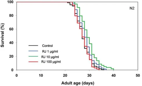
We first investigated the effects of RJ on the lifespan of the wild-type N2 strain ofC. elegans. RJ treatment was begun at the young adult stage with concentrations ranging from 1 to 100mg/ml. RJ treatment at 10mg/ml extended the mean lifespan by 7–9%, whereas either 1 or 100mg/ml RJ had little or no effects on the lifespan (Fig. 1, Table S1), indicating that there is an optimal dose of RJ for lifespan extension. In contrast, pRJ prolonged the mean lifespan at all concentrations tested (1, 10 and 100mg/ml). The maximal effect was observed at 10mg/ml, at which concentration the mean lifespan was increased by 7–18% (Fig. 2, Table S1). These results suggest that both RJ and pRJ contain the lifespan-extending agents and that these agents are not proteinaceus.
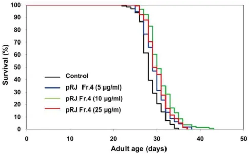
To gain insights into the nature of the lifespan-extending agents, we performed fractionations of both RJ and pRJ. RJ was divided into EtOH-soluble (RJ-Fr.1) and water-soluble (RJ-Fr.2) fractions. Neither of these fractions extended the lifespan (Fig. S1, Fig. S2, Table S1). pRJ was fractionated by Octadecyl-silica (ODS) column chromatography and eluted with water (pRJ-Fr.4) and subse-quently with 30% MeOH (pRJ-Fr.5). pRJ-Fr.4 at concentrations from 5 to 100mg/ml increased the mean lifespan (Fig. 3, Table S1). The maximal effect was observed at 10mg/ml, at which concentration the mean lifespan was increased by 9%. In contrast, pRJ-Fr.5 at concentrations of 10, 25 and 100mg/ml increased the mean lifespan by 8–9%, 18–19% and 17–19%, respectively (Fig. 4, Table S1). These results indicated that the lifespan-extending agents in pRJ were enriched in the 30% MeOH-eluted fraction more than in the water-eluted fraction.
Gene expression changes during pRJ-Fr.5 treatment To understand the mechanism underlying the lifespan extension by pRJ-Fr.5, we analyzed genome-wide changes in gene expression during treatment ofC. elegansN2 with pRJ-Fr.5. Using the Agilent C. elegans (V2) Gene Expression Microarray, we monitored the expression of 20,000 genes. To identify differen-tially regulated genes, we eliminated all probes with absent or marginal flags and then performed at-test with the significance level set atp,0.05. Of these genes, 733 were further selected using the criterion of at least a 1.8-fold change (Table S2). Further analysis of these 733 genes revealed that pRJ-Fr.5 down-regulated
ins-9 and up-regulated ins-20 and ins-23, all of which encode insulin-like peptides (Table S2). Among these insulin-like peptide
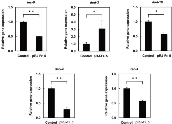
genes, real-time RT-PCR confirmed down-regulation ofins-9gene expression after pRJ-Fr.5 treatment (Fig. 5). These results are consistent with previous findings implicating reduced insulin/IGF-1 signaling (IIS) in lifespan extension [insulin/IGF-1]. pRJ-Fr.5 also down-regulateddod-19,dao-4,and fkb-4 and up-regulated dod-3(Table S2). These expression changes were all verified by real-time RT-PCR analysis (Fig. 5) and, more importantly, correlated with the changes observed when IIS is reduced in C. elegans [52], [53]. Certain DNA motifs were previously reported to be associated with the FOXO transcription factor DAF-16 [53], [54], the activation of which is known to promote longevity inC. elegans
upon reduction of IIS [1]. The DAF-16-binding element (DBE: TTGTTTAC) [54] and the DAF-16-associated element (DAE: CTTATCA) [53] were overrepresented in the upstream regions of
ins-9,dod-3,dod-19,dao-4andfkb-4(Table 1), suggesting that their gene expression is controlled by DAF-16 activity.
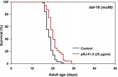
Effects of pRJ-Fr.5 on lifespan indaf-16deletion mutants To clarify whether the IIS-DAF-16 pathway is involved in Fr.5-induced extension of lifespan, we examined the effects of pRJ-Fr.5 on the lifespan of adaf-16deletion mutant. The findings that pRJ-Fr.5 extended the mean lifespan of this mutant by 8–12% (Fig. 6, Table S1) and that this effect was smaller than that observed in wild-type N2 (18–19%) (Fig. 4, Table S1) indicated that pRJ-Fr.5 extends the lifespan by both IIS-DAF-16 pathway-dependent and -inpathway-dependent mechanisms.
Effects of pRJ-Fr.5 on DAF-16 nuclear translocation To ascertain whether pRJ-Fr.5 acts on the IIS-DAF-16 pathway, we examined the effects of pRJ-Fr.5 treatment on DAF-16 nuclear localization, which has been shown to be augmented when IIS is abrogated [55], [56]. We found that pRJ-Fr.5 treatment induced DAF-16 nuclear localization (Fig. S3), suggesting that pRJ-Fr.5 acts on the IIS-DAF-16 pathway.
Analysis of pRJ-Fr.5 components
Next, we analyzed the components of pRJ-Fr.5. The amount of sugars contained in pRJ-Fr.5 was estimated to be 20%(w/w) in terms of glucose. Peptides accounted for more than 60%(w/w) of Fr.5. The molecular weight measurement indicated that pRJ-Fr.5 contained low-molecular weight peptides, such as dipeptides and tripeptides, as well as 16.5% (w/w) 10-HDA. We also measured the 10-HDA content in RJ, pRJ and the other fractions derived from them. The 10-HDA concentrations were as follows: RJ: 1.7%, pRJ: 5.3%, RJ-Fr.1: 8.9%, RJ-Fr.2: 2.1% and pRJ-Fr.4: ,0.1%. These results showed that 10-HDA was enriched especially in pRJ-Fr.5.
Effects of 10-HDA on lifespan in N2 anddaf-16deletion mutants
To elucidate whether 10-HDA is a lifespan-extending agent, we assessed its effects on lifespan. Worms treated beginning at the young adult stage with concentrations of 10-HDA ranging from 10 to 100mM all showed extensions of the mean and maximum lifespans (Fig. 7, Table S3). The largest increase was observed at 25mM, at which concentration the mean lifespan was increased by 12%.
type N2 to similar extents clearly indicates that its lifespan-extending effect is mediated through a mechanism independent of the IIS-DAF-16 pathway.
Effects of combined treatment with pRJ-Fr.5 and 10-HDA on lifespan
To examine the contribution of 10-HDA to pRJ-Fr.5-induced lifespan extension, we tested the effect of combining pRJ-Fr.5 and 10-HDA on lifespan. The lifespan extension achieved by the combination of pRJ-Fr.5 and 10-HDA was greater than that induced by each treatment alone, but the effect was less than additive (Fig. 9, Table S3). These results suggest that pRJ-Fr.5 and 10-HDA do not extend lifespan independently of each other. Therefore, part of the lifespan extension by pRJ-Fr.5 is probably due to its 10-HDA component.
Discussion
The present study demonstrates that RJ has the ability to prolong the lifespan ofC. elegans (Fig. 1), as it is known to do in
Drosophila [9] and mice [29], suggesting that RJ may contain longevity-promoting factors that can act in diverse species across phyla. This lifespan-extending activity of RJ inC. eleganswas not diminished by protease treatment of RJ (Fig. 2), indicating that proteins in RJ are not responsible for the lifespan extension. The water-eluted fraction of pRJ (pRJ-Fr.4) had some lifespan-extending activity (Fig. 3), suggesting that water-soluble com-pounds, such as sugars, amino acids, vitamins or peptides including protein-proteolysis products, may have such activity. Although RJ could extend lifespan (Fig. 1, Table S1), neither the EtOH-soluble (RJ-Fr.1) nor the water-soluble (RJ-Fr.2) fraction of RJ exhibited lifespan-extending activity (Fig. S1, Fig. S2). It is Figure 1. The effects of RJ on the lifespan ofC. elegans.The survival curves of N2 hermaphrodites treated with RJ (0 (control), 1, 10 or 100mg/
ml) are shown. These substances were administered at 20uC, from the young adult stage until death. Day 0 corresponds to the L4 molt. The percentage of live worms is plotted against adult age. Detailed parameters are presented in Table S1.
doi:10.1371/journal.pone.0023527.g001
Figure 2. The effects of pRJ on the lifespan ofC. elegans.The survival curves of N2 hermaphrodites treated with pRJ (0 (control), 1, 10 or 100mg/ml) are shown. The experiment was performed as described in Figure 1 Legend. Detailed parameters are presented in Table S1.
unclear why this activity was not found in either Fr.1 or RJ-Fr.2. One possibility is that the concentrations of the lifespan-extending agents in RJ-Fr.1 and RJ-Fr.2 used in this study may be above or below the narrow dose range that can extend lifespan.
We found that 10-HDA extended the lifespan of C. elegans
(Fig. 7). This is the first evidence that 10-HDA, a defined natural component of RJ, can extend organismal lifespan. 10-HDA is known to have several pharmacological activities such as antibacterial [57], antitumor [58], anti-inflammatory [59], and anti-angiogenic [60] as well as the ability to promote neurogenesis [61] and collagen production [62]. Additionally, 10-HDA is known to possess growth-inhibitory activity in honeybee queens [63]. The present observations demonstrate that 10-HDA can also perform more integrative functions, such as extending organismal lifespan.
The 30% MeOH-eluted fraction of pRJ (pRJ-Fr.5) generated by ODS column chromatography exhibited higher lifespan-extending activity than did pRJ-Fr.4 (Fig. 3, Fig. 4, Table S1). This result can be partly explained by the higher concentration of 10-HDA in pRJ-Fr.5. Furthermore, the finding that the lifespan extension induced by both pRJ-Fr.5 and 10-HDA was greater than that induced by each treatment alone but was less than additive (Fig. 9, Table S3) suggests that part of the lifespan extension by pRJ-Fr.5 was likely due to the 10-HDA contained in pRJ-Fr.5.
A variety of intricate regulatory networks have been shown to control lifespan [2]. Among them, IIS has been well established as a fundamental pathway that regulates the lifespan of C. elegans, Drosophilaand mice [64]. It has been suggested that this pathway is a key determinant of the lifespan differences between honeybee queens and workers [65]. Reduced IIS extends the lifespan through Figure 3. The effects of pRJ-Fr.4 on the lifespan ofC. elegans.The survival curves of N2 hermaphrodites treated with pRJ Fr.4 (0 (control), 5, 10 or 25mg/ml) are shown. The experiment was performed as described in Figure 1 Legend. Detailed parameters are presented in Table S1.
doi:10.1371/journal.pone.0023527.g003
Figure 4. The effects of pRJ-Fr.5 on the lifespan ofC. elegans.Survival curves of N2 hermaphrodites treated with pRJ-Fr.5 (0 (control), 10, 25 or 100mg/ml). The experiment was performed as described in Figure 1 Legend. Detailed parameters are presented in Table S1.
doi:10.1371/journal.pone.0023527.g004
DAF-16, a FOXO transcription factor inC. elegans [66-68]. We found that pRJ-Fr.5 induced nuclear localization of DAF-16 (Fig. S3), indicating that pRJ-Fr.5 activated DAF-16. However, our results showed that the mean-lifespan extension by pRJ-Fr.5 in N2 was greater than that in thedaf-16deletion mutant (Fig. 3, Fig. 4, Table S1), indicating that pRJ-Fr.5 extended the lifespan by both DAF-16-dependent and DAF-16-independent mechanisms. This finding is consistent with the notion that pRJ-Fr.5 extends the lifespan in part through the IIS-DAF-16 pathway and in part through some other mechanism.
We performed DNA microarray and real-time RT-PCR analyses to identify pRJ-Fr.5-regulated genes. In these genes,ins-9was down-regulated by pRJ-Fr.5. Among the 40 known insulin-like peptides in
C. elegans, INS-1 [69], INS-7 [53], INS-11 [70], INS-18 [71] and
DAF-28 [72], [73] have been reported to be regulators of lifespan. Similar toins-1anddaf-28, ins-9is also expressed in chemosensory neurons such as ASI [74], which plays an important role in lifespan determination [74], [75]. Interestingly, the expression ofins-7has been reported to be regulated by IIS-DAF-16 [76]. We also suggested thatins-9 expression is also controlled by IIS-DAF-16 from the finding that the DBE and DAE are overrepresented in the upstream regions ofins-9(Table 1).
We also found that pRJ-Fr. 5 down-regulateddod-19,dao-4and
fkb-4 and up-regulateddod-3 (Fig. 5, Table S2), gene expression changes that are also observed when IIS is reduced [52], [53]. The
dod-19gene encodes an unknown protein; however, intriguingly, it is one of the known determinants of lifespan [53]. It is also interesting to note thatfkb-4 encodes a homolog of the mammalian protein FKBP [52], which binds to the immunosuppressant FK506 and rapamycin. FKBP is involved in the mammalian target of rapamycin (TOR) pathway [77–82] and in diverse cellular functions, including protein folding and the modulation of oxidative stress [83]. FKBP also has neural roles [84], [85]. Inhibition of the TOR pathway has been found to increase lifespan in a variety of species, including yeast, nematodes, flies, and mice [86–91]. The deletion of bothfkb-4 and fkb-5, another FKBP gene, results in lethality under cold conditions [92], and it has been observed that cold conditions affect lifespan in C. elegans [3]. Interestingly, the TOR pathway works as an energy- and nutrient-sensing pathway to determine the queen/worker differentiation in honeybees [93]. Further research is necessary to determine whether these genes are actually involved in the lifespan extension mediated by pRJ-Fr.5.
Recent investigations have provided evidence of common longevity regulation pathways between nematodes, insects and Figure 5. The effects of pRJ-Fr.5 treatment on gene expression inC. elegans.Relative expression levels of genes (ins-9, dod-3, dod-19,dao-4, andfkb-4) in N2 hermaphrodites treated with pRJ-Fr.5 (0 (control) or 25mg/ml) for 24 h starting at the L4 stage. Data are expressed as the mean6S.E.
(n = 3). *: p,0.05; **: p,0.01, compared with control (Student’s t test). doi:10.1371/journal.pone.0023527.g005
Table 1.DAF-16 promoter elements in the upstream region of genes commonly regulated by reduced IIS and pRJ-Fr.5.
Gene Cosmid no.
DBE TTGTTTAC
DAE CTTATC
dod-3 C24B9.9 3 1
dod-19 ZK6.10 1 3
fkb-4 ZC455.10 1 1
dao-4 ZC373.6 1 1
ins-9 C06E2.8 2 2
The number of DAF-16-binding elements and DAF-16-associated elements in the 2kb upstream of each gene is shown.
mammals [1], [64], [87], [90], [91]. The further identification and characterization of the longevity-promoting compounds contained in RJ will broaden our understanding of the gene networks involved in longevity regulation in diverse species and may lead to the development of nutraceutical interventions in the aging process.
Materials and Methods
Nematode strains and culture conditions
The C. elegans strains were maintained at 20uC on nematode growth medium (NGM) agar with Escherichia coliOP50 as a food source, as previously described [94]. The N2 Bristol strain was used as the wild-typeC. elegans. The mutant strain used in this study was CF1038:daf-16(mu86)I and TJ356:zIs356[Ex(daf-16::gfp+rol-6)].
Royal jelly and protease treatment
Fresh RJ, which was produced by honeybees (Apis mellifera L.) foraging onBrassica sp. in China, was obtained from Api Co., Ltd., Gifu, Japan. RJ hydrolyzed by Protease N (pRJ) was prepared as previously described [95]. The following drugs and chemicals were purchased and used: 10-HDA (Alfresa Pharma Co., Ltd., Osaka, Japan) and Protease N ‘‘Amano’’ (from Bacillus subtilis; Amano Enzyme Inc. Aichi, Japan).
Fractionation of RJ
Fresh RJ (1 kg) was mixed with water (1 L), hexane (2 L) and EtOH (4 L), and then shaken slowly overnight at room temperature. This mixture was filtered through No. 2 filter paper and then the extracts were concentrated under pressure until they Figure 6. The effects of pRJ-Fr.5 on the lifespan ofdaf-16(mu86)mutants.The survival curves ofdaf-16(mu86)mutant hermaphrodites treated with pRJ-Fr.5 (0 (control) or 25mg/ml). The experiment was performed as described in Figure 1 Legend. Detailed parameters are presented in
Table S1.
doi:10.1371/journal.pone.0023527.g006
Figure 7. The effects of 10-HDA on the lifespan ofC. elegans.The survival curves of N2 hermaphrodites incubated with 10-HDA (0 (control), 10, 25, 50 or 100mM) are shown. The experiment was performed as described in Figure 1 Legend. Detailed parameters are presented in Table S3.
doi:10.1371/journal.pone.0023527.g007
became a dark yellow material (RJ-Fr.2). This residue was dried by heating under reduced pressure and then mixed with 5% EtOH. The supernatant from this suspension was then freeze-dried (RJ-Fr.1). The yields of RJ-Fr.1 and RJ-Fr.2 were 40.9% and 16.0%, respectively.
Fractionation of pRJ
pRJ (20 g) was mixed with water and then chromatographed on an ODS column. The column was eluted stepwise with water and 30% (v/v) aqueous MeOH. Each fraction (1 L each) was collected and freeze-dried. These fractions were designated as pRJ-Fr.4 (15 g in the H2O phase) and pRJ-Fr.5 (3.8 g in the 30% MeOH phase).
Determination of lifespan
Eggs that were isolated with hypochlorite were placed on fresh NGM agar plates containing UV-killedE. colistrain OP50, unless otherwise stated. UV-killing was used to avoid any effects of liveE. colion the compounds examined in this study and any effects of these compounds on growth of liveE.coli. To kill the OP50, plates covered with OP50 were UV-irradiated as previously described [96]. Worms were raised until the L4 molt and were subsequently transferred onto a new plate containing 40mM 5-fluoro-29-deoxyuridine (FUdR, Sigma Aldrich, St. Louis, MO, USA) to prevent self-fertilization. The day of transfer at the L4 molt was counted as 0-day adult in the lifespan assay. The worms were transferred to fresh plates daily, and the number of surviving Figure 8. The effects of 10-HDA on the lifespan ofdaf-16(mu86)mutants.The survival curves ofdaf-16(mu86) mutant hermaphrodites incubated with 10-HDA (0 (control), 10, 25, 50 or 100mM) are shown. The experiment was performed as described in Figure 1 Legend. Detailed
parameters are presented in Table S3. doi:10.1371/journal.pone.0023527.g008
Figure 9. The effects of 10-HDA and/or pRJ-Fr.5 on the lifespan ofC. elegans.The survival curves of N2 hermaphrodites incubated with 10-HDA (0 or 25mM) and/or pRJ-Fr.5 (0 or 25mg/ml) are shown. The experiment was performed as described in Figure 1 Legend. Detailed parameters
are presented in Table S3.
worms was monitored until death unless otherwise stated. Worms were judged to be dead when they did not respond to a mechanical stimulus. To focus on aging, worms that had become desiccated on the side of the plate after crawling off, that displayed extruded internal organs or that died because of progeny hatching inside the uterus (matricidal death) were excluded from our analysis. The results of the survival assays were analyzed using the Kaplan-Meier method, and significance was measured with the log-rank test using the statistical analysis package StatMate III (ATMS, Tokyo, Japan).
Treatment with compounds
EtOH solutions of RJ, pRJ and RJ-Fr.1, aqueous solutions of RJ-Fr.2, pRJ-Fr.4 and pRJ-Fr.5 as well as 10-HDA in DMSO, were added to liquid NGM that had been autoclaved and cooled to 50uC. The media were immediately dispensed into Petri dishes. Experiments involving RJ, pRJ and RJ-Fr.1 were performed in parallel with those involving a control group treated with 0.1% EtOH; and experiments involving 10-HDA were conducted in parallel with those involving a control group treated with 0.03% DMSO.
DNA microarray analysis
TheC. elegansN2 strains were treated with pRJ-Fr.5 (0 (control) or 25mg/ml) for 24 h beginning at the L4 stage. A total of 8,000-10,000 worms were collected for each sample. The worms were homogenized in TRIzolHReagent (InvitrogenTM, Carlsbad, CA) using a Precellys 24 (Bertin Technologies, Montigny-le-Breton-neux, France). Total RNA was extracted with a PureLinkTMRNA Mini kit (InvitrogenTM). The Agilent C. elegans (V2) Gene Expression Microarray, 4x44K (G2519F-020186) was used for global gene expression analysis. This microarray contains 43,803
C. eleganscomplementary DNA (cDNA) probes, each consisting of a single 60-oligomer oligonucleotide sequence. Target RNA labeling and hybridization were performed according to the protocol for one-color microarray-based gene expression analysis using the Quick Amp Labeling Kit (Agilent Technologies, Santa Clara, CA). In brief, 500 ng of RNA was transcribed using the oligo(dT)-based T7 promoter primer and MMLV-RT in the first-and second-strfirst-and cDNA synthesis reactions. The double-strfirst-anded cDNAs were used as templates for the preparation of fluorescent complementary RNAs (cRNAs) in the presence of T7 RNA polymerase and cyanine 3-CTP dye in an in vitro transcription reaction. The labeled cRNAs were purified, fragmented, and hybridized to microarrays in a rotating hybridization oven at 10 rpm for 17 h at 65uC. After hybridization, the microarrays were washed according to the manufacturer’s instructions and scanned using an Agilent DNA Microarray Scanner with Scan Control software (Agilent Technologies). The resulting images were processed, and the raw data were collected using the Agilent Feature Extraction software. The gene expression data were analyzed using GeneSpring GX 11 (Agilent Technologies). The signal intensity of each probe was normalized by a percentile shift, in which each value was divided by the 75th percentile of all the values in its array. The microarray data discussed in this publication have been deposited in NCBI’s Gene Expression Omnibus (GEO) and are accessible through the GEO Series accession number GSE26094 (http://www.ncbi.nlm.nih.gov/ geo/query/acc.cgi?acc=GSE26094). To identify the genes with biological significance, we applied flags attributed to the signal intensity of each probe, the fold change, and the Student’st-test values. All the data are MIAME compliant.
Real-time RT-PCR analysis
Total RNA was reverse-transcribed to cDNA using a High Capacity cDNA Reverse Transcription kit (Applied Biosystems), and subjected to real-time PCR using the SYBR Premix Ex Taq II (Perfect Real Time) (TaKaRa) and the Thermal Cycler Dice Real Time System (TaKaRa). The following primers were used:ins-9
forward, 59-GGCGAGAAGAACCTTGGAAAC-39;ins-9reverse, 59-ACAGCACAGCTTAGAGAGATCCTG-39; ins-20 forward, 59-TCATCATCACAGGCACAAAGG-39;ins-20reverse, 59-GC-AAAATATCATCATCCGTCAGG-39; ins-23forward, 59-CAG-AGCTTCACGTTCGTAGGG-39;ins-23 reverse, 59-GAACAG-TACTCGGTTGGACTTGG-39;dod-3forward, 59-AAGCCAT-GTTCCCGAATGAG-39; dod-3 reverse, 59-GCTGCGAAAAG-CAAGAAAATG-39;dod-19forward, 59-ACCGTTCCCAGTTT-TACAGTCC-39;dod-19reverse, 59-TATTTTGAGGCGCGGA-TACAC-39; dao-4 forward, 59-GCACATTACAAATGCTTCA-AGGAC-39; dao-4 reverse, 59 -TGACACCCTCATCCCCATA-AC-39; fkb-4 forward, 59 -CTATGCGAGGAATGTGTATTG-GAG-39;fkb-4reverse, 59 -TGGACAGTGTAATAGAGTGGCT-GAC-39;rla-1forward, 59-ACCGGCGAGAAGATCGCTAC-39;
rla-1reverse, 59-CGGAAGAGACAGAAGTGATGAGG-39. The relative expression level of each gene was calculated using the comparative Ct method. Ribosomal protein, Large subunit, Acidic (P1) family member (rla-1) was used as an internal control gene. Analysis of pRJ-Fr.5 components
Sugar content was estimated by orcinol/sulfuric acid analysis. Peptide content was estimated by the Lowry method. The molecular weights of peptides were estimated by HPLC analysis on a Superdex Peptide HR 10/30 column (Pharmacia Biotech, Uppsala, Sweden). The 10-HDA content was determined using HPLC with a Consmosil 5C18-MS-II column (Nacalai Tesque, Tokyo, Japan) at 40uC. The column was eluted with a mobile phase of 10 mM phosphate buffer (pH 2.5) and MeOH (1960:1540, v/v) at a flow rate of 1.0 ml/min.
Supporting Information
Figure S1 The effect of RJ-Fr.1 on the lifespan ofC. elegans. The survival curves of N2 hermaphrodites incubated with RJ-Fr.1 (0 (control), 10, 25 or 100mg/ml) are shown. The RJ-Fr.1 was given at 20uC from 0-day adult until death. Day 0 corresponds to the L4 molt. The percentage of live worms is plotted against adult age. Detailed parameters are presented in Table S1.
(TIF)
Figure S2 The effect of RJ-Fr.2 on the lifespan ofC. elegans. The survival curves of N2 hermaphrodites incubated with RJ-Fr.2 (0 (control), 10, 25 or 100mg/ml) are shown. The RJ-Fr.2 was given at 20uC from 0-day adult until death. Day 0 corresponds to the L4 molt. The percentage of live worms is plotted against adult age. Detailed parameters are presented in Table S1.
(TIF)
Figure S3 The effect of pRJ-Fr.5 treatment on DAF-16 nuclear translocation. DAF-16 nuclear translocation was examined in a
intestinal cells is shown. N = 58 (untreated) and 102 (pRJ-Fr.5 treated).
(TIF)
Table S1 Effects of RJ or pRJ on the lifespan. (XLSX)
Table S2 Differentially regulated genes by pRJ-Fr.5 treatment. (XLSX)
Table S3 Effects of 10-HDA and/or pRJ-Fr.5 on the lifespan. (XLSX)
Acknowledgments
We are grateful to Ms. Mami Kishima (RIKEN OSC) for her technical advice. Some strains were provided by the Caenorhabditis Genetics Center funded by the National Institutes of Health National Center for Research Resources (NCRR).
Author Contributions
Conceived and designed the experiments: SH YH YF HM YA KI MI. Performed the experiments: YH YF HM SH YA AS TK MI. Analyzed the data: YH SH YF HM YA KI AS TK MT YN MI. Wrote the paper: SH YH MI YF HM YA KI TK.
References
1. Kenyon C (2005) The plasticity of aging: insights from long-lived mutants. Cell 120: 449–460.
2. Kenyon C (2010) The genetics of ageing. Nature 464: 504–512.
3. Klass MR (1977) Aging in the nematodeCaenorhabditis elegans: major biological and environmental factors influencing life span. Mech Ageing Dev 6: 413–429. 4. Lithgow GJ, White TM, Melov S, Johnson TE (1995) Thermotolerance and extended life-span conferred by single-gene mutations and induced by thermal stress. Proc Natl Acad Sci USA 92: 7540–7544.
5. Philpott DE, Bensch KG, Miquel J (1974) Life span and fine structural changes in oxygen-poisonedDrosophila melanogaster. Aerosp Med 45: 283–289. 6. Honda S, Ishii N, Suzuki K, Matsuo M (1993) Oxygen-dependent perturbation
of life span and aging rate in the nematode. J Gerontol 48: B57–B61. 7. Honda Y, Tanaka M, Honda S (2010) Redox regulation, gene expression and
longevity. Geriat Gerontol Int 10: S59–S69.
8. Bordone L, Guarente L (2005) Calorie restriction, sirt1 and metabolism: understanding longevity. Nat Rev Mol Cell Biol 6: 298–305.
9. Gardner TS (1948) The use of Drosophila melanogaster as a screening agent for longevity factors; pantothenic acid as a longevity factor in royal jelly. J Gerontol 3: 1–8.
10. Harrington LA, Harley CB (1988) Effect of vitamin E on lifespan and reproduction inCaenorhabditis elegans. Mech Ageing Dev 43: 71–78.
11. Driver C, Georgeou A (2003) Variable effects of vitamin E on Drosophila longevity. Biogerontology 4: 91–95.
12. Zou S, Sinclair J, Wilson MA, Carey JR, Liedo P, et al. (2007) Comparative approaches to facilitate the discovery of prolongevity interventions: Effects of tocopherols on lifespan of three invertebrate species. Mech Ageing Dev 128: 222–226.
13. Soukas AA, Kane EA, Carr CE, Melo JA, Ruvkun G (2009) Rictor/TORC2 regulates fat metabolism, feeding, growth, and life span inCaenorhabditis elegans. Genes Dev 23: 496–511.
14. Hashimoto T, Horikawa M, Nomura T, Sakamoto K (2010) Nicotinamide adenine dinucleotide extends the lifespan ofCaenorhabditis elegansmediated by sir-2.1anddaf-16. Biogerontology 11: 31–43.
15. Honda Y, Tanaka M, Honda S (2010) Trehalose extends longevity in the nematodeCaenorhabditis elegans. Aging Cell 9: 558–569.
16. D’Antona G, Ragni M, Cardile A, Tedesco L, Dossena M, et al. (2010) Branched-chain amino acid supplementation promotes survival and supports cardiac and skeletal muscle mitochondrial biogenesis in middle-aged mice. Cell Metab 12: 362–372.
17. Winston ML (1991) The biology of the honey bee. CambridgeMA: Harvard Univ Press. pp 56.
18. Remolina SC, Hughes KA (2008) Evolution and mechanisms of long life and high fertility in queen honey bees. Age (Dordr) 30: 177–185.
19. Page RE Jr., Peng CY (2001) Aging and development in social insects with emphasis on the honey bee,Apis melliferaL. Exp Gerontol 36: 695–711. 20. Takenaka T (1982) Chemical composition of royal jelly. Honeybee Sci 3: 69–74. 21. Hattori N, Nomoto H, Fukumitsu H, Mishima S, Furukawa S (2010) AMP N1-oxide, a unique compound of royal jelly, induces neurite outgrowth from PC12 cells via signaling by protein kinase A independent of that by mitogen-activated protein kinase. Evid Based Complement Alternat Med 7: 63–68.
22. Fontana R, Mendes MA, de Souza BM, Konno K, Cesar LM, et al. (2004) Jelleines: a family of antimicrobial peptides from the Royal Jelly of honeybees (Apis mellifera). Peptides 25: 919–928.
23. Bı´likova´ K, Hanes J, Nordhoff E, Saenger W, Klaudiny J, et al. (2002) Apisimin, a new serine-valine-rich peptide from honeybee (Apis melliferaL.) royal jelly: purification and molecular characterization. FEBS Lett 528: 125–129. 24. Gasic S, Vucevic D, Vasilijic S, Antunovic M, Chinou I, et al. (2007) Evaluation
of the immunomodulatory activities of royal jelly components in vitro. Immunopharmacol Immunotoxicol 29: 521–536.
25. Henschler D (1956) Identification of choline esters in biological material, especially acetylcholine in royal jelly of the bee. Hoppe-Seylers Z. physiol. Chem 305: 34–41.
26. Colhoun EH, Smith MV (1960) Neurohormonal properties of royal jelly. Nature 188: 854–855.
27. Wei W, Wei M, Kang X, Deng H, Lu Z (2009) A novel method developed for acetylcholine detection in royal jelly by using capillary electrophoresis coupled
with electrogenerated chemiluminescence based on a simple reaction. Electro-phoresis 30: 1949–1952.
28. Butenandt A, Rembold H (1957) Royal jelly of the honeybee. I. Isolation, constitution analysis, and incidence of 10-hydroxy-delta 2-decenoic acid. Hoppe-Seylers Z physiol Chem 203: 284–289.
29. Inoue S, Koya-Miyata S, Ushio S, Iwaki K, Ikeda M, et al. (2003) Royal Jelly prolongs the life span of C3H/HeJ mice: correlation with reduced DNA damage. Exp Gerontol 38: 965–969.
30. Fujiwara S, Imai J, Fujiwara M, Yaeshima T, Kawashima T, et al. (1990) A potent antibacterial protein in royal jelly. Purification and determination of the primary structure of royalisin. J Biol Chem 265: 11333–11337.
31. Bincoletto C, Eberlin S, Figueiredo CA, Luengo MB, Queiroz ML (2005) Effects produced by royal jelly on haematopoiesis: relation with host resistance against Ehrlich ascites tumour challenge. Int Immunopharmacol 5: 679–688. 32. Kataoka M, Arai-N, Taniguchi-Y, Kohno K, Iwaki K, et al. (2001) Analysis of
anti-allergic function of royal jelly. J Nat Med 55: 174–180.
33. Kamakura M, Mitani N, Fukuda T, Fukushima M (2001) Antifatigue effect of fresh royal jelly in mice. J Nutr Sci Vitaminol. Tokyo, 47: 394–401. 34. Fujii A, Kobayashi S, Kuboyama N, Furukawa Y, Kaneko Y, et al. (1990)
Augmentation of wound healing by royal jelly (RJ) in streptozotocin-diabetic rats. Jpn J Pharmacol 53: 331–337.
35. Kohno K, Okamoto I, Sano O, Arai N, Iwaki K, et al. (2004) Royal jelly inhibits the production of proinflammatory cytokines by activated macrophages. Biosci Biotechnol Biochem 68: 138–145.
36. Okamoto I, Taniguchi Y, Kunikita T, Kohno K, Iwaki K, et al. (2003) Major royal jelly protein 3 modulates immune responsesin vitroandin vivo. Life Sci 1: 2029–2045.
37. Erem C, Deger O, Ovali E, Barlak Y (2006) The effects of royal jelly on autoimmunity in Graves’ disease. Endocrine 30: 175–183.
38. Hattori N, Nomoto H, Fukumitsu H, Mishima S, Furukawa S (2007) Royal jelly-induced neurite outgrowth from rat pheochromocytoma PC12 cells requires integrin signal independent of activation of extracellular signal-regulated kinases. Biomed Res 28: 139–146.
39. Taniguchi Y, Kohno K, Inoue S, Koya-Miyata S, Okamoto I, et al. (2003) Oral administration of royal jelly inhibits the development of atopic dermatitis-like skin lesions in NC/Nga mice. Int Immunopharmacol 3: 1313–1324. 40. Vittek J (1995) Effect of royal jelly on serum lipids in experimental animals and
humans with atherosclerosis. Experientia 51: 927–935.
41. Hidaka S, Okamoto Y, Uchiyama S, Nakatsuma A, Hashimoto K, et al. (2006) Royal jelly prevents osteoporosis in rats: beneficial effects in ovariectomy model and in bone tissue culture model. Evid Based Complement Alternat Med 3: 339–348. 42. Narita Y, Nomura J, Ohta S, Inoh Y, Suzuki KM, et al. (2006) Royal jelly
stimulates bone formation: physiologic and nutrigenomic studies with mice and cell lines. Biosci Biotechnol Biochem 70: 2508–2514.
43. Nagai T, Inoue R, Suzuki N, Nagashima T (2006) Antioxidant properties of enzymatic hydrolysates from royal jelly. J Med Food 9: 363–367.
44. Guo H, Ekusa A, Iwai K, Yonekura M, Takahata Y, et al. (2008) Royal jelly peptides inhibit lipid peroxidation in vitro and in vivo. J Nutr Sci Vitaminol. Tokyo, 54: 191–195.
45. Matsui T, Yukiyoshi A, Doi S, Sugimoto H, Yamada H, et al. (2002) Gastrointestinal enzyme production of bioactive peptides from royal jelly protein and their antihypertensive ability in SHR. J Nutr Biochem 13: 80–86. 46. Tokunaga KH, Yoshida C, Suzuki KM, Maruyama H, Futamura Y, et al.
(2004) Antihypertensive effect of peptides from royal jelly in spontaneously hypertensive rats. Biol Pharm Bull 27: 189–192.
47. Sultana A, Nabi AH, Nasir UM, Maruyama H, Suzuki KM, et al. (2008) A dipeptide YY derived from royal jelly proteins inhibits renin activity. Int J Mol Med 21: 677–681.
48. Wilson MA, Shukitt-Hale B, Kalt W, Ingram DK, Joseph JA, et al. (2006) Blueberry polyphenols increase lifespan and thermotolerance inCaenorhabditis elegans. Aging Cell 5: 59–68.
50. Brown MK, Evans JL, Luo Y (2006) Beneficial effects of natural antioxidants EGCG and alpha-lipoic acid on life span and age-dependent behavioral declines inCaenorhabditis elegans. Pharmacol Biochem Behav 85: 620–628.
51. Abbas S, Wink M (2009) Epigallocatechin gallate from green tea (Camellia sinensis) increases lifespan and stress resistance inCaenorhabditis elegans. Planta Med 75: 216–221.
52. Yu H, Larsen PL (2001) DAF-16-dependent and independent expression targets of DAF-2 insulin receptor-like pathway inCaenorhabditis elegansinclude FKBPs. J Mol Biol 314: 1017–1028.
53. Murphy CT, McCarroll SA, Bargmann CI, Fraser A, Kamath RS, et al. (2003) Genes that act downstream of DAF-16 to influence the lifespan ofCaenorhabditis elegans.Nature 424, 277-283.
54. Furuyama T, Nakazawa T, Nakano I, Mori N (2000) Identification of the differential distribution patterns of mRNAs and consensus binding sequences for mouse DAF-16 homologues. Biochem J 349: 629–634.
55. Henderson ST, Johnson TE (2001) daf-16 integrates developmental and environmental inputs to mediate aging in the nematodeCaenorhabditis elegans. Curr Biol 11: 1975–1980.
56. Lin K, Hsin H, Libina N, Kenyon C (2001) Regulation of theCaenorhabditis eleganslongevity protein DAF-16 by insulin /IGF-1 and germline signaling. Nature Genet 28: 139–145.
57. Blum MS, Novak AF, Taber S III (1959) 10-Hydroxy-delta 2-decenoic acid, an antibiotic found in royal jelly. Science 130: 452–453.
58. Townsend GF, Morgan JF, Tolnai S, Hazlett B, Morton HJ, et al. (1960) Studies on the in vitro antitumor activity of fatty acids. I. 10-Hydroxy-2-decenoic acid from royal jelly. Cancer Res 20: 503–510.
59. Yang XY, Yang DS, Wei-Zhang, Wang JM, Li CY, et al. (2010) 10-Hydroxy-2-decenoic acid from Royal jelly: a potential medicine for RA. J Ethnopharmacol 128: 314–321.
60. Izuta H, Chikaraishi Y, Shimazawa M, Mishima S, Hara H (2009) 10-Hydroxy-2-decenoic acid, a major fatty acid from royal jelly, inhibits VEGF-induced angiogenesis in human umbilical vein endothelial cells. Evid Based Complement Alternat Med 6: 489–494.
61. Hattori N, Nomoto H, Fukumitsu H, Mishima S, Furukawa S (2007) Royal jelly and its unique fatty acid, 10-hydroxy-trans-2-decenoic acid, promote neurogen-esis by neural stem/progenitor cells in vitro. Biomed Res 28: 261–266. 62. Koya-Miyata S, Okamoto I, Ushio S, Iwaki K, Ikeda M, et al. (2004)
Identification of a collagen production-promoting factor from an extract of royal jelly and its possible mechanism. Biosci Biotechnol Biochem 68: 767–773. 63. Kinoshita G, Shuel RW (1975) Mode of action of royal jelly in honeybee
development. X Some aspects of lipid nutrition. Can J Zoology 53: 311–319. 64. Tatar M, Bartke A, Antebi A (2003) The endocrine regulation of aging by
insulin-like signals. Science 299: 1346–1351.
65. Corona M, Velarde RA, Remolina S, Moran-Lauter A, Wang Y, et al. (2007) Vitellogenin, juvenile hormone, insulin signaling, and queen honey bee longevity. Proc Natl Acad Sci U S A 104: 7128–7133.
66. Kenyon C, Chang J, Gensch E, Rudner A, Tabtiang R (1993) AC. elegans mutant that lives twice as long as wild type. Nature 366: 461–464.
67. Lin K, Dorman JB, Rodan A, Kenyon C (1997)daf-16: An HNF-3/forkhead family member that can function to double the life-span ofCaenorhabditis elegans. Science 278: 1319–1322.
68. Ogg S, Paradis S, Gottlieb S, Patterson GI, Lee L, et al. (1997) The Fork head transcription factor DAF-16 transduces insulin-like metabolic and longevity signals inC. elegans. Nature 389: 994–999.
69. Pierce SB, Costa M, Wisotzkey R, Devadhar S, Homburger SA, et al. (2001) Regulation of DAF-2 receptor signaling by human insulin andins-1, a member of the unusually large and diverseC. elegansinsulin gene family. Genes Dev 15: 672–686.
70. Kawano T, Nagatomo R, Kimura Y, Gengyo-Ando K, Mitani S (2006) Disruption of ins-11, aCaenorhabditis elegansinsulin-like gene, and phenotypic analyses of the gene-disrupted animal. Biosci Biotechnol Biochem 70: 3084–3087.
71. Kawano T, Ito Y, Ishiguro M, Takuwa K, Nakajima T, et al. (2000) Molecular cloning and characterization of a new insulin/IGF-like peptide of the nematode Caenorhabditis elegans. Biochem Biophys Res Comm 273: 431–436.
72. Malone EA, Inoue T, Thomas JH (1996) Genetic analysis of the roles ofdaf-28 andage-1 in regulatingCaenorhabditis elegans dauer formation. Genetics 143: 1193–1205.
73. Li W, Kennedy SG, Ruvkun G (2003) daf-28 encodes a C. elegansinsulin superfamily member that is regulated by environmental cues and acts in the DAF-2 signaling pathway. Genes Dev 17: 844–858.
74. Alcedo J, Kenyon C (2004) Regulation of C. elegans longevity by specific gustatory and olfactory neurons. Neuron 41: 45–55.
75. Bishop NA, Guarente L (2007) Two neurons mediate diet-restriction-induced longevity inC. elegans. Nature 447: 545–549.
76. Murphy CT, Lee SJ, Kenyon C (2007) Tissue entrainment by feedback regulation of insulin gene expression in the endoderm ofCaenorhabditis elegans. Proc Natl Acad Sci USA104: 19046–19050.
77. Brown EJ, Albers MW, Shin TB, Ichikawa K, Keith CT, et al. (1994) A mammalian protein targeted by G1-arresting rapamycin-receptor complex. Nature 369: 756–758.
78. Sabatini DM, Erdjument-Bromage H, Lui M, Tempst P, Snyder SH (1994) RAFT1: a mammalian protein that binds to FKBP12 in a rapamycin-dependent fashion and is homologous to yeast TORs. Cell 78: 35–43.
79. Chiu MI, Katz H, Berlin V (1994) RAPT1, a mammalian homolog of yeast Tor, interacts with the FKBP12/rapamycin complex. Proc Natl Acad Sci U S A 91: 12574–12578.
80. Sabers CJ, Martin MM, Brunn GJ, Williams JM, Dumont FJ, et al. (1995) Isolation of a protein target of the FKBP12-rapamycin complex in mammalian cells. J Biol Chem 270: 815–822.
81. Bai X, Ma D, Liu A, Shen X, Wang QJ, et al. (2007) Rheb activates mTOR by antagonizing Its endogenous inhibitor, FKBP38. Science 318: 977–980. 82. Dunlop EA, Dodd KM, Seymour LA, Tee AR (2009) Mammalian target of
rapamycin complex 1-mediated phosphorylation of eukaryotic initiation factor 4E-binding protein 1 requires multiple protein-protein interactions for substrate recognition. Cell Signal 21: 1073–1084.
83. Kang CB, Hong Y, Dhe-Paganon S, Yoon HS (2008) FKBP family proteins: immunophilins with versatile biological functions. Neurosignals 16: 318–325. 84. Sabatini DM, Lai MM, Snyder SH (1997) Neural roles of immunophilins and
their ligands. Mol Neurobiol 15: 223–239.
85. Sanokawa-Akakura R, Cao W, Allan K, Patel K, Ganesh A, et al. (2010) Control of Alzheimer’s amyloid beta toxicity by the high molecular weight immunophilin FKBP52 and copper homeostasis inDrosophila. PLoS One 5: e8626. 86. Kaeberlein M, Powers RW 3rd, Steffen KK, Westman EA, Hu D, et al. (2005)
Regulation of yeast replicative life span by TOR and Sch9 in response to nutrients. Science 310: 1193–1196.
87. Vellai T, Takacs-Vellai K, Zhang Y, Kovacs AL, Orosz L, et al. (2003) Influence of TOR kinase on lifespan inC. elegans. Nature 426: 620.
88. Jia K, Chen D, Riddle DL (2004) The TOR pathway interacts with the insulin signaling pathway to regulateC. eleganslarval development, metabolism and life span.Development131: 3897–3906.
89. Sheaffer KL, Updike DL, Mango SE (2008) The target of rapamycin pathway antagonizes pha-4/FoxA to control development and aging. Curr Biol 18: 1355–1364.
90. Kapahi P, Zid BM, Harper T, Koslover D, Sapin V, et al. (2004) Regulation of lifespan inDrosophilaby modulation of genes in the TOR signaling pathway. Curr Biol 14: 885–890.
91. Harrison DE, Strong R, Sharp ZD, Nelson JF, Astle CM, et al. (2009) Rapamycin fed late in life extends lifespan in genetically heterogeneous mice. Nature 460: 392–395.
92. Winter AD, Eschenlauer SC, McCormack G, Page AP (2007) Loss of secretory pathway FK506-binding proteins results in cold-sensitive lethality and associate extracellular matrix defects in the nematodeCaenorhabditis elegans. J Biol Chem 282: 12813–12821.
93. Patel A, Fondrk MK, Kaftanoglu O, Emore C, Hunt G, et al. (2007) The making of a queen: TOR pathway is a key player in diphenic caste development. PLoS One 2: e509.
94. Brenner S (1974) The genetics ofCaenorhabditis elegans.Genetics 77: 71–94. 95. Suzuki K, Yoshida C, Tokunaga K, Maruyama H, Futamura Y, et al. (2003)
Inhibition of angiotensin I-converting enzyme by protease digests from Royal jelly. J Jpn Soc Food Sci Technol 50: 286–288.
96. Honda Y, Tanaka M, Honda S (2008) Modulation of longevity and diapause by redox regulation mechanisms under the insulin-like signaling control in Caenorhabditis elegans.Exp. Gerontol 43: 520–529.