Adhesion molecule profiles of B-cell
non-Hodgkin’s lymphomas in the
leukemic phase
Departamento de Clínica Médica e Centro de Terapia Celular, Faculdade de Medicina de Ribeirão Preto, Universidade de São Paulo, Ribeirão Preto, SP, Brasil
D.M. Matos, E.G. Rizzatti, A.B. Garcia, D.A.P. Gallo and R.P. Falcão
Abstract
We evaluated the expression of 10 adhesion molecules on peripheral blood tumor cells of 17 patients with chronic lymphocytic leukemia, 17 with mantle-cell lymphoma, and 13 with nodal or splenic marginal B-cell lymphoma, all in the leukemic phase and before the beginning of any therapy. The diagnosis of B-cell non-Hodgkin’s lymphomas was based on cytological, histological, immunophenotypic, and mo-lecular biology methods. The mean fluorescence intensity of the adhesion molecules in tumor cells was measured by flow cytometry of CD19-positive cells and differed amongst the types of lymphomas. Comparison of chronic lymphocytic leukemia and mantle-cell lym-phoma showed that the former presented a higher expression of CD11c and CD49c, and a lower expression of CD11b and CD49d adhesion molecules. Comparison of chronic lymphocytic leukemia and marginal B-cell lymphoma showed that the former presented a higher expression of CD49c and a lower expression of CD11a, CD11b, CD18, CD49d, CD29, and CD54. Finally, comparison of mantle-cell lymphoma and marginal B-cell lymphoma showed that marginal B-cell lymphoma had a higher expression of CD11a, CD11c, CD18, CD29, and CD54. Thus, the CD49c/CD49d pair consistently demonstrated a distinct pattern of expression in chronic lymphocytic leukemia compared with mantle-cell lymphoma and marginal B-cell lymphoma, which could be helpful for the differential diagnosis. Moreover, the distinct profiles of adhesion molecules in these diseases may be responsible for their different capacities to invade the blood stream.
Correspondence
R.P. Falcão
Laboratório de Hematologia Departamento de Clínica Médica FMRP, USP
Av. Bandeirantes, 3900 14049-900 Ribeirão Preto, SP Brasil
Fax: +55-16-3633-1144 E-mail: [email protected]
Publication supported by FAPESP.
Received May 9, 2006 Accepted August 15, 2006
Key words
•Non-Hodgkin’s lymphoma •Adhesion molecules •Flow cytometry •CD49c/CD49d
Introduction
B-cell non-Hodgkin’s lymphomas (B-NHL) are a heterogeneous group of neoplas-tic diseases whose tumor cells are the malig-nant counterparts of normal B-lymphocytes (1). Although in most cases B-NHL arise from the lymph node (2),they can also dis-play a leukemic phase characterized by the
cases in other series (6), and in nodal and splenic marginal B-cell lymphoma, it has been reported to be present in 11 and 57% of cases, respectively (7). These differences in the clinical behavior of B-NHL are most likely related to differential expression of various adhesion molecules on the mem-brane of tumor cells (4).
Adhesion molecules are a group of ligands and receptors involved in several biological processes, particularly cell migration (8). Given that some B-NHL subtypes are con-sidered to be the malignant counterparts of distinctive steps in normal B-cell develop-ment, it is reasonable to infer that the malig-nant lymphoid cells use the same mechan-isms of lymphocyte migration as normal B-cells to disseminate from their primary sites. In fact, a number of in vitro (9) and preclini-cal experiments (10,11), as well as clinipreclini-cal observations (12-14) give support to this hypothesis.
Here we studied the expression of 10 adhesion molecules in tumor cells from the peripheral blood of patients with leukemic B-NHL (chronic lymphocytic leukemia, mantle-cell lymphoma, nodal or splenic marginal B-cell lymphoma) to determine how the down-regulation of specific adhesion markers inherently associated with malig-nant transformation contributes to the bio-genesis of the leukemic phase observed in some B-NHL subtypes.
Patients, Material and Methods
Patients
We studied the peripheral blood of 47 patients with a diagnosis of B-NHL in the leukemic phase: 17 with chronic lympho-cytic leukemia, 17 with mantle-cell lym-phoma, and 13 with nodal or splenic mar-ginal B-cell lymphoma. Some clinical and laboratory features of the patients are shown in Table 1. We defined the leukemic phase of B-NHL by the presence of a minimum of
3000 abnormal lymphoid cells/µL associ-ated with immunoglobulin light chain re-striction. The flow cytometry studies were performed before the beginning of treat-ment. The research protocol was approved by the Ethics Committee of the University Hospital, School of Medicine of Ribeirão Preto, and written informed consent was obtained from all patients.
The diagnosis of B-NHL was based on cytological, histological, immunopheno-typic, and molecular biology methods. All cases of chronic lymphocytic leukemia had a score of 4 or 5 in the Matutes scoring system (15,16), while mantle-cell lymphoma and marginal B-cell lymphoma had a score of 0 to 3. In addition, except for one patient, who showed CYCLIN D1 positivity only by immunohistochemistry, all cases of mantle-cell lymphoma showed evidence of CYCLIN D1 overexpression as measured by real-time polymerase chain reaction (data not shown).
Flow cytometry
All samples were analyzed with a FACScan flow cytometer (Becton Dickin-son, San Jose, CA, USA) equipped with an argon ion laser with a wavelength of 488 nm, by collecting 10,000 events per tube. The Cell Quest software (Becton Dickinson) was used for data acquisition and analysis. Only CD19-positive cells were gated for analysis (Figure 1A), and the expression of adhesion molecules was measured by the mean fluo-rescent intensity (MFI) obtained from histo-gram graphs on a linear scale (Figure 1B). Since the setting of an arbitrary cut-off value led to no meaningful conclusions, we pre-ferred to use the MFI as the variable to estimate the intensity of expression of the adhesion molecules. The final MFI value was obtained by subtracting the “MFI of non-specific isotypes” from the “MFI of the relevant antigen”.
density gradient centrifugation (1.077 g/mL; Becton Dickinson). The flow cytometry panel consisted of monoclonal antibodies against the following adhesion molecules (with the respective clones in parentheses): CD11a, αL integrin (HI111); CD11b, αM integrin (ICRF44); CD11c, αX integrin (B-ly6); CD18, ß2 integrin (6.7); CD29, ß1 integrin
(MAR4); CD44, H-CAM (G44-26); CD49c, α3 integrin, VLA-3 (C3II.1); CD49d, α4 in-tegrin, VLA-4 (9F10); CD54, ICAM-1 (HA58); CD62L, L-selectin (Dreg-56). All monoclonal antibodies were conjugated with fluorescein isothiocyanate, phycoerythrin, or perydin chlorophyl protein and all were pur-chased from Becton Dickinson. To exclude T lymphocytes and monocytes from analy-sis, CD19 was added to all tubes: tube 1, CD18 x CD11a x CD19; tube 2, CD11b x CD19; tube 3, CD11c x CD19; tube 4, CD44 x CD29 x CD19; tube 5, CD62 x CD54 x CD19; tube 6, CD49c x CD19; tube 7, CD49d x CD19.
Statistical analysis
Comparison of the expression of adhe-sion molecules between the three groups was performed by the Kruskal-Wallis test using GraphPad Prism and Statistical Anal-ysis System (SAS) softwares, with the level of significance set at P < 0.05. When a P value of 0.05 or less was found in the Krus-kal-Wallis test, the Dunn post-test was ap-plied to analyze pairs of groups.
Results
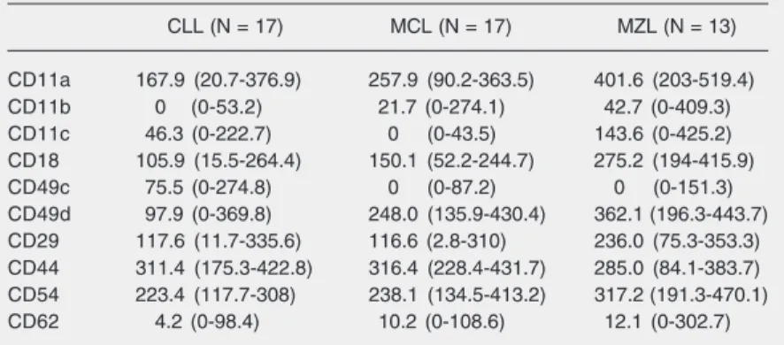
The expression of the adhesion molecules of malignant lymphoid cells was measured by flow cytometry in CD19-positive cells. The MFI of the adhesion molecules CD11a, CD11b, CD11c, CD18, CD49c, CD49d, CD29, and CD54 was different in the three groups, as shown in Table 2. The Dunn post-test was applied when the P value was 0.05 or less. The results of pair comparisons are
Table 1. Clinical and laboratory features of the patients according to diagnosis.
CLL MCL MZL
Sex (M/F) 11/6 14/3 10/3
Age (years) 62 (45-78) 67 (34-81) 66 (28-95)
Lymphadenomegaly 9/17 (53%) 11/17 (65%) 4/13 (31%)
Splenomegaly 8/17 (47%) 14/17 (82%) 13/13 (100%)
Lymphocytes (x 109/L) 42.7 (6.0-122.4) 62.4 (6.7-256.0) 18.7 (3.6-67.6)
Data are reported as median (range) or number (percent). CLL = chronic lymphocytic leukemia; MCL = mantle-cell lymphoma; MZL = marginal B-cell lymphoma.
Figure 1. Flow cytometry graphs. A, A gate strategy graph: only both CD19-positive and low side scatter-SSC (internal cellular granularity) cells were selected for analysis (square). B, A histogram graph: a single-parameter histogram in linear scale shows the expression of the adhesion molecule CD49d.
Table 2. Adhesion molecule on lymphoma cells according to diagnosis.
CLL (N = 17) MCL (N = 17) MZL (N = 13)
CD11a 167.9 (20.7-376.9) 257.9 (90.2-363.5) 401.6 (203-519.4)
CD11b 0 (0-53.2) 21.7 (0-274.1) 42.7 (0-409.3)
CD11c 46.3 (0-222.7) 0 (0-43.5) 143.6 (0-425.2) CD18 105.9 (15.5-264.4) 150.1 (52.2-244.7) 275.2 (194-415.9)
CD49c 75.5 (0-274.8) 0 (0-87.2) 0 (0-151.3)
CD49d 97.9 (0-369.8) 248.0 (135.9-430.4) 362.1 (196.3-443.7) CD29 117.6 (11.7-335.6) 116.6 (2.8-310) 236.0 (75.3-353.3) CD44 311.4 (175.3-422.8) 316.4 (228.4-431.7) 285.0 (84.1-383.7) CD54 223.4 (117.7-308) 238.1 (134.5-413.2) 317.2 (191.3-470.1) CD62 4.2 (0-98.4) 10.2 (0-108.6) 12.1 (0-302.7)
shown in Table 3.
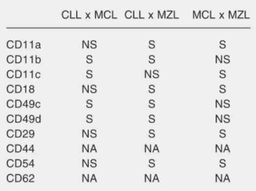
Comparison of chronic lymphocytic leu-kemia and mantle-cell lymphoma showed that chronic lymphocytic leukemia presented a higher expression of CD11c and CD49c, and a lower expression of CD11b and CD49d. Comparison of chronic lymphocytic leuke-mia and marginal B-cell lymphoma showed that the former presented a higher expres-sion of CD49c and a lower expresexpres-sion of CD11a, CD11b, CD18, CD49d, CD29, and CD54. Finally, comparison of mantle-cell lymphoma and marginal B-cell lymphoma showed that the latter had a higher expres-sion of CD11a, CD11c, CD18, CD29, and CD54.
Discussion
We analyzed the expression of 10 adhe-sion molecules in the peripheral blood of 47 patients with a diagnosis of B-NHL. We expected that the higher frequency of blood invasion by tumor cells in chronic lympho-cytic leukemia, when compared with the other B-NHL, could be explained by specif-ic profiles of adhesion molecule expression. Our finding of the lower expression of
CD11a and CD49d in chronic lymphocytic leukemia suggests that these adhesion mol-ecules are probably responsible for the dif-ferent compartment the disease infiltrates compared to its related entity, the small lym-phocytic lymphoma (17,18). Actually, since the structure of normal lymphoid follicles in lymph nodes depends on the appropriate association between B lymphocytes and den-dritic follicular cells through the interaction of CD11a and ICAM-1 (an intercellular ad-hesion molecule), and of CD49d and VCAM-1 (a vascular cell adhesion molecule), re-spectively (19), the lower expression of CD11a and CD49d on tumor cells of chronic lymphocytic leukemia could facilitate their detachment from the lymph node, while the expression of these adhesion molecules is preserved in small lymphocytic lymphoma (17,18). In fact, the down-regulation of CD11a and CD49d in chronic lymphocytic leukemia could also explain the peripheral blood invasion by these malignant cells, as compared with the occasional infiltration observed in mantle-cell lymphoma and mar-ginal B-cell lymphoma.
Our findings of a greater expression of CD49c and a lower expression of CD49d in chronic lymphocytic leukemia as compared with the other B-NHL have been previously observed (20,21). The combination of strong expression of CD49c and weak expression of CD49d is the hallmark of chronic lym-phocytic leukemia and can be used in the differentiation between this disease and the other B-NHL in the leukemic phase. In fact, it has been shown that imbalance in CD49c/ CD49d expression in chronic lymphocytic leukemia also contributes to the peripheral blood invasion (22). Moreover, a recent study has demonstrated prognostic relevance for the combination of high CD49c and low CD49d expression in chronic lymphocytic leukemia (23).
CD29 is the common ß1 chain of α sub-units of integrins α3/CD49c and α4/CD49d (8). Since the CD29 and α subunits are
Table 3. Pairwise comparison of B-cell non-Hodg-kin’s lymphomas for surface antigens by the Dunn post-test.
CLL x MCL CLL x MZL MCL x MZL
CD11a NS S S
CD11b S S NS
CD11c S NS S
CD18 NS S S
CD49c S S NS
CD49d S S NS
CD29 NS S S
CD44 NA NA NA
CD54 NS S S
CD62 NA NA NA
expressed together by a non-covalent bond, the higher expression of this adhesion mole-cule observed in marginal B-cell lymphoma is probably the result of the concomitant higher expression of CD49d in this subtype of B-NHL.
The presence of the myelomonocytic antigens CD11b and CD11c on cells of chronic lymphocytic leukemia has been pre-viously reported (24,25). CD11b has a het-erogeneous expression in chronic lympho-cytic leukemia. Whereas some publications described a high expression of this adhesion molecule in chronic lymphocytic leukemia (20), others reported a low expression (21,25). The data presented in the current study are in agreement with these latter re-ports, and since the mantle-cell lymphoma and the marginal B-cell lymphoma showed a higher expression of CD11b compared to chronic lymphocytic leukemia, this adhe-sion molecule could eventually be used to help differentiate between chronic lympho-cytic leukemia and the other B-NHL in the leukemic phase. Moreover, CD11b may have other biological functions in chronic lym-phocytic leukemia since it has been reported to be associated with a higher probability of disease progression and poor survival (26), as well as to prevent the induction of apopto-sis in this disease (27). Our findings add more information about the expression of CD11b in mantle-cell lymphoma and in mar-ginal B-cell lymphoma since there are only a few studies in the literature regarding this adhesion molecule in these diseases (28).
The role of CD11c in the dissemination of B-NHL is still controversial. Angelopou-lou et al. (28) and Bairey et al. (29) found a higher frequency of splenic involvement in cases of chronic lymphocytic leukemia with strong CD11c positivity. Moreover, this ad-hesion molecule is usually over-expressed in splenic marginal B-cell lymphoma (28) and in hairy cell leukemia (30,31), both dis-eases characterized by large splenomegaly, which could suggest a role for CD11c in the
infiltration of the spleen. However, our data do not support this hypothesis, since 14 of 17 of our patients with the diagnosis of mantle-cell lymphoma presented an enlarged spleen, but showed a very low expression of CD11c. In agreement with our results, An-gelopoulou et al. (32), in a study of 11 patients with mantle-cell lymphoma, ob-served that the CD11c was negative in all patients, including a subgroup of 6 patients with exclusively splenic disease.
CD54 is a member of the immunoglobu-lin superfamily that normally has a lower expression in peripheral blood mononuclear cells when compared with the lymph node (33). In our series, marginal B-cell lym-phoma presented the highest expression of this adhesion molecule. Similarly, Csanaky et al. (34), studying 7 patients with this disease, found a higher level of CD54 ex-pression when compared with chronic lym-phocytic leukemia. Stauder et al. (35) and Horst et al. (36) showed that the lower ex-pression of CD54 in indolent B-NHL corre-lates with a higher frequency of invasion of peripheral blood. In addition, Terol et al. (37) showed that low expression or absence of CD54 was associated with a higher fre-quency of disseminated disease (stage IV), extranodal involvement and bone marrow infiltration. Down-regulation of CD54 seems to favor the dissemination of tumor cells by a still unknown biological pathway and little information is available about the expres-sion of this adheexpres-sion molecule in splenic marginal B-cell lymphoma.
molecule expression in tumor cells. Further-more, we demonstrated that the CD49c/CD49d pair consistently presented a distinct pattern of expression in chronic lymphocytic leukemia compared to mantle-cell lymphoma and mar-ginal B-cell lymphoma, which could be
help-References
1. Kuppers R. Mechanisms of B-cell lymphoma pathogenesis. Nat Rev Cancer 2005; 5: 251-262.
2. Muller AM, Ihorst G, Mertelsmann R, Engelhardt M. Epidemiology of non-Hodgkin’s lymphoma (NHL): trends, geographic distribution, and etiology. Ann Hematol 2005; 84: 1-12.
3. Müller-Hermelink HK, Montserrat E, Catovsky D, Harris NL. Chronic lymphocytic leukemia/small lymphocytic lymphoma. In: Jaffe ES, Harris NL, Stein H, Vardiman JW (Editors), World Health Organiza-tion ClassificaOrganiza-tion of Tumours. Pathology and genetics of tumours of haematopoietic and lymphoid tissues. Lyon: IARC Press; 2001. p 127-130.
4. Criel A, Pittaluga S, Verhoef G, Wlodarska I, Meeus P, Mecucci C, et al. Small B cell NHL and their leukemic counterpart: differences in subtyping and assessment of leukemic spread. Leukemia 1996; 10: 848-853.
5. Bain BJ, Catovsky D. The leukaemic phase of non-Hodgkin’s lym-phoma. J Clin Pathol 1995; 48: 189-193.
6. Cohen PL, Kurtin PJ, Donovan KA, Hanson CA. Bone marrow and peripheral blood involvement in mantle cell lymphoma. Br J Haematol 1998; 101: 302-310.
7. Berger F, Felman P, Thieblemont C, Pradier T, Baseggio L, Bryon PA, et al. Non-MALT marginal zone B-cell lymphomas: a description of clinical presentation and outcome in 124 patients. Blood 2000; 95: 1950-1956.
8. Drillenburg P, Pals ST. Cell adhesion receptors in lymphoma dis-semination. Blood 2000; 95: 1900-1910.
9. Stauder R, Hamader S, Fasching B, Kemmler G, Thaler J, Huber H. Adhesion to high endothelial venules: a model for dissemination mechanisms in non-Hodgkin’s lymphoma. Blood 1993; 82: 262-267. 10. Roos E. Adhesion molecules in lymphoma metastasis. Cancer
Me-tastasis Rev 1991; 10: 33-48.
11. Roossien FF, de Rijk D, Bikker A, Roos E. Involvement of LFA-1 in lymphoma invasion and metastasis demonstrated with LFA-1-defi-cient mutants. J Cell Biol 1989; 108: 1979-1985.
12. Pals ST, Drillenburg P, Radaszkiewicz T, Manten-Horst E. Adhe-sion molecules in the dissemination of non-Hodgkin’s lymphomas. Acta Haematol 1997; 97: 73-80.
13. Freedman AS. Expression and function of adhesion receptors on normal B cells and B cell non-Hodgkin’s lymphomas. Semin Hematol 1993; 30: 318-328.
14. Timens W. Cell adhesion molecule expression and homing of hema-tologic malignancies. Crit Rev Oncol Hematol 1995; 19: 111-129. 15. Matutes E, Owusu-Ankomah K, Morilla R, Garcia MJ, Houlihan A,
Que TH, et al. The immunological profile of B-cell disorders and proposal of a scoring system for the diagnosis of CLL. Leukemia 1994; 8: 1640-1645.
16. Moreau EJ, Matutes E, A’Hern RP, Morilla AM, Morilla RM,
Owusu-Ankomah KA, et al. Improvement of the chronic lymphocytic leuke-mia scoring system with the monoclonal antibody SN8 (CD79b). Am J Clin Pathol 1997; 108: 378-382.
17. Inghirami G, Wieczorek R, Zhu BY, Silber R, la-Favera R, Knowles DM. Differential expression of LFA-1 molecules in non-Hodgkin’s lymphoma and lymphoid leukemia. Blood 1988; 72: 1431-1434. 18. Nadkarni JJ, Perambakam SM, Rathore VB, Amin KM, Parikh PM,
Naresh KN, et al. Expression of adhesion molecules in B-cell chronic lymphocytic leukaemia: an analysis in lymphoid compartments -peripheral blood, bone marrow and lymph node. Cancer Biother Radiopharm 1998; 13: 269-274.
19. Koopman G, Parmentier HK, Schuurman HJ, Newman W, Meijer CJ, Pals ST. Adhesion of human B cells to follicular dendritic cells involves both the lymphocyte function-associated antigen 1/intercel-lular adhesion molecule 1 and very late antigen 4/vascular cell adhesion molecule 1 pathways. J Exp Med 1991; 173: 1297-1304. 20. Baldini L, Cro L, Calori R, Nobili L, Silvestris I, Maiolo AT. Differential
expression of very late activation antigen-3 (VLA-3)/VLA-4 in B-cell non-Hodgkin lymphoma and B-cell chronic lymphocytic leukemia. Blood 1992; 79: 2688-2693.
21. Sembries S, Pahl H, Stilgenbauer S, Dohner H, Schriever F. Re-duced expression of adhesion molecules and cell signaling recep-tors by chronic lymphocytic leukemia cells with 11q deletion. Blood 1999; 93: 624-631.
22. Baldini LG, Cro LM. Structure and function of VLA integrins: differen-tial expression in B-cell leukemia/lymphoma. Leuk Lymphoma 1994; 12: 197-203.
23. Zucchetto A, Sonego P, Degan M, Bomben R, Dal Bo M, Russo S, et al. Surface-antigen expression profiling (SEP) in B-cell chronic lym-phocytic leukemia (B-CLL): Identification of markers with prognostic relevance. J Immunol Methods 2005; 305: 20-32.
24. De la Hera A, Alvarez-Mon M, Sanchez-Madrid F, Martinez C, Durantez A. Co-expression of Mac-1 and p150,95 on CD5+ B cells. Structural and functional characterization in a human chronic lym-phocytic leukemia. Eur J Immunol 1988; 18: 1131-1134.
25. De Rossi G, Zarcone D, Mauro F, Cerruti G, Tenca C, Puccetti A, et al. Adhesion molecule expression on B-cell chronic lymphocytic leukemia cells: malignant cell phenotypes define distinct disease subsets. Blood 1993; 81: 2679-2687.
26. Tassies D, Montserrat E, Reverter JC, Villamor N, Rovira M, Rozman C. Myelomonocytic antigens in B-cell chronic lymphocytic leukemia. Leuk Res 1995; 19: 841-848.
27. Plate JM, Long BW, Kelkar SB. Role of beta2 integrins in the prevention of apoptosis induction in chronic lymphocytic leukemia B cells. Leukemia 2000; 14: 34-39.
28. Angelopoulou MK, Kontopidou FN, Pangalis GA. Adhesion mol-ecules in B-chronic lymphoproliferative disorders. Semin Hematol
ful for the differential diagnosis.
1999; 36: 178-197.
29. Bairey O, Zimra Y, Rabizadeh E, Shaklai M. Expression of adhesion molecules on leukemic B cells from chronic lymphocytic leukemia patients with predominantly splenic manifestations. Isr Med Assoc J 2004; 6: 147-151.
30. Lucio PJ, Faria MT, Pinto AM, da Silva MR, Correia Junior ME, da Costa RJ, et al. Expression of adhesion molecules in chronic B-cell lymphoproliferative disorders. Haematologica 1998; 83: 104-111. 31. Marotta G, Raspadori D, Sestigiani C, Scalia G, Bigazzi C, Lauria F.
Expression of the CD11c antigen in B-cell chronic lymphoprolifera-tive disorders. Leuk Lymphoma 2000; 37: 145-149.
32. Angelopoulou MK, Kapiris E, Kourtis I, Siakantaris MP, Kittas Ch, Pangalis GA. The adhesion molecule profile of mantle cell lym-phoma (MCL) vs B-chronic lymphocytic leukemia (B-CLL). Blood 1997; 90: 522 (Abstract).
33. Boyd AW, Dunn SM, Fecondo JV, Culvenor JG, Duhrsen U, Burns GF, et al. Regulation of expression of a human intercellular adhe-sion molecule (ICAM-1) during lymphohematopoietic differentiation. Blood 1989; 73: 1896-1903.
34. Csanaky G, Matutes E, Vass JA, Morilla R, Catovsky D. Adhesion receptors on peripheral blood leukemic B cells. A comparative study on B cell chronic lymphocytic leukemia and related lymphoma/ leukemias. Leukemia 1997; 11: 408-415.
35. Stauder R, Greil R, Schulz TF, Thaler J, Gattringer C, Radaskiewicz T, et al. Expression of leucocyte function-associated antigen-1 and 7F7-antigen, an adhesion molecule related to intercellular adhesion molecule-1 (ICAM-1) in non-Hodgkin lymphomas and leukaemias: possible influence on growth pattern and leukaemic behaviour. Clin Exp Immunol 1989; 77: 234-238.
36. Horst E, Radaszkiewicz T, Hooftman-den Otter A, Pieters R, van Dongen JJ, Meijer CJ, et al. Expression of the leucocyte integrin LFA-1 (CD11a/CD18) and its ligand ICAM-1 (CD54) in lymphoid malignancies is related to lineage derivation and stage of differentia-tion but not to tumor grade. Leukemia 1991; 5: 848-853.