review
Copyright
© ABE&M todos os dir
eitos r
eser
vados.
Adipose tissue at the crossroads
in the development of the
metabolic syndrome, inflammation
and atherosclerosis
Tecido adiposo na encruzilhada no desenvolvimento da síndrome metabólica, inflamação e aterosclerose
Bernardo Léo Wajchenberg1, 2, Marcia Nery1,
Maria Rosaria Cunha1, Maria Elizabeth Rossi da Silva1
ABSTRACT
The authors analyze insulin resistance, the metabolic syndrome and endothelial dysfunction as consequence of a common antecedent, a low grade inflammation, indicating that in obesity the-re is a chronically activated inflammatory state of the adipose tissue. Furthermothe-re, the inflam-matory signaling is discussed according to the adipose tissue depot, visceral or subcutaneous. Arq Bras Endocrinol Metab. 2009;53(2):145-150.
Keywords
Obesity; visceral and subcutaneous adipose tissue; low grade inflammation; atherosclerosis; metabolic syndrome
RESUMO
Os autores analisam a resistência à insulina, a síndrome metabólica e a disfunção endotelial como consequência de um antecedente comum, a inflamação de baixo nível, o que mostra que a obesidade é um estado inflamatório cronicamente ativado do tecido adiposo. Discute-se, aqui, a sinalização inflamatória de acordo com a localização do tecido adiposo subcutâneo ou visceral. Arq Bras Endocrinol Metab. 2009;53(2):145-150.
Descritores
Obesidade; tecido adiposo subcutâneo ou visceral; inflamação de baixo nível; aterosclerose; síndrome metabólica
1 Serviço de Endocrinologia, Hospital das Clínicas da Faculdade de Medicina da Universidade de São Paulo (HC-FMUSP), São Paulo, SP, Brasil; Centro de Diabetes e Coração, Instituto do Coração (InCor), HC-FMUSP, São Paulo, SP, Brasil
2 Serviço de Endocrinologia, Hospital das Clínicas da Faculdade de Medicina da Universidade de São Paulo (HC-FMUSP), São Paulo, SP, Brasil
Correspondence to: Bernardo Léo Wajchenberg Av. Dr. Arnaldo, 455
01246-903 – São Paulo, SP, Brasil [email protected]
Received in Feb/02/2009 Accepted in Feb/17/2009
T
he clustering of dyslipidemia, hypertension and glucose intolerance, predominantly in overweight individuals, at risk of heart disease, has been received many names, including syndrome X and metabolic syndrome. In Reaven’s original description of the syn-drome, a central etiological role was attributed to in-sulin resistance and/or hyperinin-sulinemia, in part deter-mined by obesity (1), and this assumption has become the dominant paradigm for the metabolic syndrome (2). However, there is a substantial contradiction in such as-sociation. In part because of measurement problems and the fact that though “insulin resistance” may be important, it provides an insecure foundation for the metabolic syndrome, to a level in which it is no longerCopyright © ABE&M todos os dir
eitos r
eser
vados.
of a reverse etiology, whereby insulin resistance is a con-sequence of endothelial dysfunction, both insulin resis-tance and endothelial dysfunction are consequences of a common etiological mechanism (4). In a population of healthy adults, Yudkin and cols. (5) reported good correlations with metabolic syndrome variables, includ-ing insulin resistance and endothelial dysfunction, with a score of low grade inflammation derived from circu-lating concentrations of cytokines TNF-α and interleu-kin-6 (IL-6) and the acute-phase markers C-reactive protein (CRP) and fibrinogen, suggesting that 35% of the variance of the measures comprising the metabolic syndrome could be “explained by a state of low-grade inflammation”. The findings brought, for the first time, the possibility that the association between insulin resis-tance and the components of the metabolic syndrome could be a consequence of their common outcomes of a low-grade inflammation state.
Furthermore, there were strong and consistent rela-tionships with anthropometric measures of obesity and central fat distribution, as about 20% of the variance of the acute-phase markers could be explained, statisti-cally, on the basis of adiposity (5). Many prospective studies have shown that excess of body fat in the upper (central or abdominal) part of the body is more often correlated to the features of the metabolic syndrome. In contrast, individuals with fat stored in gluteal-femoral or peripheral depots (lower-body obesity) or female-type of fat distribution have a lower risk of morbidity from these metabolic disturbances (6). As reviewed by Karelis and cols. (7), approximately 20% of the general population can be categorized as obese but metaboli-cally healthy, presenting low visceral fat with high body mass index (BMI) and high insulin sensitivity. Quite the opposite, 18% of the population were found to have a normal body weight or were slightly overweight (meta-bolically obese normal weight), with high visceral fat, low BMI, low insulin sensitivity and high liver fat dis-playing severe metabolic abnormalities. Recently, Stefan and cols. (8), found, in a large population, that while the measurement of visceral fat provides a powerful tool to discriminate between insulin-sensitive and insulin-re-sistant, subjects within normal weight and overweight range. In the obese spectrum, the predictive effect of visceral fat was relatively weak. Visceral fat was lower in obese-insulin sensitive group than the obese-insulin resistant group, but the difference was not statistically significant, maybe unexpected considering the data col-lected by Wajchenberg and cols. (6). Nevertheless, in
agreement with our study, Stefan and cols. (8) agree that excess of fat, particularly visceral fat, when in-flamed, affects insulin sensitivity in large scale; however, with increased total adiposity, factors beyond excess of visceral fat may become more important for regulating insulin sensitivity, such as the liver and the muscle.
As suggested by Yudkin and cols. (2,5), a more likely paradigm for the metabolic syndrome seems to be that of adipose tissue-generated molecules initiat-ing a state of low-grade inflammation, with the known actions of these pro-inflammatory cytokines resulting in the combined metabolic, hemodynamic and vascu-lar consequences of this state. In this paradigm, insulin resistance merely becomes another consequence of this low grade inflammatory state.
Hotamisligil and cols. (9) were the first to describe the molecular connection between inflammation and obesity when TNF-α, an inflammatory cytokine, was found to be expressed in adipose tissue in obese ani-mal models, contributing to inhibition of insulin sig-naling pathways which has led to the exploration and characterization of several other adipokines with similar metabolic functions (10). The fact that these cytokines inhibit the effect of insulin on endothelial cells (2)may additionally contribute to insulin resistance by limiting nutrient-induced increase in nutritive capillary flow to muscle, as well as muscle glucose uptake.
Adipokines, the bioactive mediators produced in the adipose tissue, include not only adipocytes but also other cells that are present in fat tissue. Although adipocytes can produce almost all known adipokines, pre-adipocytes, as well as macrophages and endothelial cells of adipose tissue also contribute to adipokine pro-duction. Some of these substances are specifically, or abundantly, produced by fat tissue (leptin, adiponectin, visfatin, apelin, resistin) whereas others are clearly not specific for adipose tissue since they are not exclusively or preferentially expressed there and have been previ-ously identified and characterized as belonging to other families of substances [MCP-1, TNF-α, IL-6, PAI-1, angiotensinogen, 11β-hydroxysteroid dehydrogenase (11β-HSD-1, which converts inactive cortisone into active cortisol) and endocannabinoids] (11).
The adipokines, they are more than 50 different types currently recognized, act in a paracrine, autocrine and endocrine way influencing the metabolism of lipid, glucose homeostasis, in some cardiovascular risk fac-tors, such as hypertension, as well as thrombotic and inflammatory processes. The functions of adipokines
Copyright © ABE&M todos os dir
eitos r
eser
vados.
were recently extended far beyond metabolism to areas such as immunity, tumoral processes, angiogenesis and bone formation.
It is important to distinguish visceral from subcu-taneous adipose tissue in connection with the expres-sion of the different adipokines by their implications in the development of the metabolic syndrome. The visceral fat plays an important role in the pathogenesis of cardiovascular diseases since it expresses many com-ponents strongly involved as cardiovascular risk factors, such as IL-6, PAI-1 and glucocorticoids, while the sub-cutaneous fat produces mostly protective substances such as leptin and adiponectin and is less sensitive to glucocorticoids (12).Furthermore, adipokines released from visceral fat have, via portal vein, a direct access to the liver, and, therefore, a huge impact on the inflam-matory process.
The primary mechanisms of action of adipose tissue-produced inflammatory adipokines can be referred to according to the anatomic location of fat depot in which the adipokines are produced. Thus, adipokines released by the visceral depot would exert a greater effect on hepatic carbohydrate and lipid metabolism, stimulating hepatic release of acute phase response proteins in the liver (as CRP, an unspecific acute phase reactant that serves as an indicator of systemic inflammation) besides the autocrine/paracrine effects on the depot. Adipok-ines produced by the subcutaneous depot would mainly affect locally the adipose cell develop ment and function (autocrine/paracrine effects) and exert systemic effects
on, for example, skeletal muscle. The former may repre-sent the mechanism whereby inflammatory adipokines induce hepatic insulin resistance and chronic systemic inflammation, as the latter diminish adipose tissue stor-age of lipids, leading to ectopic fat accumulation in the liver and skeletal muscle (13).
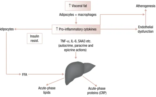
Many adipokines such as MCP-1, also known as C-C motif chemokine ligand 2 (CCL2), TNF-α and resistin (which function in humans is not clear yet), se-creted by adipocytes from obese subjects can promote macrophage infiltration and accumulation in adipose tissue and subendothelial space. It has been confirmed by recent studies that showed the association of abdomi-nal obesity with an increased amount of macrophages in adipose tissue (14). These macrophages are also im-plicated in the secretion of a panel of inflammatory cy-tokines (TNF-α, IL-6, MCP-1, PAI-1) acting in a para-crine and endopara-crine manner, finally causing a state of permanent low-grade inflammation in obese subjects. The MCP-1 (CCL2) receptor, C-C motif chemokine receptor 2 (CCR2), from other CC chemokine recep-tors present in the adipose tissue, plays a more notewor-thy role in the regulation of monocyte and macrophage recruitment, besides being necessary for macrophage-dependent inflammatory responses. This inflammatory state is implicated in cardiovascular disease, favoring endothelial insults and atheromatous changes (15). In Figure 1, the association between visceral adipose mass, secretion of inflammatory adipokines, insulin resistance and the metabolic syndrome is shown.
Figure 1. Association between visceral adipose mass, secretion of inflammatory cytokines, insulin resistance and the metabolic syndrome.
↑ Visceral fat Atherogenesis
Endothelial dysfunction Adipocytes
FFA
Acute-phase lipids
Acute-phase proteins (CRP)
Based on Schmidt and Duncan. Clin Chem LAb Med. 2003;41:1120. Insulin
resist.
↑ Pro-inflammatory cytokines
TNF-α, IL-6, SAA3 etc. (autocrime, paracrine and
epicrine actions) Adipocytes + macrophages
Copyright © ABE&M todos os dir
eitos r
eser
vados.
Adipokines are involved in every step of the ather-omatous process which begins with inflammatory changes of the vascular wall. The vascular insult is first caused by cytokines like TNF-α and IL-6, leading to increased expression of adhesion molecules such as in-tercellular adhesion molecule 1 (ICAM-1) and vascular cell adhesion molecule-1 (VCAM-1) which enhance monocyte adhesion to the endothelium. At the same time, MCP-1 (CCL2) secreted by adipocytes and by in-jured endothelial cells not only increases the migration of monocytes but also favors their transformation into foam cells. These foam cells secrete metalloproteinases that may lead to plaque rupture. Because of its low levels in obesity, the protective effects of adiponectin against plaque formation are decreased. Adiponectin is a specific white adipose tissue-derived protein, with anti-inflammatory/antiatherogenic properties such as decreasing the expression of adhesion molecules, creasing monocyte adhesion to endothelial cells, de-creasing uptake of oxidized low-density lipoprotein
(LDL), decreasing foam cell formation, and decreasing proliferation and migration of vascular smooth muscle (11). Adiponectin increases insulin sensitivity, increases smooth muscle glucose uptake and free fatty acid oxi-dation, decreases hepatic glucose production and de-creases intracellular triglycerides (16). Furthermore, obesity is also linked to a hypercoagulable state caused by increase in circulating levels of procoagulant factors such as tissue factor, fibrinogen, von Willebrand factor and factor VII. In addition, many circulating cytokines elevated in obesity will cause endothelial activation, leading to platelet activation and plug formation (17). The inhibition of fibrinolysis caused by an increased level of PAI-1 (particularly high in obesity and diabe-tes) is another component of this hypercoagulable pro-cess, favoring thrombus formation upon ruptured ath-erosclerotic plaques (18).
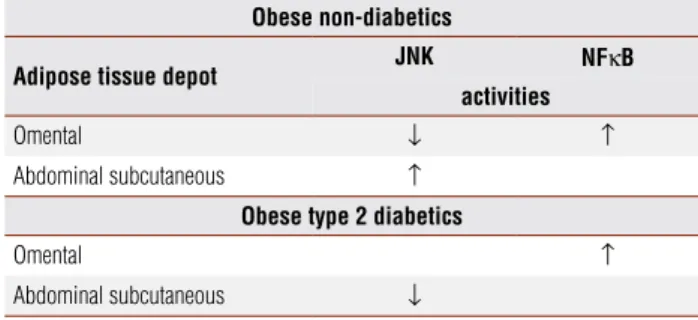
Hence, the state of chronic low-grade inflammation of obesity is probably the link between the different com-ponents of the metabolic syndrome and atherosclerosis. It has been shown that distinct inflammatory path-ways that occur in the adipocyte can be initiated by ex-tracellular mediators, such as cytokines and lipids, or in-tracellular stresses, such as endoplasmic reticulum stress or excessive production of reactie oxygen species by the mitochondria. Signals from these mediators converge to inflammatory signaling pathways, including the ki-nases JNK and inhibitor of NF-κβ kinase (IKKβ). These pathways induce the production of additional
inflam-matory mediators through transcriptional regulation as well as the inhibition of insulin signaling (19).Recently it has been found that different inflammatory pathways occur in the different adipose tissue depots, the omen-tal tissue with increased NF-κβ and increased JNK ac-tivity in subcutaneous abdominal adipose tissue. Addi-tionally, NF-κβ was shown to be influenced by obesity and diabetes status. These data suggest that NF-κβ may play a more predominant central role in inflammatory-related metabolic disease in comparison to JNK (20) (Table 1). These findings are in accordance to the well known observations that visceral adipose tissue, which includes the omental tissue, is closely associated with sub-clinical inflammation, insulin resistance and type 2
diabetes mellitus (12).
Table 1. Distinct inflammatory signalling pathways according to the adipose tissue depot
Obese non-diabetics
Adipose tissue depot JNK NFκB activities
Omental ↓ ↑
Abdominal subcutaneous ↑
Obese type 2 diabetics
Omental ↑
Abdominal subcutaneous ↓
Source: Kusminski and cols. Diabetologia. 2007;50 Suppl 1: Abstract 153. (20)
Copyright © ABE&M todos os dir
eitos r
eser
vados.
salycilates reverse hyperglycemia, insulin resistance and dyslipidemia by sensitizing insulin signaling through inhibition of IKKβ (in liver, but not in muscle) and other possible kinases (21).
Further research is needed to fully understand the pathophysiology of the adipokines and to determine potential targets for new therapeutic approaches. For now, it has been shown that lifestyle changes leading to weight loss are associated with an improvement of en-dothelial functions and reduction in circulating markers of inflammation (22,23). Interestingly, weight loss by surgical removal of visceral fat, but not by subcutaneous liposuction, is associated with significant decrease in cir-culating CRP, IL-6, interleukin-18 (IL-18) and TNF-α (24). Moreover, the thiazolidinediones (TZDs), high affinity ligands of PPARγ, which are given clinically as insulin-sensitizing agents, likely improve insulin action through multiple mechanisms, including both activat-ing lipid metabolism and reducactivat-ing production of in-flammatory mediators such as TNF-α. Because of the abundant expression of PPARγ in adipose tissue, pre-dominantly in subcutaneous fat, it is generally thought that the induction of adipogenesis to recruit new small adipocytes, and thus improve adipose tissue lipid ac-commodation and adiponectin secretion, accounts for most of the credible metabolic outcomes of thiazoli-dinediones (TZDs) (13), though they also have anti-inflammatory actions and inhibit the transcriptional activity of TNF-α promoter as well as antagonize the effects of exogenous administration of TNF-α in vivo
and in vitro, independently of the adipogenic effect of
the PPARγagonist (25). It was shown that short-term pioglitazone therapy (45 mg/day for three weeks in obese with type 2 diabetes) improved insulin sensitivity in part by reducing adipose tissue macrophage content through lowering CCL2 mRNA and CCR2 mRNA ex-pression in subcutaneous adipose tissue, which led to a decrease of macrophage content in 69% in comparison to control, resulting in less inflammation and, then, im-proved insulin sensitivity (26).
In addition to these cardiovascular and metabolic ef-fects, the chronic low grade inflammation of obesity may also play a role in other inflammatory illnesses such as joint diseases and inflammatory bowel disease. It appears to be that adipose tissue and adipokines can no longer be regarded as completely innocent in arthritis because they modulate the expression of local synovial cytokines and matrix-degrading enzymes. They may, therefore, become an attractive target for new treatment (27).
In conclusion, the adipose tissue is a highly active metabolic endocrine organ, being at the crossroads in the development of the metabolic syndrome, inflam-mation and atherosclerosis. Obesity and particularly visceral fat excess is associated with insulin resistance, hyperglycemia, atherogenic dyslipidemia, hypertension as well as prothrombotic and pro-inflammatory states.
Disclosure: No potential conflict of interest relevant to this article was reported.
REFERENCES
Reaven GM. Banting Lecture 1988. Role of insulin resistance in 1.
human disease. Diabetes. 1988;37(12):1595-607.
Yudkin JS. Insulin resistance and and the metabolic syndrome – 2.
or the pitfalls of epidemiology. Diabetologia. 2007;50(8):1576-86. Gale EA. The myth of the metabolic syndrome. Diabetologia. 3.
2005;48(9):1679-83.
Pinkney J, Coppack SW, Yudkin JS. Endothelial dysfunction: 4.
cause of the insulin resistance syndrome? Diabetes. 1997;46 Suppl 2:S9-S13
Yudkin JS, Stehouwer CDA, Emeis JJ, Coppack SW. C-reactive 5.
protein in healthy subjects: associations with obesity, insulin re-sistance, and endothelial dysfunction: a potential role of cytok-ines originating from adipose tissue? Arterioscler Thromb Vasc Biol. 1999;19(4):972-8.
Wajchenberg BL, Giannella-Neto D, Silva MER, Santos RF. Depot-6.
specific hormonal characteristics of sub-cutaneous and visceral adipose tissue and their relation to the metabolic syndrome. Horm Metab Res. 2002;34(11-12):616-21.
Karelis AD, St-Pierre DH, Conus F, Rabasa-Lhoret R, Poehlman ET. 7.
Metabolic and body composition factors in subgroups of obesity: what do we know? J Clin Endocrinol Metab. 2004;89(6):2569-75. Stefan N, Kantartzis K, Schick F, Thamer K, Rittig K, Balletshofer 8.
B, et al. Identification and characterization of Metabolically be-nign obesity in humans. Arch Intern Med. 2008;168(15):1609-16. Hotamisligil GS, Shargill NS, Spiegelman BM. Adipose expres-9.
sion of tumor necrosis factor-alpha: direct role in obesity-linked insulin resistance. Science. 1993;259(5091):87-91.
Scherer P. Adipose tissue: from lipid storage compartment to en-10.
docrine organ. Diabetes. 2006;55(6):1537-45.
Scherer PE. Adipose tissue: from lipid storage compartment to 11.
endocrine organ. Diabetes. 2006;55:1537-45
Wajchenberg BL. Subcutaneous and visceral adipose tissue: their re-12.
lation to the metabolic syndrome. Endocr Rev. 2000;21(6):697-738. Yang X, Smith U. Adipose tissue distribution and risk of metabolic 13.
disease: does thiazolidinedione-induced adipose tissue redistribu-tion provide a clue to the answer? Diabetologia. 2007;50(6):1127-39. Kamei N, Tobe K, Suzuki R, Ohsugi M, Watanabe T, Kubota N, 14.
et al. Overexpression of monocyte chemoattractant protein-1 in adipose tissues causes macrophage recruitment and insulin re-sistance. J Biol Chem. 2006;281(36):26602-14.
Berg AH, Scherer PE. Adipose tissue, inflammation, and cardio-15.
vascular disease. Circulation Res. 2005;96(9):939-49. Bloomgarden ZT. Inflammation, ather
16. osclerosis and aspects of
insulin action. Diabetes Care. 2005;28(9):2312-9.
Davi G, Guagnano MT, Ciabattoni G, Basili S, Falco A, Marinopic-17.
Copyright © ABE&M todos os dir
eitos r
eser
vados.
Sobel BE. Increased plasminogen activator inhibitor-1 and vas-18.
culopathy. A reconcilable paradox. Circulation. 1999;99(19): 2496-8.
Wellen KE, Hotamisligil GS. Inflammation, stress and diabetes. J 19.
Cin Invest. 2005;115:1111-9.
Kusminski CM, da Silva NF, Fowler AE, Creely JS, Harte AL, Baker 20.
AR, et al. The effects of adiposity, diabetic status and depot-spec-ificity on the activation of NF-κβ on JNK in human abdominal adi-pose tissue. Diabetologia. 2007;50 Suppl 1: Abstract 0153, S69. Wajchenberg BL, Nery M. Adipose tissue and inflammation. Obe-21.
sity and Metabolism. 2008;4:96-8.
Nicoletti G, Giugliano MT, Pontillo A, Cioffi M, D’Andrea F, Giugli-22.
ano D. Esposito K. Effect of a multidisciplinary program of weight reduction on endothelial function in obese women. J Endocrinol Invest. 2003;26(3):RC5-8.
Heilbroon LK, Noakes M, Clifton PM. Energy restriction and wei-23.
ght loss on very-low-fat diets reduce C-reactive protein
concen-trations in obese, healthy women. Arterioscl Thromb Vasc Biol. 2001;21(6):968-70.
Klein S, Fontana L, Young VL, Coggan AR, Kilo C, Patterson BW, 24.
Mohammed BS. Absence of effect of liposuction on insulin ac-tion and risk factors for coronary heart disease. N Engl J Med. 2004;350(25):2549-57.
Lee CH, Olson P, Evans RM. Mini-review: lipid metabolism, meta-25.
bolic diseases, and peroxisome proliferators-activated receptors. Endocrinology. 2003;144(6):2201-7.
Lee D-E, Kishore P, Li W, Zhang K, Schiwek, Hawkins M, Saper. 26.
The effect of short-term pioglitazone therapy on CCR-1 expres-sion in subcutaneous adipose tissue in subjects with T2DM. Pro-gram & Abstracts. The Endocrine Society 89th Annual Meeting,
2007 June 2-5; OR 28, p. 111.
Schaffler A, Muller-Ladner U, Scholmerich J, Bucher C. Role of 27.