Copyright © ABE&M todos os dir
eitos r
eser
vados.
Metabolic syndrome and polycystic
ovary syndrome… and vice versa
Síndrome metabólica e síndrome dos ovários policísticos... e vice-versa
Eleni Kandaraki1, Charikleia Christakou2, Evanthia Diamanti-Kandarakis2
ABSTRACT
The metabolic syndrome (MS) and the polycystic ovary syndrome (PCOS) appear to be inter-related, although they are distinct entities. Women with PCOS appear to be commonly affected by MS, while women with MS may display reproductive or endocrine features of PCOS. These clinical observations appear to be only partly attributable to the association of both syndro-mes with obesity and imply a reciprocal pathophysiologic relationship between PCOS and MS with potentially significant clinical sequelae. Adult women with MS are at a greater risk of de-veloping cardiovascular disease; women with PCOS also appear to carry such an increased risk in their postmenopausal life. Conversely, women with MS may experience reproductive disturbances, reminiscent of PCOS, more commonly than their counterparts from the general population. This review presented the current epidemiology of MS in adults and adolescents with PCOS, as well as the limited amount of data on the prevalence of features of PCOS among women with MS or MS features. We also discuss the potential pathophysiologic mechanisms underlying the relationship between these interweaving, but distinct, syndromes. Arq Bras Endocri-nol Metab. 2009;53(2):227-237.
Keywords
Polycystic ovary syndrome; metabolic syndrome; epidemiology; pathophysiology
RESUMO
A síndrome metabólica (SM) e a síndrome dos ovários policísticos (SOP) parecem estar relacio-nadas, embora constituam duas diferentes entidades. Mulheres com SOP são comumente afe-tadas pela SM, enquanto mulheres com SM podem apresentar as características reprodutivas e hormonais típicas da SOP. Essas observações clínicas podem ser atribuídas apenas parcialmen-te à presença da obesidade em ambas as síndromes. Isso se deve a uma relação fisiopatológica recíproca com potenciais sequelas de grande significado clínico. Mulheres adultas com SM estão mais propensas ao desenvolvimento de doenças cardiovasculares; mulheres com SOP também convivem com esse risco durante a pós-menopausa. Por outro lado, as mulheres com SM podem apresentar distúrbios reprodutivos, característicos da SOP, numa frequência maior do que as mulheres não portadoras de SM da população geral. Desse modo, este artigo de re-visão abordou dados epidemiológicos atuais sobre a SM em adultos e adolescentes com SOP, bem como as informações, que são limitadas, sobre a prevalência de características da SOP em mulheres com SM ou com as características desta. Também foram discutidos os mecanismos fi-siopatológicos fundamentais da relação entre essas duas síndromes interligadas, mas distintas. Arq Bras Endocrinol Metab. 2009;53(2):227-237.
Descritores
Síndrome do ovário policístico; síndrome metabólica; epidemiologia; fisiopatologia
1 York District Hospital,
York, England
2 Endocrine Section of the
1st Department of Internal
Medicine of Laiko General Hospital of University of Athens Medical School, Greece
Correspondence to:
Evanthia Diamanti-Kandarakis Medical School, University of Athens
1A Zefyrou str., Ekali 17562, Athens, Greece
Received in Feb/16/2009 Accepted in Feb/17/2009
INTRODUCTION
T
he metabolic syndrome (MS) is a constellation of cardiovascular risk factors, including impaired fast-ing glucose, central obesity, dyslipidaemia and raisedre-Copyright © ABE&M todos os dir
eitos r
eser
vados.
sistance has been recognized as a common link between these coexisting abnormalities.
During the last decade, the prevalence of MS has increased in the general population – and this increase has been steeper in women, particularly in young ones (1,2). This incremental trend comprises a major con-cern on account of its potential implications for cardio-vascular morbidity and mortality in women.
The MS is commonly present in obese women. How-ever, obesity is only one of the putative denominators of MS, as the others are linked to the individual’s metabolic susceptibility (3). Insulin resistance appears to be the main indicator of this metabolic susceptibility (3).
The polycystic ovary syndrome (PCOS), the com-monest endocrinopathy of women, has been associated with such a metabolic susceptibility, mainly attributable to the recognized association of the syndrome with insu-lin resistance. Insuinsu-lin resistance, as well as central adipos-ity, appear to affect not only obese but also lean PCOS women (4,5). Therefore, these women may be predis-posed to develop MS, independently of obesity per se.
The contributors to the PCOS/MS linkage appear to correspond particularly – but not only – to insulin resis-tance and central adiposity. The role of androgen excess in this linkage is currently under investigation (6).
This review present the current epidemiology of MS in adults and adolescents with PCOS, as well as the limit-ed amount of data on the prevalence of features of PCOS among women with MS or MS features. We also discuss the potential pathophysiologic mechanisms underlying the relationship between these interweaving, but distinct, syndromes.
DEFINITION AND EPIDEMIOLOGY
Although there is a general agreement regarding the main components of the MS, at least six diagnostic
defi-nitions have been proposed. This variation requires dif-ferent cut-off points and inclusion criteria (7).
The definition proposed by the National Cholesterol Education Program Adult Treatment Panel III (NCEP ATPIII) (8) is the most commonly used for clinical and research purposes. The International Diabetes Federation (IDF) (9) has proposed the most recent criteria, which resemble the NCEP one, with the exception that central obesity, assessed according to ethnicity-specific cut-offs, is an integral part of the IDF definition (Table 1).
The prevalence of MS differs across ethnic groups and it is highly age-dependent. It varies within indi-vidual cohorts according to the definition used. In a sizeable sample of otherwise healthy, reproductive-aged women from the National Health and Nutrition Examination Survey (NHANES) studied as a separate group, the prevalence of MS was estimated to be 26.5% (2). This study used both the NCEP ATP III and the IDF definitions of the MS. Remarkably, the compara-tive analysis of NHANES data collected from 1988 to 1994 and from 1999 to 2004 showed that the preva-lence of MS increased by 8.7% (from 17.8% to 26.5%) over the interval between the two phases of data collec-tion (2). Nevertheless, premenopausal women appear to have a lower incidence of MS if compared with men of the same age. Thus, premenopausal females, when compared to similarly aged males, seem to sustain a car-diometabolic privilege, which, however, appears to be abrogated within the pathologic context of PCOS.
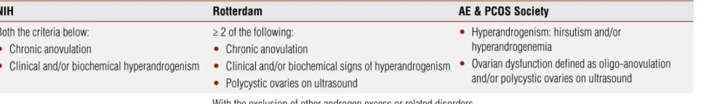
An analogy between MS and PCOS lies in the fact that the definitions of both syndromes have generated intense debate among experts in the field. In particular, there is still no universally accepted definition of PCOS, despite the fact that three different sets of criteria have been proposed from 1990 till today (Table 2) (10,11). In an attempt to reconcile the two earlier definitions of PCOS proposed by the National Institute of Health
Table 1. Definitions of MBS for women, according to WHO, NCEP ATP III and IDF criteria
WHO NCEP ATP III IDF
T2D or IFG or IGT or insulin resistance plus ≥ 2 of the following:
BMI > 30 kg/m
• 2 or WHR > 0.85
HDL < 1.0 mmol/L (< 40 mg/dL)
•
TG ≥ 1.7 mmol/L (150 mg/dL)
•
BP ≥ 140/90 mmHg or use of blood pressure
•
medication
microalbuminuria > 20 pg/min
•
Alb/Crea ratio ≥ 30 mg/g
•
≥ 3 of the following: WC ≥ 88 cm
•
HDL < 1.3 mmol/L (< 50 mg/dL)
•
TG ≥ 1.7 mmol/L (150 mg/dL)
•
BP ≥ 135/85 mmHg or use of blood pressure
•
medication
Central obesity defined as WC above the
ethnicity-specific cut-offplus ≥ 2 of the following:
TG ≥ 1.7 mmol/L (150 mg/dL) or specific treatment
•
HDL < 1.3 mmol/L (< 50 mg/dL) or specific
•
treatment
BP ≥ 135/85 mmHg or use of blood pressure
•
medication
fasting plasma glucose ≥ 5.6 mmol/L (100 mg/dL)
•
or previously diagnosed T2D
Copyright © ABE&M todos os dir
eitos r
eser
vados.
(NIH) and the Rotterdam expert conferences, respec-tively, the Androgen Excess-PCOS Society (AE-PCOS Society) has recently convened a task force (12
)
. Ac-cording to this task force, PCOS should be defined by the presence of hyperandrogenism (clinical and/or bio-chemical) and ovarian dysfunction (oligo-anovulation and/or polycystic ovaries), after the exclusion of related disorders (Table 2).Although a universal definition of PCOS has not been established as yet, there is agreement that this syndrome is the commonest endocrinopathy of re-productive aged women, with a prevalence of 6.6%-6.8% (13,14). Three consecutive studies conducted from 1999 to 2004 have established the prevalence of PCOS among the general population of premenopausal women (13-15). A Greek study published in 1999 by Diamanti-Kandarakis and cols. was the first to report the currently accepted prevalence of PCOS (13), which was confirmed by two subsequent studies – the first from Spain (14) and the second from the United States (15). All these studies have applied the NIH criteria for the diagnosis of PCOS.
However, under the Rotterdam criteria, the pheno-typic spectrum of PCOS has been broadened and also the prevalence of PCOS is expected to be higher by this PCOS definition (16,17). Among a selected population of women with normogonadotropic anovulation, the group of women diagnosed with PCOS by Rotterdam criteria appeared to be more than 1.5 times as large as the group classified as NIH-PCOS (16)
PREVALENCE OF MS IN WOMEN WITH PCOS
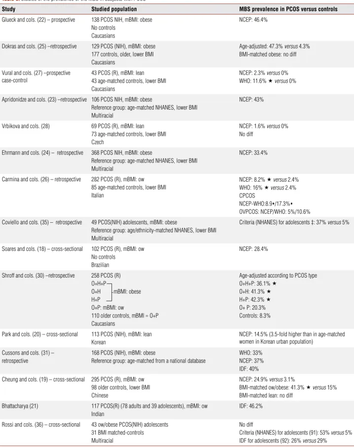
Overall, it is obvious that MS and PCOS are common disorders in reproductive-aged women in the general population. Of most interest is the substantial overlap between MS and PCOS, when the two syndromes are studied in combination. More specifically, the preva-lence of MS in women with PCOS appears to be sig-nificantly higher than the one estimated in their age-matched counterparts from the general population
(Table 3). The PCOS-MS interrelationship is not re-stricted to Caucasian women with PCOS. A high MS prevalence has been documented in Brazilian, Chinese, Korean, Indian and in multiracial PCOS populations, at least in overweight/obese patients, although the re-ported prevalence rates of MS vary across ethnic/racial regions (Table 3) (18-25).
Four US studies in predominantly obese women with PCOS have reported that 33.4% to 47.3% of these women fulfil the NCEP ATPIII criteria for MS (22-25) (Table 3). Despite being obviously higher than the US population-based estimates, these prevalence rates may, to some extent, reflect the impact of the high prevalence of obesity in the above populations – independently of PCOS per se (22-25).
Moreover, three of the above US studies did not involve specifically selected control groups (22-24). In the studies of Apridonidze and cols. (23) and by Eh-rmann and cols. (24), the relative increase of MS in PCOS was evaluated by comparing the study results to the data obtained from the Third National Health and Nutrition Examination Survey (NHANES III). This indirect way of assessment should be considered as a limitation of these studies.
In another US study (25), the apparent preponder-ance of MS in the PCOS group was eliminated, when non-obese PCOS patients were compared with age and body mass index (BMI)- matched controls, while the difference between the obese subgroups of patients and controls was reduced to non-significant levels. Thus, the actual impact of MS in women with PCOS as compared to controls, evenly matched for age and BMI, awaits further investigation.
Moreover, European studies among PCOS popula-tions with lower BMI have reported significantly lower prevalence rates of MS than the ones reported by the US studies (26-28) (Table 3). Despite the disparities between European and US investigators, some studies (26,27), albeit not others (28), have shown that the prevalence of MS is still higher in European PCOS pa-tients than in ethnicity-matched controls of similar age. Table 2. Definitions of PCOS according to available criteria
NIH Rotterdam AE & PCOS Society
Both the criteria below: Chronic anovulation
•
Clinical and/or biochemical hyperandrogenism
•
≥ 2 of the following: Chronic anovulation
•
Clinical and/or biochemical signs of hyperandrogenism
•
Polycystic ovaries on ultrasound
•
Hyperandrogenism: hirsutism and/or
•
hyperandrogenemia
Ovarian dysfunction defined as oligo-anovulation
•
and/or polycystic ovaries on ultrasound
Copyright © ABE&M todos os dir
eitos r
eser
vados.
Table 3. Studies of the prevalence of the MBS in subjects with PCOS
Study Studied population MBS prevalence in PCOS versus controls
Glueck and cols. (22) – prospective 138 PCOS NIH, mBMI: obese
No controls Caucasians
NCEP: 46.4%
Dokras and cols. (25) –retrospective 129 PCOS (NIH), mBMI: obese
177 controls, older, lower BMI Caucasians
Age-adjusted: 47.3% versus 4.3% BMI-matched obese: no diff
Vural and cols. (27) –prospective case-control
43 PCOS (R), mBMI: lean 43 age-matched controls, lower BMI Caucasians
NCEP: 2.3% versus 0%
WHO: 11.6% versus 0%
Apridonidze and cols. (23) –retrospective 106 PCOS NIH, mBMI: obese
Reference group: age-matched NHANES, lower BMI Multiracial
NCEP: 43%
Vrbikova and cols. (28) 69 PCOS (R), mBMI: lean
73 age-matched controls, lower BMI Czech
NCEP: 1.6% versus 0% No diff
Ehrmann and cols. (24) – retrospective 368 PCOS NIH, mBMI: obese
Reference group: age-matched NHANES, lower BMI Multiracial
NCEP: 33.4%
Carmina and cols. (26) – retrospective 282 PCOS (R), mBMI: ow
85 age-matched controls, lower BMI Italian
NCEP: 8.2% versus 2.4%
WHO: 16% versus 2.4%
CPCOS
NCEP-WHO:8.9/17.3%
OVPCOS: NCEP/WHO: 5%/10.6%
Coviello and cols. (35) – retrospective 49 PCOS(NIH) adolescents, mBMI: obese
Reference group: age/ethnicity-matched NHANES, lower BMI Multiracial
Criteria (NHANES) for adolescents ‡: 37% versus 5%
Soares and cols. (18) – cross-sectional 102 PCOS (R), mBMI: ow
No controls Brazilian
NCEP:28.4%
Shroff and cols. (30) –retrospective 258 PCOS (R)
O+H+P
O+H mBMI: obese H+P
O+P: mBMI: ow
110 older controls, mBMI ≈ O+P Caucasians
Age-adjusted according to PCOS type
O+H+P: 36.1%
O+H: 41.3%
H+P: 42.3%
O+ P: 20.3% Controls: 8.3%
Park and cols. (20) – cross-sectional 113 PCOS (NIH), mBMI: lean
Korean
NCEP: 14.5% (3.5-fold higher than in age-matched women in Korean urban population)
Cussons and cols. (31) – retrospective
168 PCOS (NIH), mBMI: obese
Reference group: age-matched from a national database
WHO: 33% NCEP: 37% IDF: 40%
Cheung and cols. (19) – cross-sectional 295 PCOS (R), mBMI: ow
98 older controls, lower BMI Chinese
NCEP: 24.9% versus 3.1%
BMI-matched ow/obese: 41.3% versus 15%
BMI-matched lean: no diff
Bhattacharya (21) 117 PCOS(R) (78 adults and 39 adolescents), mBMI: ow
Indian
IDF: 46.2%
Rossi and cols. (36) – cross-sectional 43 ow/obese PCOS(NIH) adolescents
31 BMI matched-controls Multiracial
No diff
Criteria (NHANES) for adolescents (91): 53% versus 5% IDF for adolescents (92): 26% versus 29%
The above studies were conducted in adult women unless else stated. In those studies that included mixed BMI groups of patients, the characterization of PCOS subjects as lean (BMI < 25 kg/m2), overweight (BMI: 25-30 kg/m2) or obese (BMI > 30 kg/m2) was based on the mean BMI value (mBMI) of each studied population.
: significantly higher than the respective value in controls; : significantly higher than the respective value in OVUL PCOS.
Copyright © ABE&M todos os dir
eitos r
eser
vados.
However, none of these studies have included patients and controls properly matched for age and BMI (26-28). Thus, not only the presence of PCOS but also BMI differences may have contributed to the above findings.
The ethnic variations in the rates of MS reported by different studies may be attributable not only to anthropometric differences between diverse ethnicity, but also to differences in the criteria used for PCOS diagnosis. The selection of PCOS patients was based on NIH criteria in studies from the United States, but mostly on Rotterdam criteria in European ones (Table 3). The patients fulfilling the NIH definition of PCOS are expected to be more severely metabolically affected than the patients selected by Rotterdam criteria (29). Thus, MS is expected to be less common in the pheno-typically heterogenous group of PCOS patients diag-nosed by Rotterdam criteria. Accordingly, women with hyperandrogenism and polycystic ovarian morphology (PCOM), but normal ovulation, as well as those with anovulation and PCOM, but normal androgen levels, had lower MS prevalence rates when compared to the respective rate in women with classic PCOS (30). Most importantly, in the group of anovulatory but normoan-drogenemic PCOS women, the age-adjusted preva-lence of MS was not significantly higher than the one in BMI-matched controls (30) (Table 3).
The majority of PCOS studies worldwide have implemented the NCEP-ATP III criteria for the diag-nosis of MS (Table 3). Nevertheless, one study, that has used both the World Health Organization (WHO) and the NCEP ATP III criteria, has suggested that the former set of criteria may be more discriminating and more appropriate for the assessment of MS in PCOS women with lower degrees of obesity (26). This sug-gestion sounds reasonable, since the MS definition by WHO criteria is less contingent on obesity and places more weight on the presence of insulin resistance or glucose intolerance (Table 1). Accordingly, in another study (27), classification according to the WHO cri-teria unveiled a significantly higher MS prevalence in PCOS women than in control ones, while the NCEP criteria identified MS in a significantly lower percentage of PCOS women, thus failing to show any difference between PCOS women and controls (27). Neverthe-less, a more recent study among a mixed population of obese and non-obese Caucasian PCOS women report-ed comparable rates of MS, whichever of the NCEP, the WHO or the IDF criteria were used for MS diag-nosis (31).
Another important limitation to the majority of the available studies is their retrospective design (Table 3). Differences in the exclusion criteria of each study should be also considered in the interpretation of their vary-ing results. For example, diabetes comprised an exclu-sion criterion in some studies (24), but not in others (23,31). In addition, one study has included patients on current treatment with oral contraceptives or insulin sensitizers (31), which are known to significantly affect metabolic parameters (32,33).
Not only adults but also adolescents with PCOS are exposed to the adverse metabolic milieu characterizing the syndrome (34), a fact which is expected to change into a higher prevalence of MS in this age group of pa-tients, as well. To explore this intuitively plausible con-cept, few studies have addressed the prevalence of MS in adolescents with PCOS (Table 3). One study based on a national database (NHANES) observed that adolescents with PCOS were found to be at increased risk for MS in comparison with a reference group (35). However, in the reference group, the mean BMI was lower and the PCOS status could not be accurately ascertained. A more recent study involving a group of obese PCOS adoles-cents and a control group well matched for age and BMI has indicated that, in obese adolescents, obesity, rather than PCOS per se, predicts MS (36). Although limited by
the small sample size, the latter study has further shown that the effect of obesity is attributable mainly to visceral adiposity rather than to overall obesity (36).
Copyright © ABE&M todos os dir
eitos r
eser
vados.
PREVALENCE OF FEATURES OF PCOS IN WOMEN
WITH MS
The relationship between PCOS and MS is possibly mutual. Thus, not only MS is prevalent among PCOS women, but also women with MS may commonly pres-ent the reproductive/endocrine hallmarks of PCOS.
Obesity, particularly visceral adiposity, and insulin resistance commonly go hand-in-hand with the devel-opment of reproductive and endocrine abnormalities, which characterize PCOS (44,45). Generally obese women present anovulation (44). In addition, simple obesity (46), particularly visceral fat excess (47), has been associated with increased production of potent androgens by the adipose tissue.
Furthermore, bariatric surgery has been reported to be successful in the treatment of anovulation, hirsut-ism, and insulin resistance associated with PCOS. In one cohort study, 24 severely obese (initial mean BMI: 50 kg/m2), young women with PCOS were treated
with Roux-en-Y gastric bypass. One year after surgery, a mean excess weight loss of 57% was paralleled with resumption of normal menses and improvement of hir-sutism in the studied population (48). Similar results have been reported in another small cohort of mor-bidly obese women with PCOS submitted to bariatric surgery (49).
Although the presence of reproductive abnormali-ties in obese is common, insulin resistant women has been the topic of several recent reviews (44,50,51
)
. Data regarding the actual prevalence of PCOS diag-nosed by available criteria in women with MS (or MS components) are limited. A prospective study (14) re-ported that 28.3% of premenopausal Spanish women observer that dietary treatment of obesity fulfilled the NIH criteria for PCOS. This percentage appears to be markedly higher than the 5.5% of prevalence of PCOS in lean women from the same ethnic region. Never-theless, within the studied population, the prevalence of PCOS was not different when considering the de-gree of obesity (52). Accordingly, in a reanalysis of combined data from two prevalence studies (15,53), obesity was shown to have only a minimal exacerbat-ing effect on the risk for PCOS, since the prevalence rates of the syndrome were slightly but not signifi-cantly different between BMI groups (underweight, normal-weight,overweight, and obese women, re-spectively) (54).Another cross-sectional population-based study has compared the presence and the frequency of
PCOS-like reproductive/endocrine features in premenopausal women with and without MS. The MS was diagnosed if three of the following eight criteria were present: first-degree relative with type 2 diabetes, BMI above 30 kg/ m2, waist/hip ratio above 0.88, blood pressure higher
than 160/95 mmHg or antihypertensive medication, fasting triglyceride levels above 1.7 nmol/L, HDL-C lower than 1.20 nmol/L, abnormal glucose metabo-lism, and fasting serum insulin 13 microU/mL. Of women with MS, 46.2% reported oligomenorrhea, as compared to 25.4% of obese controls and only 15.1% of lean controls (55). In addition, a hyperandrogenic hormone profile, defined by an increased free androgen index, appeared to be a typical feature of premenopaus-al fempremenopaus-ale MS, even without PCOS (56).
Although PCOS and MS often coexist, this associ-ation is neither causative nor inseparable. Obesity and insulin resistance/compensatory hyperinsulinemia, the two major denominators of MS, are neither nec-essary nor sufficient for the development of PCOS. Thus, women with a significant degree of insulin resis-tance/hyperinsulinemia can have regular menses and normal androgen levels (57). These clinical observa-tions are compatible with the fact that PCOS women harbour an intrinsic theca cell defect, which leads to increased ovarian androgen production, indepen-dently of other extraovarian factors. In the absence of intrinsic defects in thecal steroidogenesis, extraovar-ian factors cannot induce androgen overproduction by theca cells.
However, hyperinsulinemia superimposes an addi-tional burden upon the inherent ovarian dysfunction characterizing PCOS. Thus, in susceptible individu-als, the degree of hyperandrogenemia increases along with the exacerbation of insulin resistance/hyperinsu-linemia. These parallel courses may possibly describe the association between testosterone and insulin re-sistance/hyperinsulinemia, but do not appear to per-tain to androstenedione levels, the latter being more dependent on the stable steroidogenic defect of the PCOS ovary.
Copyright © ABE&M todos os dir
eitos r
eser
vados.
STRIKING PARALLELS IN THE PATHOPHYSIOLOGY
OF PCOS AND MS
The concept that the MS is causally an endocrine dis-ease is gaining ground. In addition to insulin, the main pathophysiologic actor of MS, an abnormal sex steroid milieu is increasingly considered for its potential con-tributing role (58). In particular, within the context of PCOS, androgen excess may serve as an endocrine modulator of MS. Thus, in the shared pathophysiologic spectrum of PCOS and MS, androgen excess possibly acts as a triggering factor in the development of MS, playing another key role beyond the one involved in ovulatory dysfunction. This role may pertain not only to systemic but also to local tissue androgen excess.
More spectifically, hyperandrogenism may amplify the adverse metabolic phenotype of PCOS in a dual mode, through the aggravation of central adiposity and the per-petuation of insulin resistance. Administration of testos-terone in female to male transsexuals has been reported to promote the deposition of visceral fat (59). Likewise, androgen administration to obese postmenopausal wom-en preferwom-entially increased visceral fat mass (60), whereas antiandrogen administration to premenopausal women with PCOS preferentially decreased visceral fat (61).
Furthermore, several observations directly impli-cate androgen excess in the perpetuation of insulin re-sistance in women. In PCOS patients, suppression of ovarian androgens via gonadotropin-releasing hormone (GnRH) administration (62) or antiandrogen therapy (63) has been associated with increased insulin sensitiv-ity assessed by the minimal model method. Moreover, in female-to-male transsexuals, androgen administra-tion has been linked with reduced insulin sensitivity in one study (64), albeit not in a subsequent report (65).
The potential linkage between insulin resistance and ovarian hyperandrogenism is also reflected in the ben-eficial metabolic effects of laparoscopic ovarian electro-cautery (LOE) in women with PCOS. A relevant study showed improved insulin sensitivity in parallel with the reduction of androgen levels in patients who underwent LOE, possibly indicating a causal interrelationship be-tween androgens and insulin resistance. In support of clinical findings, the same investigators provided mo-lecular evidence of partial reversal of insulin signaling defects in visceral fat of patients undergoing LOE (66). Accordingly, insulin responsiveness in adipocytes from amenorrhoeic women with PCOS was found to be sig-nificantly enhanced along with the reduction of testos-terone levels after an ovulatory cycle (67).
Androgens may act directly upon the signaling cas-cade, contributing to the impairment of insulin action. In cultured skeletal myotubes androgens were shown to induce insulin resistance via increased phosphoryla-tion in AKT, mammalian target of rapamycin (mTOR) and ribosomal S6-kinase (S6K), leading to increased Serine 636/639 phosphorylation of insulin receptor substrate-1 (IRS-1) (68). In another experiment in human subcutaneous adipocytes, chronic testosterone treatment was shown to induce metabolic insulin re-sistance acting via the androgen receptor. The signal-ing defect selectively affected the metabolic pathway of insulin was independent of phosphatidyloinositol-3 kinase and involved the impaired phosphorylation of protein kinase Cζ (69). It is also possible that andro-gens via the stimulation of subcutaneous lipolysis con-tribute to the excessive release of free fatty acids (70) in systemic circulation, which can act to induce IRS-1 serine phosphorilation (71).
Androgen exposure during intrauterine life in non-human primates results in a phenotype of the meta-bolic features of PCOS (72). Recently, it was shown that transient prenatal androgen exposure produces the full-blown MS in adult female rats (73
).
In this animal model, prenatal androgen exposure was shown to di-rectly induce hyperinsulinemia, despite unaltered insu-lin sensitivity, while dyslipidemia appeared to be medi-ated by prenatal androgenization-induced increases in adiposity (parametrial and subcutaneous fat). Not only prenatal but also peripubertal exposure to an androgen-ic milieu was shown to promote central fat accumula-tion, derange the metabolic/endocrine function of the adipose tissue and impair insulin sensitivity (74,75).Furthermore, experimental studies in adipocyte cell cultures (76) and clinical studies in women with PCOS (77,78) have implicated androgen excess in the direct modulation of adipocytokine secretion by the adipose tissue.
tis-Copyright © ABE&M todos os dir
eitos r
eser
vados.
sue and the liver via the hepatic portal system (46,47
)
. In the setting of obesity, increased production of highly potent androgens by the adipose tissue results from the overactivation of 17b-hydroxysteroid dehydroge-nase (17beta-HSD), which converts androstenedione to testosterone and of 5α-reductase, which converts testosterone to dihydrotestosterone. Strikingly, the ra-tio of mRNA levels of 17beta-HSD to aromatase (that converts testosterone to estradiol), in visceral adipose tissue, obtained from women undergoing elective ab-dominal surgery, was positively correlated with BMI and waist circumference (47)
. Thus, androgen excess and visceral adiposity appear to participate in a relation-ship of reciprocal enhancement.Reinforcing the bidirectional linkage between PCOS and MS, several factors, known for their car-diometabolic role, are under investigation with regard to their role in reproductive aspects. In addition to in-sulin resistance/hyperinin-sulinemia, which has a recog-nized role in the direct stimulation of ovarian andro-gen production and the distortion of follicular growth (80), the list of cardiometabolic factors with a putative reproductive role is currently expanding. Included in this list are: adipokines (81,82), plasminogen activator inhibitor-1 (PAI-1) (83), vascular endothelial growth factor (84) and AGEs (43,85,86). The paradigm of AGEs seems intriguing, since these prooxidative, proin-flammatory and proatherogenic molecules are not only endogenously produced, but also exogenously derived. Westernized diets, especially foods cooked at high tem-peratures for short time periods, are major sources of exogenous AGEs. Hence, high-AGE diets were to be significantly raise serum AGEs levels in interventional studies among nondiabetic humans (87,88). Consider-ing that AGEs and their receptor (RAGE) have been localized in human ovaries (89) and that circulating AGEs levels are increased and positively correlated with androgen levels in normoglycaemic lean PCOS women (43), AGEs may be an environmental risk fac-tor for both cardiometabolic and reproductive aberra-tions in PCOS.
CONCLUSIONS AND PERSPECTIVES
The substantial overlap between PCOS and MS seems to bear major clinical implications, since women diagnosed with PCOS appear to be more susceptible to develop MS. Moreover, even if it is not clear whether women with MS are actually more likely to develop PCOS, the
role of metabolic abnormalities in aggravating the clinical presentation of PCOS should be considered.
Overall, of major concern is to what extent the PCOS-related metabolic abnormalities translate into increased cardiovascular (CV) morbidity and mortality in these women. Recent data in postmenopausal wom-en show that PCOS is associated with an increased rate of CV events, which is partly independent of the pres-ence of obesity, diabetes and MS, and partly dependent on the degree of hyperandrogenemia (90). Thus, the burdens of CV risk conferred by PCOS and by MS, respectively, appear to be not identical, but additive. Since PCOS emerges as an independent predictor of CV complications after menopause, clinicians should be alert to minimize all the superimposable factors which may further compound the CV risk. Prevention or treatment of MS, based, primarily, on lifestyle and, secondarily, on pharmaceutical modalities, may prove to be mandatory in modifying, if not preventing, car-diometabolic as well as reproductive consequence in this susceptible population of young women, suffering from PCOS.
Disclosure: No potential conflict of interest relevant to this article was reported.
REFERENCES
R
1. egitz-Zagrosek V, Lehmkuhl E, Mahmoodzadeh S. Gender as-pects of the role of the metabolic syndrome as a risk factor for cardiovascular disease. Gend Med. 2007;4 Suppl B:S162-77. Ramos R, Olden K. The prevalence of metabolic syndrome 2.
among US women of childbearing age. Am J Publ Health. 2008;98(6):1122-7.
Grundy SM. Metabolic syndrome: a multiplex cardiovascular risk 3.
factor. J Clin Endocrinol Metab. 2007;92(2):399-404. Review. Dunaif A. Insulin resistance and the polycystic ovary syndro-4.
me: mechanism and implications for pathogenesis. Endocr Rev. 1997;18(6):774-800. Review.
Escobar-Morreale HF, San Millán JL. Abdominal adiposity and 5.
the polycystic ovary syndrome. Trends Endocrinol Metab. 2007;18(7):266-72.
Christakou C, Diamanti-Kandarakis E. Role of androgen excess 6.
on metabolic aberrations and cardiovascular risk in women with polycystic ovary syndrome. Women’s Health (Lond Engl). 2008;4(6):583-94.
Day C. Metabolic syndrome, or What you will: definitions and epi-7.
demiology. Diabetes Vasc Dis Res. 2007;4(1):32-8.
Grundy SM, Brewer B, Cleeman JI, Smith Jr SC, Lenfant C. De-8.
finition of the metabolic syndrome. Report of the National He-art, Lung and Blood Institute/American Heart Association Con-ference on Scientific Issues Related to Definition. Circulation. 2004;109:433-8.
Alberti KG, Zimmet P, Shaw J. IDF Epidemiology Task Force Con-9.
Copyright © ABE&M todos os dir
eitos r
eser
vados.
Za
10. wadski JK, Dunaif A. Diagnostic criteria for polycystic ovary syndrome: towards a rational approach. In: Dunaif A, Givens JR, Haseltine FP, Merriam GR, eds. Polycystic ovary syndrome. Bos-ton, MA: Blackwell Scientific Publications, 1992:37784.
The Rotterdam ESHRE/ASRM-Sponsored PCOS Consensus 11.
Workshopgroup. Revised 2003 consensus on diagnostic criteria and long-term health risks related to polycystic ovary syndrome (PCOS). Hum Reprod. 2004;19(1):41-7.
Azziz R, Carmina E, Dewailly D, Diamanti-Kandarakis E, Esco-12.
bar-Morreale HF, Futterweit W, Janssen OE, Legro RS, Norman RJ, Taylor AE, Witchel SF; Task Force on the Phenotype of the Polycystic Ovary Syndrome of The Androgen Excess and PCOS Society. The Androgen Excess and PCOS Society criteria for the polycystic ovary syndrome: the complete task force report. Fertil Steril. 2009;91(2):456-88.
Diamanti-Kandarakis E, Kouli CR, Bergiele AT, Filandra FA, Tsia-13.
nateli TC, Spina GG, et al. A survey of the polycystic ovary syn-drome in the Greek island of Lesbos: hormonal and metabolic profile. J Clin Endocrinol Metab. 1999;84(11):4006-11.
Asunción M, Calvo RM, San Millán JL, Sancho J, Avila S, Esco-14.
bar-Morreale HF. A prospective study of the prevalence of the polycystic ovary syndrome in unselected. Caucasian women from Spain. J Clin Endocrinol Metab. 2000;85(7):2434-8. Azziz R, Woods KS, Reyna R, Key TJ, Knochenhauer ES, Yildiz BO. 15.
The prevalence and features of the polycystic ovary syndrome in an unselected population. J Clin Endocrinol Metab. 2004;89(6)2745-9. Broekmans FJ, Knauff EA, Valkenburg O, Laven JS, Eijkemans 16.
MJ, Fauser BC. PCOS according to the Rotterdam consensus cri-teria: Change in prevalence among WHO-II anovulation and asso-ciation with metabolic factors. BJOG. 2006;113(10):1210-7. Hsu MI, Liou TH, Chou SY, Chang CY, Hsu CS. Diagnostic crite-17.
ria for polycystic ovary syndrome in Taiwanese Chinese women: comparison between Rotterdam 2003 and NIH 1990. Fertil Steril. 2007;88(3):727-9.
Soares EM, Azevedo GD, Gadelha RG, Lemos TM, Maranhão 18.
TM. Prevalence of the metabolic syndrome and its components in Brazilian women with polycystic ovary syndrome. Fertil Steril. 2008;89(3):649-55.
Cheung LP, Ma RC, Lam PM, Lok IH, Haines CJ, So WY, et al. 19.
Cardiovascular risks and metabolic syndrome in Hong Kong Chinese women with polycystic ovary syndrome. Hum Reprod. 2008;23(6):1431-8.
Park HR, Choi Y, Lee HJ, Oh JY, Hong YS, Sung YA. The metabolic 20.
syndrome in young Korean women with polycystic ovary syndro-me. Diabetes Res Clin Pract. 2007;77 Suppl 1:S243-6.
Bhattacharya SM. Metabolic syndrome in females with polycystic 21.
ovary syndrome and International Diabetes Federation criteria. J Obstet Gynaecol Res. 2008;34(1):62-6.
Glueck CJ, Papanna R, Wang P, Goldenberg N, Sieve-Smith L. In-22.
cidence and treatment of metabolic syndrome in newly referred women with confirmed polycystic ovarian syndrome. Metabo-lism. 2003;52(7):908-15.
Apridonidze T, Essah P, Iuorno M, Nestler JE. Prevalence and 23.
characteristics of metabolic syndrome in women with polycystic ovary syndrome. J Clin Endocrinol Metab. 2005;90(4):1929-35. Ehrmann D, Liljenquist D, Kasza K, Azziz R, Legro R, Ghazzi M, et 24.
al. Prevalence and predictors of the metabolic syndrome in wo-men with polycystic ovary syndrome. J Clin Endocrinol Metab. 2006;91(1):48-53.
Dokras A, Bochner M, Hollinrake E, Markham S, Vanvoorhis B, 25.
Jagasia DH. Screening women with polycystic ovary syndrome for metabolic syndrome. Obstet Gynecol. 2005;106(1):131-7. Carmina E, Napoli N, Longo RA, Rini GB, Lobo RA. Metabolic syn-26.
drome in polycystic ovary syndrome (PCOS): lower prevalence in southern Italy than in the USA and the influence of criteria for the diagnosis of PCOS. Eur J Endocrinol. 2006;154(1):141-5.
Vural B, Caliskan E, Turkoz E, Kilic T, Demirci A. Evaluation of 27.
metabolic syndrome frequency and premature carotid atheros-clerosis in young women with polycystic ovary syndrome. Hum Reprod. 2005;20(9):2409-13.
Vrbikova J, Vondra K, Cibula D, Dvorakova K, Stanicka S, Sra-28.
mkova D, et al. Metabolic syndrome in young Czech women with polycystic ovary syndrome. Hum Reprod. 2005;20(12):3328-32. Diamanti-Kandarakis E, Panidis D. Unravelling the phenotypic 29.
map of polycystic ovary syndrome (PCOS): a prospective study of 634 women with PCOS. Clin Endocrinol (Oxf). 2007;67(5):735-42. Shroff R, Syrop CH, Davis W, Van Voorhis BJ, Dokras A. Risk of 30.
metabolic complications in the new PCOS phenotypes based on the Rotterdam criteria. Fertil Steril. 2007;88(5):1389-95.
Cussons A, Watts G, Burke V, Shaw J, Zimmet P, Stuckey B. 31.
Cardiometabolic risk in polycystic ovary syndrome: a compari-son of different approaches to defining the metabolic syndrome. Hum Reprod. 2008;23(10):2352-8.
Diamanti-Kandarakis E, Nestler JE, Panidis D. Pharmaceuti-32.
cal intervention in metabolic and cardiovascular risk factors in polycystic ovary syndrome in insulin resistance and polycystic ovarian syndrome: pathogenesis, evaluation and treatment. Dia-manti-Kandarakis E, Pasquali E, Nestler JE, eds. Insulin resistan-ce and polycystic ovarian syndrome: pathogenesis, evaluation, and treatment. New Jersey: Humana Press, 2007. p. 367-87. Nader S, Diamanti-Kandarakis E. Polycystic ovary syndrome, oral 33.
contraceptives and metabolic issues: new perspectives and a uni-fying hypothesis. Hum Reprod. 2007;22(2):317-22.
Diamanti-Kandarakis E, Christakou C, Palioura E, Kandaraki E, 34.
Livadas S. Does polycystic ovary syndrome start in childhood? Pediatr Endocrinol Rev. 2008;5(4):904-11.
Coviello AD, Legro RS, Dunaif A. Adolescent girls with polycys-35.
tic ovary syndrome have an increased risk of the metabolic syndrome associated with increasing androgen levels indepen-dent of obesity and insulin resistance. J Clin Endocrinol Metab. 2006;91(2):492-7.
Rossi B, Sukalich S, Droz J, Griffin A, Cook S, Blumkin A, et al. 36.
Prevalence of metabolic syndrome and related characteristics in obese adolescents with and without polycystic ovary syndrome. J Clin Endocrinol Metab. 2008;93(12):4780-6.
Diamanti-Kandarakis E, Papavassiliou AG, Kandarakis SA, Chrou-37.
sos GP. Pathophysiology and types of dyslipidemia in PCOS. Trends Endocrinol Metab. 2007;18(7):280-5.
Conway GS, Agrawal R, Betteridge DJ, Jacobs HS. Risk factors 38.
for coronary artery disease in lean and obese women with the polycystic ovary syndrome. Clin Endocrinol (Oxf). 1992;37:119-5. Rajkhowa M, Neary RH, Kumpatla P, Game FL, Jones PW, Obhrai 39.
MS, et al. Altered composition of high density lipoproteins in women with the polycystic ovary syndrome. J Clin Endocrinol Metab. 1997;82(10):3389-94.
Diamanti-Kandarakis E, Paterakis T, Kandarakis HA. Indices of 40.
low-grade inflammation in polycystic ovary syndrome. Ann N Y Acad Sci. 2006;1092:175-86. Review.
Diamanti-Kandarakis E, Palioniko G, Alexandraki K, Bergiele A, 41.
Koutsouba T, Bartzis M. The prevalence of 4G5G polymorphism of plasminogen activator inhibitor-1 (PAI-1) gene in polycystic ovarian syndrome and its association with plasma PAI-1 levels. Eur J Endocrinol. 2004;150(6):793-8.
González F, Rote NS, Minium J, Kirwan JP. Reactive oxygen 42.
species-induced oxidative stress in the development of insulin resistance and hyperandrogenism in polycystic ovary syndrome. J Clin Endocrinol Metab. 2006;91(1):336-40.
Diamanti-Kandarakis E, Katsikis I, Piperi C, Kandaraki E, Piouka 43.
Copyright © ABE&M todos os dir
eitos r
eser
vados.
Pasquali R. Obesity, fat distribution and infertility. Maturitas. 44.
2006;54(4):363-71. Review.
Pasquali R. Obesity and androgens: facts and perspectives. Fertil 45.
Steril. 2006;85(5):1319-40.
Quinkler M, Sinha B, Tomlinson JW, Bujalska IJ, Stewart PM, Arlt 46.
W. Androgen generation in adipose tissue in women with simple obesity--a site-specific role for 17beta-hydroxysteroid dehydro-genase type 5. J Endocrinol. 2004;183(2):331-42.
Corbould AM, Bawden MJ, Lavranos TC, Rodgers RJ, Judd SJ. 47.
The effect of obesity on the ratio of type 3 17beta-hydroxysteroid dehydrogenase mRNA to cytochrome P450 aromatase mRNA in subcutaneous abdominal and intra-abdominal adipose tissue of women. Int J Obes Relat Metab Disord. 2002;26(2):165-75. Eid GM, Cottam DR, Velcu LM, Mattar SG, Korytkowski MT, Gos-48.
man G, et al. Effective treatment of polycystic ovary syndrome with Roux-en-Y gastric bypass. Surg Obes Relat Dis. 2005;1(2):77-80. Escobar-Morreale HF, Botella-Carretero JI, Alvarez-Blasco F, San-49.
cho J, San Millan JL. The polycystic ovary syndrome associated with morbid obesity may resolve after weight loss induced by bariatric surgery. J Clin Endocrinol Metab. 2005;90(12):6364-9. Nelson SM, Fleming RF. The preconceptual contraception para-50.
digm: obesity and infertility. Hum Reprod. 2007;22(4):912-5. Norman RJ, Noakes M, Wu R, Davies MJ, Moran L, Wang JX. 51.
Improving reproductive performance in overweight/obese wo-men with effective weight managewo-ment. Hum Reprod Update. 2004;10(3):267-80. Review.
Alvarez-Blasco F, Botella-Carretero JI, San Millán JL, Escobar-52.
Morreale HF. Prevalence and characteristics of the polycystic ovary syndrome in overweight and obese women. Arch Intern Med. 2006;166(19):2081-6.
Knochenhauer ES, Key TJ, Kahsar-Miller M, Waggoner W, Boots 53.
LR, Azziz R. Prevalence of the polycystic ovary syndrome in unse-lected black and white women of the southeastern United States: a prospective study. J Clin Endocrinol Metab. 1998;83(9):3078-82. Yildiz BO, Knochenhauer ES, Azziz R. Impact of obesity on the 54.
risk for polycystic ovary syndrome. J Clin Endocrinol Metab. 2008;93(1):162-8.
Korhonen S, Hippelsinen M, Niskanen L, Vanhala M, Saarikoski S. 55.
Relationship of the metabolic syndrome and obesity to polycys-tic ovary syndrome: a controlled, population-based study. Am J Obstet Gynecol. 2001;184(3):289-96.
Korhonen S, Hippeläinen M, Vanhala M, Heinonen S, Niskanen 56.
L. The androgenic sex hormone profile is an essential feature of metabolic syndrome in premenopausal women: a controlled community-based study. Fertil Steril. 2003;79(6):1327-34. Asagami T, Holmes TH, Reaven G. Differential effects of insulin 57.
sensitivity on androgens in obese women with polycystic ovary syndrome or normal ovulation. Metabolism. 2008;57(10):1355-60. Phillips G, Jing T, Heymsfield S. Does insulin resistance, visce-58.
ral adiposity, or a sex hormone alteration underlie the metabolic syndrome? Studies in women. Metabolism. 2008;57(6):838-44. Elbers JMH, Asscheman H, Seidell JC, Megens JAJ, Gooren 59.
LJG. Long-term testosterone administration increases visce-ral fat in female to male transsexuals. J Clin Endocrinol Metab. 1997;82(7):2044-7.
Lovejoy JC, Bray GA, Bourgeois MO, Macchiavelli R, Rood JC, 60.
Greeson C, et al. Exogenous androgens influence body composi-tionand regional body fat distribution in obese postmenopausal women – a clinical research center study. J Clin Endocrinol Me-tab. 1996;81(6):2198-203.
Gambineri A, Pelusi C, Genghini S, Morselli-Labate AM, Caccia-61.
ri M, Pagotto U, et al. Effect of flutamide and metformin admi-nistered alone or in combination in dieting obese women with polycystic ovary syndrome. Clin Endocrinol. 2004;60(2):241-9.
Cagnacci A, Paoletti AM, Arangino S, Melis GB, Volpe A. Effect of 62.
ovarian suppression on glucose metabolism of young lean wo-men with and without ovarian hyperandrogenism. Hum Reprod. 1999;14(4):893-7.
Diamanti-Kandarakis E, Mitrakou A, Hennes MM, Platanissiotis 63.
D, Kaklas N, Spina J, et al. Insulin sensitivity and antiandrogenic therapy in women with polycystic ovary syndrome. Metabolism. 1995;44(4):525-31.
Polderman KH, Gooren LJ, Asschermann H, Bakker A, Heine RJ. 64.
Induction of insulin resistance by androgens and estrogens. J Clin Endocrinol Metab. 1994;79(1):265-71.
Elbers JM, Giltay EJ, Teerlink T, Scheffer PG, Asscheman H, Sei-65.
dell JC, et al. Effects of sex steroids on components of the insu-lin resistance syndrome in transsexual subjects. Cinsu-lin Endocrinol (Oxf). 2003;58(5):562-71.
Seow KM, Juan CC, Hsu Y, Hwang JL, Huang LW, Ho LT. Ameliora-66.
tion of insulin resistance in women with PCOS via reduced insulin receptor substrate-1 Ser312 phosphorylation following laparos-copic ovarian electrocautery. Hum Reprod. 2007;22(4):1003-10. Marsden PJ, Murdoch AP, Taylor R. Adipocyte insulin action follo-67.
wing ovulation in polycystic ovarian syndrome. Hum Reprod. 1999;14(9): 2216-22.
Allemand M, Asmann Y, Klaus K, Nair K. An model for PCOS rela-68.
ted insulin resistance: the effects of testosterone on phosphoryla-tion of intracellular insulin signaling proteins in rat skeletal mus-cle primary culture. Fertil Steril. 2005;84 (Suppl 1):S30-31. Corbould A. Chronic testosterone treatment induces selective 69.
insulin resistance in subcutaneous adipocytes of women. J En-docrinol. 2007;192(3):585-94.
Arner P. Effects of testosterone on fat cell lipolysis. Species diffe-70.
rences and possible role in polycystic ovarian syndrome. Biochi-mie. 2005;87(1):39-43.
Draznin B. Molecular mechanisms of insulin resistance: se-71.
rine phosphorylation of insulin receptor substrate-1 and in-creased expression of p85: the two sides of a coin. Diabetes. 2006;55:2392-7.
Dumesic DA, Abbott DH, Padmanabhan V. Polycystic ovary syn-72.
drome and its developmental origins. Rev Endocr Metab Disord. 2007;8(2):127-41. Review.
Demissie M, Lazic M, Foecking EM, Aird F, Dunaif A, Levine JE. 73.
Transient prenatal androgen exposure produces metabolic syn-drome in adult female rats. Am J Physiol Endocrinol Metab. 2008;295(2):E262-8.
Mannerås L, Cajander S, Holmäng A, Seleskovic Z, Lystig T, Lönn 74.
M, et al. A new rat model exhibiting both ovarian and metabo-lic characteristics of polycystic ovary syndrome. Endocrinology. 2007;148(8):3781-91.
Perello M, Castrogiovanni D, Giovambattista A, Gaillard RC, Spi-75.
nedi E. Impairment in insulin sensitivity after early androgenisa-tion in the post-pubertal female rat. Life Sci. 2007;80(19):1792-8. Nishizawa H, Shimomura I, Kishida K, Maeda N, Kuriyama H, 76.
Nagaretani H, et al. Androgens decrease plasma adiponec-tin, an insulin-sensitizing adipocyte–derived protein. Diabetes. 2002;51(9):2734–41.
Panidis D, Kourtis A, Farmakiotis D, Mouslech T, Rousso D, Ko-77.
liakos G. Serum adiponectin levels in women with polycystic ova-ry syndrome. Hum Reprod. 2003;18(9):1790-6.
Kowalska I, Straczkowski M, Nikolajuk A, Adamska A, Karczewska-78.
Kupczewska M, Otziomek E, et al. Serum visfatin in relation to insulin resistance and markers of hyperandrogenism in lean and obese women with polycystic ovary syndrome Hum Reprod. 2007;22(7):1824-29.
Q
Copyright © ABE&M todos os dir
eitos r
eser
vados.
obesity--a site-specific role for 17beta-hydroxysteroid dehydro-genase type 5. J Endocrinol. 2004;183(2):331-42.
Diamanti-Kandarakis E. Polycystic ovarian syndrome: pathophy-80.
siology, molecular aspects and clinical implications. Expert Rev Mol Med. 2008;10(2):e3. Review.
Gosman GG, Katcher HI, Legro RS. Obesity and the role of gut 81.
and adipose hormones in female reproduction Hum Reprod Update. 2006;12(5):585-601.
Mitchell M, Armstrong DT, Robker RL, Norman RJ. Adipoki-82.
nes: implications for female fertility and obesity. Reproduction. 2005;130(5):583-97.
Devin JK, Johnson JE, Eren M, Gleaves LA, Bradham WS, Bloo-83.
dworth JR Jr, et al. Transgenic overexpression of plasminogen activator inhibitor-1 promotes the development of polycystic ova-rian changes in female mice. J Mol Endocrinol. 2007;39(1):9-16. Ferrara N, Frantz G, LeCouter J, Dillard-Telm L, Pham T, Drakshara-84.
pu A, et al. Differential expression of the angiogenic factor genes vascular endothelial growth factor (VEGF) and endocrine gland-de-rived VEGF in normal and polycystic human ovaries. Am J Pathol. 2003;162(6):1881-93.
Diamanti-Kandarakis E, Piperi C, Korkolopoulou P, Kandaraki E, 85.
Levidou G, Papalois A, et al. Accumulation of dietary glycotoxins in the reproductive system of normal female rats. J Mol Med. 2007;85(12):1413-20.
Tatone C, Amicarelli F, Carbone MC, Monteleone P, Caserta D, 86.
Marci R, et al. Cellular and molecular aspects of ovarian follicle ageing. Hum Reprod Update. 2008;14(2):131-42.
Vlassara H. Advanced glycation in health and disease: role of the 87.
modern environment. Ann NY Acad Sci. 1043:452-60.
Uribarri J, Stirban A, Sander D, Cai W, Negrean M, Buenting CE, 88.
et al. Single oral challenge by advanced glycation end products acutely impairs endothelial function in diabetic and nondiabetic subjects. Diabetes Care. 2007;30(10):2579-82.
Diamanti-Kandarakis E, Piperi C, Patsouris E, Korkolopoulou P, 89.
Panidis D, Pawelczyk L, et al. Immunohistochemical localization of advanced glycation end-products (AGEs) and their receptor (RAGE) in polycystic and normal ovaries. Histochem Cell Biol. 2007;127(6):581-9.
Shaw LJ, Bairey Merz CN, Azziz R, Stanczyk FZ, Sopko G, Brauns-90.
tein GD, et al. Postmenopausal women with a history of irregular menses and elevated androgen measurements at high risk for worsening cardiovascular event-free survival: results from the National Institutes of Health – National Heart, Lung, and Blood Institute sponsored Women’s Ischemia Syndrome Evaluation. J Clin Endocrinol Metab. 2008;93(4):1276-84.
Zimmet P, Alberti G, Kaufman F, Tajima N, Silink M, Arslanian S, 91.
Wong G, Bennett P, Shaw J, Caprio S; International Diabetes Fe-deration Task Force on Epidemiology and Prevention of Diabetes. The metabolic syndrome in children and adolescents. Lancet. 2007;369(9579):2059-61.
Cook S, Weitzman M, Auinger P, Nguyen M, Dietz WH. 2003 Pre-92.