Detection of Core2
β-1,6-
N
-Acetylglucosaminyltransferase in Post-Digital
Rectal Examination Urine Is a Reliable
Indicator for Extracapsular Extension of
Prostate Cancer
Yuta Kojima1, Tohru Yoneyama2, Shingo Hatakeyama1, Jotaro Mikami1, Tendo Sato1, Kazuyuki Mori1, Yasuhiro Hashimoto1, Takuya Koie1, Chikara Ohyama1,2, Minoru Fukuda3, Yuki Tobisawa1*
1Department of Urology, Hirosaki University Graduate School of Medicine, Hirosaki, Japan,2Department of Advanced Transplant and Regenerative Medicine, Hirosaki University Graduate School of Medicine, Hirosaki, Japan,3Glycobiology Unit, Tumor Microenvironment Program, Cancer Center, Sanford-Burnham Medical Research Institute, La Jolla, CA 92037, United States of America
Abstract
To identify appropriate candidates for aggressive treatment such as radical prostatectomy or radiation therapy of localized prostate cancer (PCa), novel predictive biomarkers of PCa aggressiveness are essential. Core2β-1,6-N-acetylglucosaminyltransferase-1 (GCNT1) is a key enzyme that forms core 2-branchedO-glycans. Its expression is associated with the progression of several cancers. We established a mouse IgG monoclonal antibody (mAb) against GCNT1 and examined the relationship of GCNT1 expression to the clinicopatholog-ical status of PCa. Paraffin-embedded PCa specimens were analyzed by immunohis-tochemistry for GCNT1 expression using a newly established mouse anti-GCNT1 mAb by ourselves. GCNT1-positive tumor showed significantly higher Gleason score and larger tumor volume. The number of GCNT1-positive cases was significantly lower in cases of organ-confined disease than in cases of extracapsular extension. GCNT1-negative tumors were associated with significantly better prostate-specific antigen (PSA)-free survival com-pared with GCNT1-positive tumors. Multivariate analysis revealed that detection of GCNT1 expression was an independent risk factor for PSA recurrence. We established new meth-ods for GCNT1 detection from PCa specimens. Immunoblotting was used to examine post-digital rectal examination (DRE) urine from PCa patients. Over 90% of GCNT1-positive PCa patients with high concentrations of PSA showed extracapsular extension. In conclu-sion, GCNT1 expression closely associates with the aggressive potential of PCa. Further research aims to develop GCNT1 detection in post-DRE urine as a marker for PCa aggressiveness.
a11111
OPEN ACCESS
Citation:Kojima Y, Yoneyama T, Hatakeyama S, Mikami J, Sato T, Mori K, et al. (2015) Detection of Core2β-1,6-N-Acetylglucosaminyltransferase in Post-Digital Rectal Examination Urine Is a Reliable Indicator for Extracapsular Extension of Prostate Cancer. PLoS ONE 10(9): e0138520. doi:10.1371/ journal.pone.0138520
Editor:Ming Tat Ling, Queensland University of Technology, AUSTRALIA
Received:April 27, 2015
Accepted:September 1, 2015
Published:September 21, 2015
Copyright:This is an open access article, free of all copyright, and may be freely reproduced, distributed, transmitted, modified, built upon, or otherwise used by anyone for any lawful purpose. The work is made available under theCreative Commons CC0public domain dedication.
Data Availability Statement:All relevant data are within the paper and its Supporting Information files.
Introduction
In the United States and Europe, prostate cancer (PCa) is the most common malignancy in men and the second-leading cause of cancer-related death [1,2]. The incidence of PCa is reportedly low in Asian countries [3]. However, its incidence is rapidly increasing in the Asia-Pacific region [4]. Some of the most critical issues related to PCa in clinical practice are overdi-agnosis and overtreatment [5]. The lack of specificity of prostate-specific antigen (PSA) testing has resulted in a debate on the usefulness of PSA-based PCa screening [6,7]. Moreover, unnec-essary treatment for indolent PCa with a low malignant potential is a major issue, as aggressive PCa treatment is sometimes associated with adverse events. A promising alternate modality to prevent overtreatment is active surveillance [8]. However, the identification of suitable patients who are good candidates for aggressive treatment is associated with difficulties. To date, there are no perfect tools for precise detection of good candidate for active surveillance [9]. There-fore, the identification and validation of biomarkers of PCa aggressiveness are important in preventing PCa overtreatment.
Preoperative serum PSA levels and biopsy Gleason scores are conventional and powerful predictors of biological outcomes after radical prostatectomy for PCa [10,11]. To improve the risk stratification for PCa recurrence after primary treatment in patients with localized PCa, many investigators have sought biomarkers that reflect the aggressive potential of PCa [12]. However, the majority of reported biomarkers have not been validated to provide information that is more useful than that provided by conventional clinicopathological parameters. With a novel biomarker representing the malignant potential of PCa, more accurate prediction of PSA recurrence and appropriate treatment selection may be possible.
Cell surface carbohydrate structures are altered during carcinogenesis and play important roles in cancer metastasis [13,14]. The presence of sialyl Lewis X, which is one of the functional terminal structure, on the cell surface of colorectal cancer is positively correlated with poor prognosis [15]. In a similar way, high Gleason score prostate cancer specimens expressed sialyl Lewis X [16]. Branching glycan have increased a functional terminal structure, and a binding affinity for specific lectins [17]. In previous study, mannosyl (alpha-1,6-)-glycoproteinβ
-1,6-N-acetyl-glucosaminyltransferase (MGAT5) and Core 2β-1, 6-N -acetylglucosaminyltransfer-ase-1 (GCNT1) formed GlcNAcβ1,6 branching glycan increased PCa aggressiveness [18,19].
GCNT1 [20,21] is a key enzyme that synthesizes core 2-branchedO-glycans by catalyzing the transfer ofN-acetylglucosamine from uridine diphosphate-N-acetylglucosamine with aβ1, 6-linkage toα-N-acetylgalactosamine of a core 1O-glycan (Fig 1A). A previous study analyzed
GCNT1mRNA expression in fresh colorectal tumor samples and showed that expression of core 2-branchedO-glycans is closely correlated with the malignant potential of colorectal can-cer [22]. This is also true for pulmonary adenocarcinoma [23]. In immunohistochemistry using a polyclonal antibody [21], we demonstrated that GCNT1 expression is closely related with the aggressive potential of PCa [18], testicular cancer [24], and bladder cancer [25]. In recent study, a gene expression array showed GCNT1 was overexpressed in prostate cancer tis-sue [26]. However, the establishment of a monoclonal anti-GCNT1 antibody is essential for further research, including validation studies to elucidate the clinical utility of GCNT1 as a biomarker.
Here, we raised a monoclonal antibody (mAb) against GCNT1 by immunization of a mouse with GCNT1 specific peptide (S1 File) to evaluate the potential of the latter as an indicator of PCa aggressiveness. In this study, we demonstrated that the anti-GCNT1 mAb showed high specificity against human GCNT1 and that GCNT1 expression in PCa specimens from radical prostatectomy correlates with PCa aggressiveness. In addition, detection of GCNT1 in post-digital rectal examination (DRE) urine by the anti-GCNT1 mAb predicted extracapsular
Institutes Health Grants UO1 CA168924 (MF); The Suzuki Urological Research Foundation, research grant 2014 (YT). The funders had no role in study design, data collection and analysis, decision to publish, or preparation of the manuscript.
Competing Interests:The authors have declared
that no competing interests exist.
Abbreviations:DRE, digital rectal examination; GCNT1, core2β-1,6-N
extension of PCa. Therefore, detection of GCNT1 in post-DRE urine may serve as a minimally invasive method to predict PCa aggressiveness.
Materials and Methods
Materials
ISOGEN II Reagent was purchased from Nippon Gene (Japan). A purified rabbit anti-mouse IgG antibody (γ-chain specific) was purchased from Zymed. A horseradish peroxidase (HRP)-conjugated goat anti-rabbit IgG (H+L) antibody and an HRP-(HRP)-conjugated goat anti-mouse IgG antibody were acquired from Cell Signaling Technology. An HRP-conjugated goat anti-mouse IgG antibody was acquired from Millipore. Purified mouse myeloma protein from MOPC 21 (IgG,κ), 2-mercaptoethanol, and bovine serum albumin (BSA) were purchased from Sigma-Aldrich. Tween-20 was purchased from Wako Pure Chemicals (Japan), as were DMEM and Ham’s F12 medium. Penicillin G/streptomycin solution was from Hyclone. Precision Plus Pro-tein standards Dual Color were from Bio-Rad, and skim milk was from Yukijirushi (Japan).
Cells
Chinese hamster ovary (CHO) cells were maintained in the alpha modification of Eagle's mini-mum essential medium (α-MEM) supplemented with 100 U/mL of penicillin, 100μg/mL of
streptomycin, and 10% fetal bovine serum (FBS).
Immunohistochemical analysis of PCa specimens
Between 2005 and 2011, 250 PCa patients were treated with radical prostatectomy at the Department of Urology, Hirosaki University Graduate School of Medicine, Hirosaki, Japan. The tumor specimens were formalin-fixed and embedded in paraffin. Deparaffinized speci-mens were incubated with 5μg/mL of mouse anti-human GCNT1 mAb (clone HU127),
fol-lowed by incubation with HRP-conjugated goat anti-mouse IgG antibody (H+L; Millipore).
Immunoblotting analysis of post-DRE urine specimens
Post-DRE urine was filled into 50 mL conical tubes, frozen immediately, and stored at -80°C until analysis. Post-DRE urine specimens were collected from 35 patients who underwent radi-cal prostatectomy from 2010 to 2013 at the Department of Urology, Hirosaki University Grad-uate School of Medicine, Hirosaki, Japan. Frozen samples were thawed overnight at 4°C and briefly centrifuged (5000 xg, 5 min) to separate the supernatant and solids. Fifty microliters of the supernatant were spotted on to a nitrocellulose membrane. The membrane fixed with post-DRE urine protein was incubated with an anti-GCNT1 mAb (HU127), followed by an HRP-conjugated secondary antibody. Signals representing GCNT1 were enzymatically detected using the Novex1ECL Chemiluminescent Substrate Reagent Kit (Life Technologies) and visu-alized in a ChemiDocXRS+ System (Bio-Rad). Signal mean values were measured by Image Lab software (Bio-Rad). Amount of GCNT1 expression was calculated based on the signal mean values of recombinant human GCNT1 (R&D systems, 7248-GT).
Auto-chemiliminescent signals were subtracted from total signals. Total protein concentration of post-DRE urine samples were measured by a BCA Protein Assay Kit (Pierce). Informed con-sent was obtained from all patients. All patients provided their written informed concon-sent to
compared between GCNT1-positive and GCNT1-negative specimens. Survival was analyzed using Kaplan-Meier curves.
participate in this study. The ethical committee of Hirosaki University approved the protocol of this study (The study about carbohydrate structure change in urological disease; Approval number: 2014–195). The study was performed in accordance with the ethical standards of the Declaration of Helsinki.
Staging and grading of tumors
All patients were preoperatively evaluated using DRE, serum PSA testing, bone scanning, pel-vic computed tomography, and transrectal ultrasonography. Using an 18-G needle, 6–12 pros-tate needle biopsy samples were obtained under ultrasound guidance. Staging was performed using the 2002 American Joint Committee on Cancer Staging Manual [27], while the Gleason grading system was used for tumor grading [28].
PSA measurement and patient follow-up
Serum PSA levels were determined using IMx (Abbott Laboratories, Abbott Park, IL). Postop-erative PSA levels were considered to be increased (PSA recurrence) if they were0.2 ng/mL during two consecutive visits in a 1-month interval. Time zero was defined as the day of sur-gery. Patients with constantly detectable PSA levels (<0.001 ng/mL) after surgery were
recorded as recurrences at time zero. Follow-up intervals were calculated from the date of sur-gery to the last recorded follow-up (median, 48.4 months; range, 13.2–82.9 months).
Statistical analysis
The chi-squared test was used to analyze the association of GCNT1 status with clinical and his-topathological parameters. PSA-free survival was evaluated using Kaplan–Meier curves and differences between groups were assessed using the log-rank test. We used the SPSS 21.0 soft-ware package (SPSS, Chicago, IL) for all statistical analyses. Optimal PSA and GCNT1 expres-sion cut-off values were calculated the using the following formula: cut-off = (1−sensitivity)2
+ (1−specificity)2[29,30].
Results
GCNT1 expression in PCa positively correlates with cancer progression
and PSA recurrence
To confirm antibody specificity for GCNT1, a mouse anti-human GCNT1 antibody was puri-fied from hybridoma supernatant. Dose-dependent binding of the mouse anti-human GCNT1 mAb (clone HU127) to immobilized recombinant human GCNT1 (rhGCNT1) was detected using HRP-conjugated goat anti-mouse IgG in an enzyme-linked immunosorbent assay (ELISA) (S1A Fig). In the absence of immobilized rhGCNT1, no antibody binding was observed (S1B Fig). In immunoblotting analysis, the HU127 anti-human GCNT1 mAb bound specifically to rhGCNT1, but not to rhGCNT3 (S1C Fig). HU127 anti-human GCNT1 mAb also specifically detected transient expression of GCNT1 in CHO cells (S1D and S1E Fig). In the case of HU127 anti-human GCNT1 mAb premixed with GCNT1 peptide antigen prior to incubation on immunohistochemistry and immunoblotting, the GCNT1 signals were dimin-ished (S1F and S1G Fig). The results also suggested that HU127 anti-human GCNT1 mAb held high specificity against GCNT1.
specimens from 250 patients exhibited different clinical parameters (S1 Table). GCNT1 expres-sion in prostatectomy specimens positively correlated with the postoperative Gleason score. Over 80% of tumor specimens with extracapsular extension of PCa (pT3 and pT4) expressed GCNT1, and GCNT1-positive tumors were significantly larger than GCNT1-negative tumors (S2 Table).
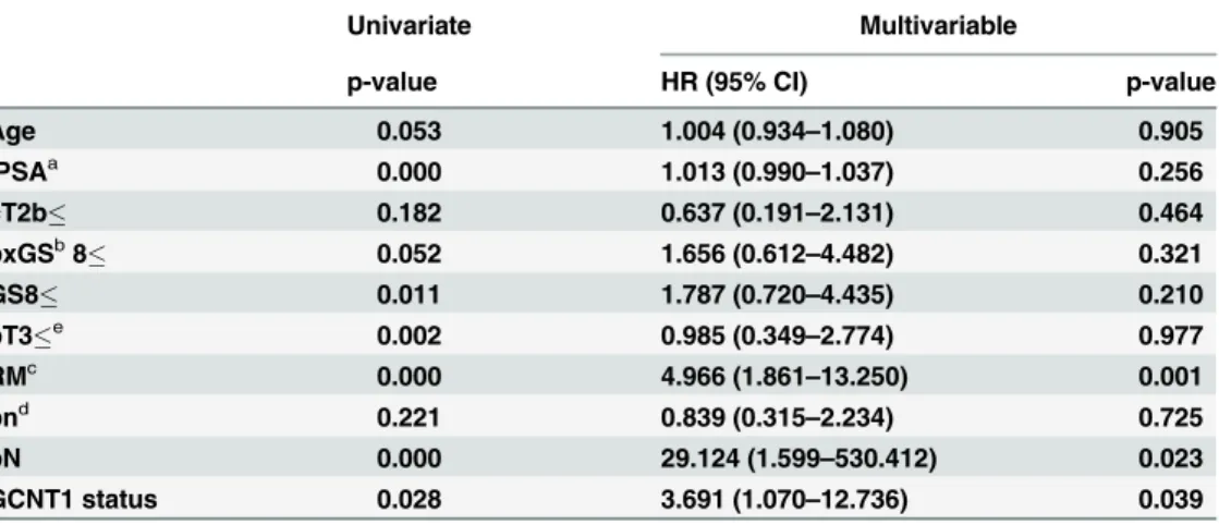
As shown inFig 1C, GCNT1-positive patients were at significantly higher risk of PSA recur-rence after radical prostatectomy. According to multivariate analysis, PSA levels, margin status, and GCNT1 expression in the tumor were independent risk factors for PSA recurrence (Table 1).
Detection of GCNT1 in post-DRE urine of PCa patients allows prediction
of extracapsular extension of PCa
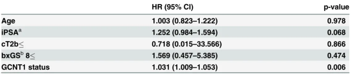
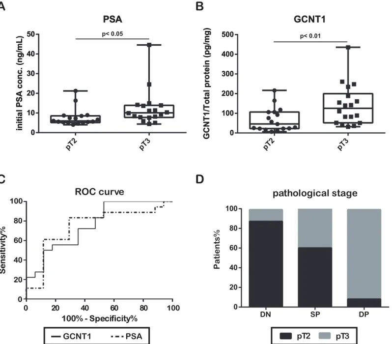
To establish a semi-quantitative high-throughput screen for GCNT1 expression, post-DRE urine specimens, which contain high concentrations of PCa proteins, were analyzed by the dot-blotting method using the anti-human GCNT1 antibody (Fig 2). Prediction of extracapsu-lar extension of PCa is a good predictor of PSA recurrence (S2 Fig). The initial PSA level and GCNT1 expression were highly correlated to extracapsular extension of PCa in a logistic regression analysis (Table 2). The optimal cut-off values for PSA and GCNT1 expression levels were determined to be 7.52 ng/mL and 79.36 pg/mg by the receiver-operator characteristic curve for prediction of extracapsular extension of PCa using the following formula: cut-off = (1
−sensitivity)2+ (1−specificity)2[29,30] (Fig 3A–3C). Based on these clinicopathological
parameters, we established following recurrence risk stratifications: double negative-risk (PSA<7.52 ng/mL, GCNT1<79.36 pg/mg), single positive-risk (PSA>7.52 ng/mL or
GCNT1>79.36 pg/mg) and double positive-risk (PSA>7.52 ng/mL and GCNT1>79.36 pg/
mg). Over 90% of double positive-risk patients had extracapsular extension of PCa in this risk
Table 1. Univariate and multivariable analyses of risk factors for prostate-specific antigen recurrence.
Univariate Multivariable
p-value HR (95% CI) p-value
Age 0.053 1.004 (0.934–1.080) 0.905
iPSAa 0.000 1.013 (0.990–1.037) 0.256
cT2b 0.182 0.637 (0.191–2.131) 0.464
bxGSb8 0.052 1.656 (0.612
–4.482) 0.321
GS8 0.011 1.787 (0.720–4.435) 0.210
pT3e 0.002 0.985 (0.349–2.774) 0.977
RMc 0.000 4.966 (1.861
–13.250) 0.001
pnd 0.221 0.839 (0.315
–2.234) 0.725
pN 0.000 29.124 (1.599–530.412) 0.023
GCNT1 status 0.028 3.691 (1.070–12.736) 0.039
a, pre-treatment prostate-speci
fic antigen b, Gleason score
c, cancer existence at the resected margin d, perineural invasion
e, extracapsular extension
CI, confidence interval; GCNT1, core2β-1,6-N-acetylglucosaminyltransferase-1; HR, hazard ratio; PSA, prostate-specific antigen.
stratification (Fig 3D). These results indicate that a high PSA concentration and GCNT1 expression in post-DRE urine are good predictors of extracapsular extension of PCa.
Discussion
Aberrant glycosylation of cell surface glycoproteins plays an important role in cancer initiation, proliferation, invasion, and metastasis [31–33]. Biosynthesis of oligosaccharides on glycopro-teins is performed in concert by several glycosyltransferases. The functional terminal structure
Fig 2. Detection of core2β-1,6-N-acetylglucosaminyltransferase-1 in post-digital rectal examination urine specimens.(A) Post-digital rectal examination urine specimens were collected and (B) centrifuged. (C) Supernatants were collected and spotted onto a nitrocellulose membrane. (D) Core2β -1,6-N-acetylglucosaminyltransferase-1 (GCNT1) was detected by an anti-GCNT1 monoclonal antibody, followed by a horseradish peroxidase (HRP)-conjugated antibody. (E) After treatment with a chemiluminescence reagent, the GCNT1 signal was recorded by a ChemiDoc+ system.
doi:10.1371/journal.pone.0138520.g002
Table 2. Logistic regression analyses of risk factors for extracapsular extension of prostate cancer.
HR (95% CI) p-value
Age 1.003 (0.823–1.222) 0.978
iPSAa 1.252 (0.984
–1.594) 0.068
cT2b 0.718 (0.015–33.566) 0.866
bxGSb8 1.569 (0.457–5.385) 0.474
GCNT1 status 1.031 (1.009–1.053) 0.006
a; pre-treatment prostate-speci
fic antigen b; Gleason score
CI, confidence interval; GCNT1, core2β-1,6-N-acetylglucosaminyltransferase-1; HR, hazard ratio; PSA, prostate-specific antigen
such as sialy lewis X (sLeX) and sialyl lewis A (sLeA) is also formed by glycosyltransferases [16,
34]. The sLeX and sLeA, which were widely known ligand of carbohydrate-binding proteins, are closely related to metastasis [35]. Not only sLe antigens, but also internal structures, partic-ularly GlcNAc beta1,6 branching structures and polylactosamines, are closely related to cancer malignancy [22,31]. GCNT1 is one of the glycosyltransferases that forms the core 2O-glycans
Fig 3. Prostate-specific antigen concentration and core2β-1,6-N-acetylglucosaminyltransferase-1 expression predict extracapsular extension of prostate cancer.(A) Prostate-specific antigen (PSA) concentration and (B) Core2β-1,6-N-acetylglucosaminyltransferase-1 (GCNT1) expression levels were significantly higher in prostate cancer (PCa) patients with extracapsular extension than in patients with organ-confined disease. (C) Receiver-operator characteristic curve analysis of PSA and GCNT1 revealed that the area under the curve of PSA was 0.7455 and GCNT1 was 0.7614. (D) Risk stratification was established using PSA and GCNT1 to predict the outcome of local PCa. Double negative (DN)-risk (PSA<7.52 ng/mL, GCNT1<79.36 pg/mg), single positive (SP)-risk (PSA>7.52 ng/mL or GCNT1>79.36 pg/mg) and double positive (DP)-risk (PSA>7.52 ng/mL and GCNT1>79.36 pg/mg) patients are compared.
on the surface of lymphocytes and various cancer cells [18,24,36,37]. Previously, it was reported that GCNT1 expression is associated with the metastatic potential of colorectal cancer [22], lung cancer [23], testicular cancer [24] and PCa [18]. It has also been reported that GCNT1-expressing cancers can escape the host immune response [25,38], especially from host natural killer cells that bind to galectin-3 on core 2-branchingO-glycans [25,38].
This study established a mAb against GCNT1 and evaluated its potential as an indicator of PCa aggressiveness. Immunohistochemical analysis of radical prostatectomy specimens showed that GCNT1 expression on PCa cells closely related to extracapsular extension of PCa (Fig 1). Moreover, patients with GCNT1-positive PCa exhibited worse PSA-free survival com-pared with patients with GCNT1-negative tumors (Fig 1). These results indicate that GCNT1 expression strongly correlates with the malignant potential of PCa.
Although immunohistochemistry provided much information on protein expression in PCa, the analysis was not quantitative. Using post-DRE urine, we established a
semi-quantitative and high-throughput screening for the malignant potential of PCa (Figs2and3). Recent studies reported that prostate cancer antigen 3 and a TMPRSS2:ERG fusion were two of the most useful indicators of PCa. These markers were focused on prospective PCa screening and early detection of PCa [39]. Prostate cancer antigen 3 and the TMPRSS2:ERG fusion almost reported PCR-based study and did not present sufficient biological evidence of PCa aggressiveness. We also reported that detection of aberrant glycosylation of PSA improved PCa screening but did not predict PCa aggressiveness [40]. In this study, GCNT1 expression in post-DRE urine was correlated with extracapsular extension of PCa. Moreover, a combination of initial PSA concentration and GCNT1 expression could predict extracapsular extension in over 90% of PCa. Therefore, GCNT1 detection in post-DRE urine improved prediction of PCa invasiveness.
Because GCNT1 is not a cancer-specific protein, its expression was unsuitable for PCa screening. Therefore, a combination of reported PCa screening markers and GCNT1 may improve the development of therapeutic strategies of PCa. Although the mechanism of GCNT1-driven regulation in cancer progression is poorly understood, our study demonstrates that GCNT1 can be a predictor of the malignant potential of PCa. Further clinical research is necessary to determine the utility of GCNT1 as a biomarker of PCa.
Supporting Information
S1 Fig. Preparation of an anti-human core 2β-1,6-N-acetylglucosaminyltransferase-1
pre-treated anti-human GCNT1 mAb. (TIF)
S2 Fig. Extracapsular extension of prostate cancer was one of the strong predictor of pros-tate-specific antigen recurrence.Prostate-specific antigen-free survival periods were com-pared between organ-confined disease (pT2) and extracapsular extension (pT3 and pT4). Survival was analyzed by Kaplan-Meier curves.
(EPS)
S1 File. Supplementary materials and methods.
(DOCX)
S1 Table. Core2β-1,6-N-acetylglucosaminyltransferase-1 status and patient data.
(DOCX)
S2 Table. Core2β-1,6-N-acetylglucosaminyltransferase-1 status and pathological
parame-ters.
(DOCX)
S3 Table. Patient data of post-digital rectal examination urine specimens.
(DOCX)
Acknowledgments
The authors thank Dr. Shigeru Tsuboi for useful discussions.
Author Contributions
Conceived and designed the experiments: CO YT. Performed the experiments: YK TY JM TS YT. Analyzed the data: SH YT. Contributed reagents/materials/analysis tools: TY SH KM YH TK CO MF YT. Wrote the paper: CO YT.
References
1. Ferlay J, Steliarova-Foucher E, Lortet-Tieulent J, Rosso S, Coebergh JW, Comber H, et al. Cancer inci-dence and mortality patterns in Europe: estimates for 40 countries in 2012. European journal of cancer. 2013; 49(6):1374–403. PMID:23485231.
2. Siegel R, Naishadham D, Jemal A. Cancer statistics, 2013. CA: a cancer journal for clinicians. 2013; 63(1):11–30. doi:10.3322/caac.21166PMID:23335087.
3. Gronberg H. Prostate cancer epidemiology. Lancet. 2003; 361(9360):859–64. doi: 10.1016/S0140-6736(03)12713-4PMID:12642065.
4. Baade PD, Youlden DR, Cramb SM, Dunn J, Gardiner RA. Epidemiology of prostate cancer in the Asia-Pacific region. Prostate international. 2013; 1(2):47–58. doi:10.12954/PI.12014PMID:24223402;
PubMed Central PMCID: PMC3814115.
5. Klotz L. Prostate cancer overdiagnosis and overtreatment. Current opinion in endocrinology, diabetes, and obesity. 2013; 20(3):204–9. PMID:23609043.
6. Andriole GL, Crawford ED, Grubb RL 3rd, Buys SS, Chia D, Church TR, et al. Mortality results from a randomized prostate-cancer screening trial. The New England journal of medicine. 2009; 360-(13):1310–9. doi:10.1056/NEJMoa0810696PMID:19297565; PubMed Central PMCID:
PMC2944770.
7. Schroder FH, Hugosson J, Roobol MJ, Tammela TL, Ciatto S, Nelen V, et al. Screening and prostate-cancer mortality in a randomized European study. The New England journal of medicine. 2009; 360-(13):1320–8. doi:10.1056/NEJMoa0810084PMID:19297566.
9. Ramsay CR, Adewuyi TE, Gray J, Hislop J, Shirley MD, Jayakody S, et al. Ablative therapy for people with localised prostate cancer: a systematic review and economic evaluation. Health technology assessment. 2015; 19(49):1–490. doi:10.3310/hta19490PMID:26140518.
10. Epstein JI, Partin AW, Sauvageot J, Walsh PC. Prediction of progression following radical prostatec-tomy. A multivariate analysis of 721 men with long-term follow-up. The American journal of surgical pathology. 1996; 20(3):286–92. PMID:8772781.
11. Kattan MW, Eastham JA, Stapleton AM, Wheeler TM, Scardino PT. A preoperative nomogram for dis-ease recurrence following radical prostatectomy for prostate cancer. Journal of the National Cancer Institute. 1998; 90(10):766–71. PMID:9605647.
12. Choudhury AD, Eeles R, Freedland SJ, Isaacs WB, Pomerantz MM, Schalken JA, et al. The role of genetic markers in the management of prostate cancer. European urology. 2012; 62(4):577–87. doi: 10.1016/j.eururo.2012.05.054PMID:22695242.
13. Miyake M, Taki T, Hitomi S, Hakomori S. Correlation of expression of H/Le(y)/Le(b) antigens with sur-vival in patients with carcinoma of the lung. The New England journal of medicine. 1992; 327(1):14–8.
doi:10.1056/NEJM199207023270103PMID:1317941.
14. Kannagi R, Izawa M, Koike T, Miyazaki K, Kimura N. Carbohydrate-mediated cell adhesion in cancer metastasis and angiogenesis. Cancer science. 2004; 95(5):377–84. PMID:15132763.
15. Nakamori S, Kameyama M, Imaoka S, Furukawa H, Ishikawa O, Sasaki Y, et al. Increased expression of sialyl Lewisx antigen correlates with poor survival in patients with colorectal carcinoma: clinicopatho-logical and immunohistochemical study. Cancer research. 1993; 53(15):3632–7. PMID:8101764.
16. Inaba Y, Ohyama C, Kato T, Satoh M, Saito H, Hagisawa S, et al. Gene transfer of alpha1,3-fucosyltransferase increases tumor growth of the PC-3 human prostate cancer cell line through enhanced adhesion to prostatic stromal cells. International journal of cancer Journal international du cancer. 2003; 107(6):949–57. doi:10.1002/ijc.11513PMID:14601054.
17. Ohyama C, Tsuboi S, Fukuda M. Dual roles of sialyl Lewis X oligosaccharides in tumor metastasis and rejection by natural killer cells. The EMBO journal. 1999; 18(6):1516–25. doi:10.1093/emboj/18.6.1516
PMID:10075923; PubMed Central PMCID: PMC1171240.
18. Hagisawa S, Ohyama C, Takahashi T, Endoh M, Moriya T, Nakayama J, et al. Expression of core 2 beta1,6-N-acetylglucosaminyltransferase facilitates prostate cancer progression. Glycobiology. 2005; 15(10):1016–24. doi:10.1093/glycob/cwi086PMID:15932919.
19. Tsui KH, Chang PL, Feng TH, Chung LC, Sung HC, Juang HH. Evaluating the function of matriptase and N-acetylglucosaminyltransferase V in prostate cancer metastasis. Anticancer research. 2008; 28-(4A):1993–9. PMID:18649738.
20. Bierhuizen MF, Fukuda M. Expression cloning of a cDNA encoding UDP-GlcNAc:Gal beta 1-3-GalNAc--R (GlcNAc to GalNAc) beta 1-6GlcNAc transferase by gene transfer into CHO cells expressing poly-oma large tumor antigen. Proceedings of the National Academy of Sciences of the United States of America. 1992; 89(19):9326–330. PMID:1329093; PubMed Central PMCID: PMC50119.
21. Skrincosky D, Kain R, El-Battari A, Exner M, Kerjaschki D, Fukuda M. Altered Golgi localization of core 2 beta-1,6-N-acetylglucosaminyltransferase leads to decreased synthesis of branched O-glycans. The Journal of biological chemistry. 1997; 272(36):22695–702. PMID:9278427.
22. Shimodaira K, Nakayama J, Nakamura N, Hasebe O, Katsuyama T, Fukuda M. Carcinoma-associated expression of core 2 beta-1,6-N-acetylglucosaminyltransferase gene in human colorectal cancer: role of O-glycans in tumor progression. Cancer research. 1997; 57(23):5201–6. PMID:9393734.
23. Machida E, Nakayama J, Amano J, Fukuda M. Clinicopathological significance of core 2 beta1,6-N-acetylglucosaminyltransferase messenger RNA expressed in the pulmonary adenocarcinoma deter-mined by in situ hybridization. Cancer research. 2001; 61(5):2226–31. PMID:11280791.
24. Hatakeyama S, Kyan A, Yamamoto H, Okamoto A, Sugiyama N, Suzuki Y, et al. Core 2
N-acetylglucosaminyltransferase-1 expression induces aggressive potential of testicular germ cell tumor. International journal of cancer Journal international du cancer. 2010; 127(5):1052–9. doi:10.1002/ijc. 25117PMID:20017138; PubMed Central PMCID: PMC2897929.
25. Tsuboi S, Sutoh M, Hatakeyama S, Hiraoka N, Habuchi T, Horikawa Y, et al. A novel strategy for eva-sion of NK cell immunity by tumours expressing core2 O-glycans. The EMBO journal. 2011; 30-(15):3173–85. doi:10.1038/emboj.2011.215PMID:21712812; PubMed Central PMCID:
PMC3160189.
26. Chen Z, Gulzar ZG, St Hill CA, Walcheck B, Brooks JD. Increased expression of GCNT1 is associated with altered O-glycosylation of PSA, PAP, and MUC1 in human prostate cancers. The Prostate. 2014; 74(10):1059–67. doi:10.1002/pros.22826PMID:24854630.
28. Epstein JI. An update of the Gleason grading system. The Journal of urology. 2010; 183(2):433–40.
doi:10.1016/j.juro.2009.10.046PMID:20006878.
29. Akobeng AK. Understanding diagnostic tests 3: Receiver operating characteristic curves. Acta paedia-trica. 2007; 96(5):644–7. doi:10.1111/j.1651-2227.2006.00178.xPMID:17376185.
30. Hatakeyama S, Amano M, Tobisawa Y, Yoneyama T, Tsuchiya N, Habuchi T, et al. Serum N-glycan alteration associated with renal cell carcinoma detected by high throughput glycan analysis. The Jour-nal of urology. 2014; 191(3):805–13. doi:10.1016/j.juro.2013.10.052PMID:24140550.
31. Dennis JW, Granovsky M, Warren CE. Glycoprotein glycosylation and cancer progression. Biochimica et biophysica acta. 1999; 1473(1):21–34. PMID:10580127.
32. Fukuda M. Possible roles of tumor-associated carbohydrate antigens. Cancer research. 1996; 56-(10):2237–44. PMID:8625291.
33. Hakomori S. Tumor malignancy defined by aberrant glycosylation and sphingo(glyco)lipid metabolism. Cancer research. 1996; 56(23):5309–18. PMID:8968075.
34. Muramatsu H, Kusano T, Sato M, Oda Y, Kobori K, Muramatsu T. Embryonic stem cells deficient in I beta1,6-N-acetylglucosaminyltransferase exhibit reduced expression of embryoglycan and the loss of a Lewis X antigen, 4C9. Glycobiology. 2008; 18(3):242–9. doi:10.1093/glycob/cwm138PMID: 18184719.
35. Ishikawa D, Kikkawa H, Ogino K, Hirabayashi Y, Oku N, Taki T. GD1alpha-replica peptides functionally mimic GD1alpha, an adhesion molecule of metastatic tumor cells, and suppress the tumor metastasis. FEBS letters. 1998; 441(1):20–4. PMID:9877157.
36. Kim J, Villadsen R, Sorlie T, Fogh L, Gronlund SZ, Fridriksdottir AJ, et al. Tumor initiating but differenti-ated luminal-like breast cancer cells are highly invasive in the absence of basal-like activity. Proceed-ings of the National Academy of Sciences of the United States of America. 2012; 109(16):6124–9. doi: 10.1073/pnas.1203203109PMID:22454501; PubMed Central PMCID: PMC3341000.
37. Lee SH, Yu SY, Nakayama J, Khoo KH, Stone EL, Fukuda MN, et al. Core2 O-glycan structure is essential for the cell surface expression of sucrase isomaltase and dipeptidyl peptidase-IV during intes-tinal cell differentiation. The Journal of biological chemistry. 2010; 285(48):37683–92. doi:10.1074/jbc. M110.162735PMID:20841351; PubMed Central PMCID: PMC2988373.
38. Okamoto T, Yoneyama MS, Hatakeyama S, Mori K, Yamamoto H, Koie T, et al. Core2 O-glycan-expressing prostate cancer cells are resistant to NK cell immunity. Molecular medicine reports. 2013; 7-(2):359–64. doi:10.3892/mmr.2012.1189PMID:23165940; PubMed Central PMCID: PMC3573034.
39. Tallon L, Luangphakdy D, Ruffion A, Colombel M, Devonec M, Champetier D, et al. Comparative evalu-ation of urinary PCA3 and TMPRSS2: ERG scores and serum PHI in predicting prostate cancer aggres-siveness. International journal of molecular sciences. 2014; 15(8):13299–316. doi:10.3390/
ijms150813299PMID:25079439; PubMed Central PMCID: PMC4159795.