Injectable Collagen Matrix Using Bifunctional Peptides
Paul T. Hamilton1, Michelle S. Jansen2, Sathya Ganesan2, R. Edward Benson3, Robin Hyde-DeRuyscher4, Wayne F. Beyer5, Joseph C. Gile6, Shrikumar A. Nair2, Jonathan A. Hodges2*, Hanne Grøn7
1Department of Microbiology, North Carolina State University, Raleigh, North Carolina, United States of America,2Affinergy, LLC, Research Triangle Park, North Carolina, United States of America,3Platform Technology and Science, GlaxoSmithKline, Research Triangle Park, North Carolina, United States of America,4Manufacturing Sciences, Biogen Idec, Research Triangle Park, North Carolina, United States of America,5QNS Group, LLC, Durham, North Carolina, United States of America,6Gile Surgical Support, Bangor, Maine, United States of America,7Haemophilia Biochemistry, Novo Nordisk, Ma˚løv, Denmark
Abstract
To promote healing of many orthopedic injuries, tissue engineering approaches are being developed that combine growth factors such as Bone Morphogenetic Proteins (BMP) with biomaterial carriers. Although these technologies have shown great promise, they still face limitations. We describe a generalized approach to create target-specific modular peptides that bind growth factors to implantable biomaterials. These bifunctional peptide coatings provide a novel way to modulate biology on the surface of an implant. Using phage display techniques, we have identified peptides that bind with high affinity to BMP-2. The peptides that bind to BMP-2 fall into two different sequence clusters. The first cluster of peptide sequences contains the motif W-X-X-F-X-X-L (where X can be any amino acid) and the second cluster contains the motif F-P-L-K-G. We have synthesized bifunctional peptide linkers that contain BMP-2 and collagen-binding domains. Using a rat ectopic bone formation model, we have injected rhBMP-2 into a collagen matrix with or without a bifunctional BMP-2: collagen peptide (BC-1). The presence of BC-1 significantly increased osteogenic cellular activity, the area of bone formed, and bone maturity at the site of injection. Our results suggest that bifunctional peptides that can simultaneously bind to a growth factor and an implantable biomaterial can be used to control the delivery and release of growth factors at the site of implantation.
Citation:Hamilton PT, Jansen MS, Ganesan S, Benson RE, Hyde-DeRuyscher R, et al. (2013) Improved Bone Morphogenetic Protein-2 Retention in an Injectable Collagen Matrix Using Bifunctional Peptides. PLoS ONE 8(8): e70715. doi:10.1371/journal.pone.0070715
Editor:Fabrizio Gelain, University of Milan-Bicocca, Italy
ReceivedSeptember 27, 2012;AcceptedJune 28, 2013;PublishedAugust 8, 2013
Copyright:ß2013 Hamilton et al. This is an open-access article distributed under the terms of the Creative Commons Attribution License, which permits unrestricted use, distribution, and reproduction in any medium, provided the original author and source are credited.
Funding:The work was supported in part by the National Institutes of Arthritis and Musculoskeletal and Skin Disease, General Medicine, and Dental and Craniofacial Research of the National Institutes of Health (NIH) under award numbers R43AR053387, R43AR051264, R44GM077753 and R44DE018071. The funders had no role in study design, data collection and analysis, decision to publish, or preparation of the manuscript. The content is solely the responsibility of the authors and does not necessarily represent the official views of the NIH.
Competing Interests:MSJ, SG, SAN, and JH are employees of Affinergy. PTH is an employee of North Carolina State University. REB is an employee of GlaxoSmithKline. RHD is an employee of Biogen Idec. WFB is an employee of QNS Group. JCG is an employee of Gile Surgical Support. HG is an employee of Novo Nordisk. The BMP-binding peptides are contained in a family of issued and pending patents (see United States Patent number 7,572,766) assigned to Affinergy. There are no marketed products to declare. This does not alter the authors’ adherence to all the PLoS ONE policies on sharing data and materials, as detailed online in the guide for authors.
* E-mail: [email protected]
Introduction
Approximately 7.9 million fractures occur each year in the United States alone, and approximately 10% of fractures exhibit delayed or impaired healing [1]. Bone morphogenetic proteins (BMPs) are osteogenic growth factors that have been shown to stimulate new bone formation and fracture healing [2,3]. In clinical trials, recombinant human BMP-2 (rhBMP-2) has been shown to accelerate healing of open tibial fractures [4], and rhBMP-7 has been used to treat tibial nonunions [5]. These clinical applications, however, require open surgical procedures to insert the BMP–loaded carrier. In addition, supraphysiological amounts of BMPs are required to promote bone formation due to the growth factor’s rapid diffusion away from its carrier [6,7]. The use of high doses, however, raises concerns about bone formation away from the site and impact on nearby tissues and organs [8]; in accordance, rhBMP-2 use has been linked to a variety of serious adverse events [9].
BMP after a few days in vivo. Calcium phosphate cement–based formulations were able to retain measurable amounts of BMP at the site of injection for 14 days [12]. These formulations, however, relied on entrapment of the BMP within the cement, raising concerns about a lack of release of BMP to the surrounding site of healing. We propose that the use of engineered peptides with high affinities for both BMP and the matrix material joined as a bifunctional peptide may provide more controlled release of BMP from the matrix and promote optimal healingin vivo.
Bifunctional peptides have been constructed by a number of groups as a way to modify materials and promote cell adhesion or growth factor binding. Gama and his colleagues [14,15] have fused cell adhesion domains such as RGD and IKVAV to a cellulose-binding domain. The 25 kD fusion proteins promote fibroblast or mesenchymal stem cell adhesion to bacterial cellulose. Murphy and colleagues have also reported the successful use of modular peptides to deliver growth factors and mesenchymal stem cells to hydroxyapatite coatings [16,17,18].
In this study, we have used phage display technology to identify a series of peptides which bind to BMPs. The BMP–binding peptides can be organized into two sequence motifs. Consensus peptides from each motif were coupled to a collagen-binding peptide to form bifunctional peptides that can bind simultaneously to BMP-2 and collagen. The bifunctional peptide therefore can bind and retain BMP-2 onto a collagen matrix, slowing the release of BMP-2 from the matrix. We tested the ability of the bifunctional peptide to improve bone formationin vivousing a rat ectopic bone formation bioassay.
Materials and Methods
Ethics statement
All procedures with animals were performed under protocols approved by Affinergy’s Institutional Animal Care and Use Committee in a facility with assurance from the Office of Laboratory Animal Welfare (A4544-01).
Materials
Horseradish peroxidase (HRP)–conjugated anti-M13 monoclo-nal antibody was from GE Healthcare (Piscataway, NJ). Tween 20, 2,29-Azino-bis(3-ethylbenzothiazoline-6-sulfonic acid) dia-mmonium salt (ABTS), streptavidin (SA) fromStreptomyces avidinii, bovine serum albumin (BSA),para-nitrophenylphosphate reagent (p-NPP) and all other chemicals were purchased from Sigma-Aldrich (St. Louis, MO). Recovered human plasma was purchased from the American Red Cross (Durham, NC). rhBMP-2 (355-BM/CF) and an anti-BMP antibody (MAB3552) were purchased from R&D Systems (Minneapolis, MN). In vivo studies were performed using rhBMP-2 (INFUSE) purchased from Medtronic (Ref. 7510600). N-a-Fmoc-amino acids (with orthogonal side chain protecting groups) were purchased from Novabiochem (Merck KGaA, Darmstadt, Germany). Alkaline phosphatase– labeled goat anti-mouse secondary antibody was purchased from Promega (Madison, WI).
Phage Display
rhBMP-2 was biotinylated using a Sulfo-NHS-Biotin reagent (Pierce EZ-link biotinylation kit) following the manufacturer’s protocol. The biotinylated rhBMP-2 was immobilized onto a streptavidin-coated 96-well microtiter plate (Immulon IV) and the plates blocked with 0.5% BSA in phosphate buffered saline, 0.05% Tween-20 (PBST). Phage display was performed as previously described [19,20]. Ten different phage display libraries were screened for peptides that bind to rhBMP-2. Each library was designed around a specific amino acid motif or amino acid bias. After 3 rounds of phage display selections, the pools of enriched phage were plated on a lawn ofE. coliDH5aF’ cells. Individual phage were picked and propagated onE. coliovernight. The cells were removed by centrifugation, and 10ml of the phage-containing supernatant was added to the wells phage-containing BMP-2 or to a control well containing buffer. After incubation and washing, phage were detected in an ELISA–type assay using an HRP-conjugated, anti-M13 monoclonal antibody (1:1000).
Generating Focused Library for BMP-2 binding peptides To generate the Motif 1–focused and Motif 2–focused libraries, oligonucleotides were synthesized to encode peptides that have a restricted set of possible amino acids in selected positions in the peptide (Table 1). A short complementary primer was annealed to the 39end of the library oligonucleotide and extended with T7 polymerase. After second strand synthesis, the DNA was digested with Xba I and Xho I and ligated into mAEK phage display vector. The ligation reaction was used to transform electro-competent E. coli DH12S. Transformed cells were grown overnight in 26YT medium. The phage-containing culture supernatant was collected and the phage concentrated by PEG precipitation. Precipitated phage were resuspended in phosphate buffered saline containing 20% glycerol, aliquoted and stored at
220uC.
DNA sequence analysis
DNA from isolated positive phage clones was amplified using the TempliPhi DNA amplification kit and DNA analysis was performed by Sequetech (Mountain View, CA).
Peptide Synthesis
Peptides were synthesized by solid-phase peptide synthesis techniques on a Rainin Symphony Peptide Synthesizer using standard Fmoc chemistry (HBTU/HOBT activation, 20% piper-idine in DMF for Fmoc removal). After all residues were coupled, simultaneous cleavage and side chain deprotection was achieved by treatment with a trifluoroacetic acid (TFA) cocktail. Crude peptide was precipitated with cold diethyl ether and purified by high-performance liquid chromatography on a Shimadzu HPLC using a Vydac C18 reversed-phase silica column (10mm, 120 A˚ , 250 mm622 mm) using a linear gradient of water/acetonitrile containing 0.1% TFA. Homogeneity of the synthetic peptides was evaluated by analytical RP-HPLC (Vydac C18 silica column, 10mm, 120 A˚ , 250 mm64.6 mm) and the identity of the peptides
Table 1.Focused library design for motif 1 and motif 2 BMP-binding peptides*.
Motif 1 Library X5-[W/L/C/Y/F/S]-X2-[W/L/C/Y/F/S]-X-[A/G/N/S/T]-[L/F/I/M/V]-X5
Motif 2 Library X3-[L/F/I/M/V]-X-[W/L/C/Y/F/S]-[P/S/T/A]-[ L/F/I/M/V]-[I/M/T/N/K/S/R]-X8
was determined by MALDI-TOF-MS. All peptides were synthe-sized with a biotin coupled to the epsilon-amino group of the C-terminal lysine.
Bifunctional Peptide Design
A consensus BMP-binding peptide was generated for each BMP-binding motif. Peptides that bind to collagen were isolated by phage display on demineralized bone matrix [21]. Bifunctional peptides (Collagen: BMP) were generated by joining a collagen-Table 2.Scoring system for histological analysis of bone growth.
GRADE BONE CROSS-SECTIONAL AREA*
0 No Evidence of Bone Formation
1.1 1–10% of Implant Shows Evidence of Bone Formation
1.2 11–25% of Implant Shows Evidence of Bone Formation
2.1 26–35% of Implant Shows Evidence of Bone Formation
2.2 36–50% of Implant Shows Evidence of Bone Formation
3 51–75% of Implant Shows Evidence of Bone Formation
4 76–100% of Implant Shows Evidence of Bone Formation
GRADE BONE MATURITY
0 No Bone
1 Immature/Unorganized
2 Immature
3 Mature
4 Mature/Well Organized
GRADE CELLULAR ACTIVITY
0 None
1 Rare
2 Few
3 Moderate
4 Dense
*Implant evaluated at 106Magnification. doi:10.1371/journal.pone.0070715.t002
Figure 1. BMP-2 Binding Peptides.Biotinylated BMP-2 was immobilized on streptavidin-coated plates and subjected to multiple rounds of phage display selections using 10 different phage display peptide libraries. Individual binding phage were isolated and the sequence of the BMP-binding peptide deduced from the phage DNA sequence. Alignment of the peptides revealed two general sequence motifs among the peptides: motif 1: W-X-X-F-X-X-L and motif 2: L-X-F-P-L-K. These motifs were used to generate 2ndgeneration focused libraries. In addition, representative synthetic peptides were made and tested for binding to BMP-2.
binding peptide sequence to each of the BMP-binding sequences through a short, flexible amino acid linker (GSSG): [peptide BC-1]
–
SWWGFWNGSAAPVWSR-GSSG-AGAWEAFSSLSGSRV-GSSGK(Biotin) and [peptide BC-2] – SWWGFWNGSAAPV-WSR-GSSG-AGALGFPLKGEVVEGWA-GSSGK(Biotin). The GSSG linker is intended to join the two binding domains and has no inherent properties.
BMP-2 peptide binding assay
To measure the ability of a BMP-binding peptide to capture its target growth factor out of buffer or a complex biological solution such as plasma, biotinylated peptides were immobilized onto a streptavidin-coated 96 well plate. Varying low nanomolar concentrations of BMP-2 were ‘‘spiked’’ into human plasma or Tris-buffered saline (0.5 M NaCl), 0.05% Tween-20 (TBST) and added to the peptide-containing plates. After 1 hr incubation at room temperature (RT), plates were washed and incubated with an anti-BMP-2 antibody followed by a goat anti-mouse secondary antibody conjugated to alkaline phosphatase. Adding p-NPP produced a colored reactant, which was quantified using a SpectroMax (Molecular Devices) plate reader at 405 nm.
Preparation of a 4% fibrillar collagen gel
Collagen stock solution (6.4 mg/ml, Inamed cat#5413, lot1387646) was neutralized overnight at room temperature using 200 mM sodium phosphate, pH 9.4. The fibrillar collagen was
pelleted by centrifugation at 17,2006g for 20 minutes at 10uC. The collagen weight percent was determined using a bicinchoninic acid protein assay (BCA assay, Pierce cat#23255).
Preparation of 1.5% injectable collagen gel for BMP-2 deliveryin vivo
The stock fibrillar collagen (4%) was diluted in PBS containing rhBMP-2 with or without the collagen-BMP bifunctional peptide, to generate a final collagen wt% of 1.5%. The amount of BMP-2 and peptide were optimized using pilot experimentsin vivo such that 200ml of the injected collagen gel contained 2mg of BMP-2
and a 50-fold molar excess of the peptide.
Injectable collagen gel binding assay
The injectable collagen gel (0.1 mL) was aliquoted into a polypropylene plate. The plate was blocked with 150ml of 1% BSA in TBS for 30 min. After spinning for 2 min, the supernatant was removed without disrupting the collagen gel. The bifunctional peptide (30mM) was mixed with rhBMP-2 (0.0017 to100 nM) in Binding Buffer (125 mM glutamic acid, 10% sucrose, 12.5% glycine, 10% polysorbate 80, 5 M NaCl) for 30 min with gentle agitation. The peptide: rhBMP-2 complex (50mL) was then added to the collagen gel and incubated at room temperature for 1 h with gentle agitation. The collagen gel was washed three times with TBST and rhBMP-2 was detected with an anti-BMP antibody (R&D Systems MAB3552). The wells were then washed, and an alkaline phosphatase labeled goat anti-mouse secondary antibody was added. After incubation and washing, binding was measured using the chromogenic reagentp-NPP, and absorbance was read at 405 nm.
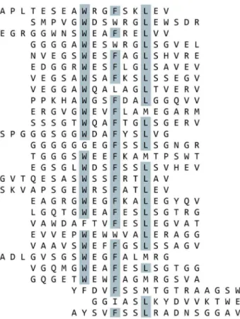
Figure 2. BMP-2 Binding Peptides Containing Motif 1 Isolated from Selections Using the Focused Libraries.Biotinylated BMP-2 was immobilized on streptavidin-coated plates and subjected to multiple rounds of phage display selections using the focused libraries. Peptides which contain motif 1 were aligned. Amino acids which are present in over half of the aligned sequences at a given position are highlighted.
doi:10.1371/journal.pone.0070715.g002
Figure 3. BMP-2 Binding Peptides Containing Motif 2 Isolated from Selections Using the Focused Libraries.Biotinylated BMP-2 was immobilized on streptavidin-coated plates and subjected to multiple rounds of phage display selections using the focused libraries. Peptides which contain motif 2 were aligned. Amino acids which are present in over half of the aligned sequences at a given position are highlighted.
In vivoectopic bone formation study
Male Sprague Dawley rats (150–175 g) were obtained from Taconic farms, Charles River Laboratories (Raleigh, NC) and acclimated prior to surgery. The surgery was performed under general anesthesia by weight-adapted intraperitoneal injection of Xylazine 2% (Medistar; 12 mg/kg body weight) and Ketamine hydrochloride (Ketaset; 100 mg/mL; 80 mg/kg body weight). The thorax and abdomen were shaved and scrubbed with Betadine and alcohol. Using aseptic technique, two 5 mm incisions were made at the midline so that two subcutaneous pouches were prepared by blunt dissection. Collagen gel (200ml)
containing 2mg rhBMP-2 with or without collagen-BMP
bifunc-tional peptide (BC-1) was injected into the left or the right side of the subcutaneous region. Each rat received two injections and a total of 20 animals were used in the study. The BMP-2 dose (2mg) used in this experiment had been determined from preliminary experiments (data not shown). In those experiments, rats were implanted with collagen gel containing either 1 or 5mg of BMP-2. Collagen gel without peptide resulted in bone formation at the 5mg dose but not at the 1mg dose. Collagen gel with peptide resulted in bone formation at both doses (1 and 5mg), and a dose of 2mg BMP-2 was selected for the experiment.
The sites containing the injected material were explanted at two weeks and fixed in 10% neutral buffered formalin. The samples were placed in Formical 2000 decalcifier (American Mastertech) for 24 hours followed by Cal-arrest neutralizing solution for one hour. The tissue samples were processed in a Thermo-Electron, Shandon Excelsior Automated Tissue Processor for 14 hours. The process includes additional fixation in 10% buffered neutral formalin, dehydration through an ethanol gradient, and clearing with xylene at 40uC. Tissue was then embedded on a Leica EF 1140 H tissue embedding center in paraffin blocks for sectioning. Paraffin blocks were sectioned at 5 microns on a Reichert-Jung RM2065 microtome using Accu-Edge High Profile disposable stainless steel microtome blades. Three serial sections at intervals of 30 microns were obtained and placed on Super-frost Plus slides for hematoxylin and eosin staining (H&E). Staining was performed using a Sakura DRS-601 automatic slide stainer with a regressive Harris hematoxylin and Eosin Y Alcoholic for histological and morphological examination.
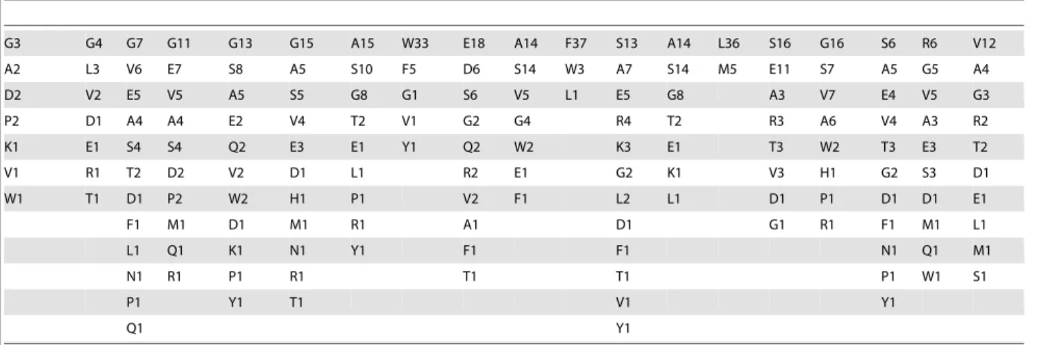
Histological sections were scored based on three criteria: Bone Cross-Sectional Area, Bone Maturity and Cellular Activity. Each segment was scored based on a 0–4 point scale by two observers Table 3.Summary of BMP-2 binding peptides containing motif 1*.
G3 G4 G7 G11 G13 G15 A15 W33 E18 A14 F37 S13 A14 L36 S16 G16 S6 R6 V12
A2 L3 V6 E7 S8 A5 S10 F5 D6 S14 W3 A7 S14 M5 E11 S7 A5 G5 A4
D2 V2 E5 V5 A5 S5 G8 G1 S6 V5 L1 E5 G8 A3 V7 E4 V5 G3
P2 D1 A4 A4 E2 V4 T2 V1 G2 G4 R4 T2 R3 A6 V4 A3 R2
K1 E1 S4 S4 Q2 E3 E1 Y1 Q2 W2 K3 E1 T3 W2 T3 E3 T2
V1 R1 T2 D2 V2 D1 L1 R2 E1 G2 K1 V3 H1 G2 S3 D1
W1 T1 D1 P2 W2 H1 P1 V2 F1 L2 L1 D1 P1 D1 D1 E1
F1 M1 D1 M1 R1 A1 D1 G1 R1 F1 M1 L1
L1 Q1 K1 N1 Y1 F1 F1 N1 Q1 M1
N1 R1 P1 R1 T1 T1 P1 W1 S1
P1 Y1 T1 V1 Y1
Q1 Y1
*Based on 41 sequences. Format is single-letter amino acid code and frequency of occurrence at that position in the peptide. doi:10.1371/journal.pone.0070715.t003
Table 4.Summary of BMP-2 binding peptides containing motif 2*.
G7 G5 A3 L9 G6 F14 P18 L17 K10 G18 E4 V6 V8 E5 G5 W4 A4
E4 D2 S3 V8 R2 L3 I1 R6 Q3 P4 I2 S3 A3 A2 D2
S2 E2 G2 G1 S2 W1 S1 D2 T4 M2 D2 V3 K2 E2
A1 K1 D2 A1 T1 I2 L3 Q2 G2 D2 P2 P2
F1 L1 P2 E1 T2 W1 A1 K1 Q1 V2 V2
M1 N1 F1 H1 A1 L1 P1 T1 G1 H1
P1 P1 R1 N1 R1 W1 T1 W1 M1 K1
V1 R1 T1 P1 S1 Y1 W1 T1 F1
S1 V1 V1 V1 G1
V1 W1 W1 W1
W1 Y1 Y1
Y1
(blinded to the study) who evaluated the entire implant under 106
magnification. The scoring system is summarized inTable 2.
Results
Isolation of BMP-binding peptides using phage display peptide libraries
Phage display was performed against immobilized rhBMP-2 with 10 different peptide libraries, representing over 10 billion peptide sequences. Biopanning with these libraries revealed a set of 16 peptides that bound to BMP-2 (Figure 1). These first-generation BMP–binding peptides fall into two different sequence clusters. The first cluster of peptide sequences contains the motif W-X-X-F-X-X-L (single letter amino acid code, where X can be any amino acid), designated motif 1, and the second cluster contains the motif F-P-L-K-G, designated motif 2. A series of truncations in which conserved amino acids were deleted in the BMP-binding peptides results in a loss of BMP-2 binding activity (data not shown). The consensus sequence among the peptides indicates that all the peptides within a sequence cluster are binding to the same site on BMP-2, and the common amino acids that make up the motif are responsible for the specific interactions with BMP-2.
Using these sequence motifs, we designed a focused phage display library around each motif (Table 1). Each of the focused libraries was screened for additional peptide sequences that would bind to BMP-2 using standard phage display techniques [22]. Combined, 59 peptide sequences were found positive for BMP-2
Table 5.Sequence of consensus BMP-binding peptides.
Synthetic Peptide# Peptide Sequence
B-17 (motif 1) GGGAWEAFSSLSGSRVGSSGK-(Biotin)
B-18 (motif 2) GGALGFPLKGEVVEGWAGSSGK-(Biotin)
doi:10.1371/journal.pone.0070715.t005
Figure 4. BMP-2 Binding to Peptide.Biotinylated peptides were immobilized on streptavidin-coated plates and incubated with a range of rhBMP-2 concentrations in TBST for 1 h. BMP-2 binding was analyzed using antibody andp-NPP detection. Peptides B-6, B-17, and B-18 had the highest binding affinities for rhBMP-2. Peptide N-1 is a negative control that binds hexokinase. Data are presented as the absorbance read at 405 nm. doi:10.1371/journal.pone.0070715.g004
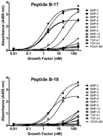
Figure 5. Peptide cross-reactivity to other growth factors. Peptides B-17 (A) and B-18 (B) were immobilized on a streptavidin-coated plate, and a range of concentrations of growth factors in the TGF-bsuperfamily were titrated onto the plate in TBST for 1 h. Growth factor binding was analyzed using antibody andp-NPP detection. Both peptide B-17 and B-18 bound to rhBMP-2, rhBMP-6, and rhBMP-7. Peptide B-17 also cross-reacted with rhBMP-3, rhBMP-5, and rhBMP-12. The peptides had lower affinities for all other growth factors tested. Data are presented as the absorbance read at 405 nm.
binding. Of these 59, 41 sequences were found around motif#1 (Figure 2) and 18 represented motif#2 (Figure 3). After aligning the peptides, the frequency of amino acid identity was scored at each position (Tables 3 & 4). Positions that have only one, two or three amino acid identities represented at a given position are likely to promote BMP–binding, therefore defining a new feature for the binding motif. From the alignment, a consensus sequence was established for the binding domain revealed by each focused library. Synthetic peptides were synthesized based on each consensus sequence: B-17 is the consensus for motif #1 and B-18 is the consensus for motif#2 (Table 5).
Relative affinity of BMP-2 binding peptides for BMP-2 To compare binding affinities between first and second generation BMP–binding peptides, we tested a range of BMP-2 concentrations (0.001 to 10 pmoles/well) for binding against immobilized peptide on streptavidin coated plates (Figure 4). The consensus peptides B-17 and B-18 bound BMP-2 with high apparent affinity with EC50 values of 1.4 and 1.9 nM, respec-tively. Peptide B-6 also had a low EC50 value of 1.4 nM but was not used in subsequent experiments.
Cross-reactivity of BMP-2–binding peptides with other growth factors
BMPs are members of the TGF-b superfamily and so we examined our peptides’ abilities to bind other family member proteins. The two BMP–binding peptides that contain the consensus motif#1 (B-17) and consensus motif #2 (B-18) were tested for binding to BMP-2, -3, -4, -5, -6, -7, -9, -12, -14, TGF-b1, TGF-b3, and PDGF-BB. Both peptides bound to BMP-2, BMP-6 and BMP-7 but showed no binding to TGF-b1, TGF-b3, or PDGF-BB (Figure 5; EC50, Peptide B-17: 1.4 nM, BMP-2; 1.3 nM, BMP-6; 17.0 nM, BMP-7. EC50, Peptide B-18: 2.8 nM, BMP-2; 6.5 nM, BMP-6; 16.2 nM, BMP-7). The lack of specific binding for TGF-b1, TGF-b3 or PDGF-BB, suggests that the peptides intended for BMP-2 binding harbor a specific interaction with a sequence or structural motif found in several BMPs but not in other growth factors. The two peptides, B-17 and B-18, however, do not show identical specificity among the BMP
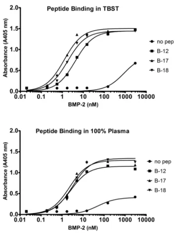
Figure 6. Peptide-mediated capture of BMP-2 from spiked TBST or plasma. The biotinylated peptides were coated on streptavidin plates and incubated in TBST (A) or plasma (B) with nanomolar concentrations of rhBMP-2 for 1 h. BMP-2 binding was analyzed using antibody andp-NPP detection. The peptides captured more rhBMP-2 from solution than the no peptide control. Data are presented as the absorbance read at 405 nm.
doi:10.1371/journal.pone.0070715.g006
Figure 7. Bifunctional peptide–mediated binding of BMP-2 to injectable collagen.A bifunctional peptide (BC-1) was synthesized containing Peptide B-17 and a collagen-binding peptide with a short amino acid linker. rhBMP-2 was mixed with or without the bifunctional peptide and added to a collagen gel. The bifunctional peptide enhanced the retention of rhBMP-2 to the collagen gel (no peptide, EC50 = 5.5 nM; BC-1, EC50 = 0.41 nM). Data are presented as the absorbance read at 405 nm.
doi:10.1371/journal.pone.0070715.g007
Figure 8. Ectopic bone formation with rhBMP-2 delivered in a collagen gel with or without the bifunctional peptide.rhBMP-2 (2mg) was delivered in a rat ectopic bone model either alone (no peptide) or in combination with a 50-fold molar excess of the bifunctional peptide (Collagen-BMP peptide). H&E stained slides were scored for osteogenic cellular activity, bone area and bone maturity by two observers and the median score for each group is shown in the figure. ****, p,.0001 vs no peptide.
proteins. B-17 also bound to BMP-3, BMP-5 and BMP-12, whereas 18 did not. Competition experiments with 17 and B-18 demonstrated that the two peptides compete for binding to BMP-2 (data not shown). This finding indicates that the peptides
bind at or near the same site on BMP-2. The cross-reactivity results indicate that B-17 binds to features on BMPs that are found in BMP-2, -3, -5, -6, -7, and -12 whereas B-18 binds to features found in only BMP-2, -6, and -7.
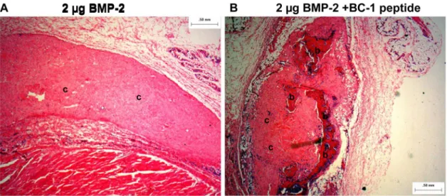
Figure 9. Representative histology image (hematoxylin and eosin stain) from the rat ectopic model obtained at a 2X magnification. A: 2mg BMP-2 in 1.5% collagen gel andB: 2mg BMP-2 with 50-fold molar excess of collagen-BMP-2 bifunctional peptide in 1.5% collagen gel.b–
Represent regions of bone;c– represent regions of collagen; cells are stained blue. The image shows only cellular activity in sample A, whereas sample B shows bone formation and increased cellular activity.
doi:10.1371/journal.pone.0070715.g009
Table 6.Histology scores (average from two observers blinded to the study) of all samples with rhBMP-2 delivered in a collagen gel with or without the bifunctional peptide.
Osteogenic Cellular activity Bone area Bone maturity
2mg rhBMP-2
2mg rhBMP-2+
bifunctional peptide 2mg rhBMP-2
2mg rhBMP-2+
bifunctional peptide 2mg rhBMP-2
2mg rhBMP-2+
bifunctional peptide
0.8 2.8 0.0 2.1 0.0 3.0
0.8 3.0 0.0 2.2 0.0 3.0
0.8 3.0 0.0 2.1 0.0 3.0
0.8 3.0 0.0 2.2 0.0 3.0
0.8 2.0 0.0 1.1 0.0 2.0
0.5 3.8 0.0 3.0 0.0 3.0
0.5 2.6 0.0 2.5 0.0 3.0
1.8 2.5 0.0 2.1 0.0 2.0
1.8 3.1 1.1 2.5 1.0 3.0
0.5 1.8 0.0 0.0 0.0 0.0
1.5 2.4 0.0 1.2 0.0 2.0
0.8 2.5 0.0 1.2 0.0 2.0
0.8 2.5 0.0 1.2 0.0 3.5
0.8 3.3 0.0 2.2 0.0 3.0
0.8 *nd 0.0 *nd 0.0 *nd
1.5 2.4 0.0 1.1 0.0 2.0
0.8 2.6 0.0 1.2 0.0 2.0
1.3 1.9 0.0 0.0 0.0 0.0
0.8 3.5 0.0 3.5 0.0 3.5
1.3 1.5 0.0 0.0 0.0 0.0
*nd –not determined.
Capture of BMP-2 from a complex biological fluid To model endogenous capture of a specific protein in vitro, picomole amounts of rhBMP-2 (0.001 to 15 pmoles/well) were mixed with TBST or human plasma for 1 h. After washing the plates in TBST, an alkaline phosphatase–conjugated antibody against BMP-2 was applied, and bound BMP-2 was measured with a colorimetric assay on a plate reader. Incubating BMP-2–binding peptides in human plasma had no effect on the peptide’s ability to bind and capture BMP-2 (Figure 6). Binding reactions in TBST controls exhibited virtually identical affinity for the target growth factor relative to the biological fluids. These data show that the peptides are not ‘‘fouled’’ by other proteins in biological fluids, suggesting that bifunctional peptide–mediated capture and con-centration of endogenously produced growth factors could also be a viable therapeutic strategy.
Bifunctional Peptides
Bifunctional peptides consisting of the BMP–binding sequences and the collagen-binding sequences were synthesized and tested for the ability to bind BMP-2 to a collagen gel. Both 1 and BC-2 bound BMP-BC-2, but to minimize the size and cost of the animal study, only BC-1 was used in thein vivomodel. BC-1 was chosen because the BMP-binding sequence in BC-1, the B-17 sequence, bound a wider range of BMPs than the B-18 sequence which might be useful in future experiments. BMP-2 was incubated with the bifunctional peptide BC-1 and then added to plates containing the injectable collagen gel. BMP-2 is known to bind collagen weakly [23], but the bifunctional peptide increased binding of BMP-2 to the collagen gel more than 10-fold (no peptide: EC50 = 5.5 nM; bifunctional peptide: EC50 = 0.41 nM;
Figure 7).
In vivomodel – Ectopic Bone Formation
Ectopic bone formation at two weeks was evaluated by histological scoring of the H&E stained slides by two observers. The slides were scored for infiltration of osteogenic cells (fibroblasts, osteoblasts, osteogenic progenitors and cells of the cartilage), each of which were scored on a scale of 1–4: 1-rare, 2-few, 3-moderate and 4- dense. Osteogenic cellular activity was obtained by an average of the scores for each cell type for every animal. Comparison between groups was performed using medians. As shown in Figure 8, the median cellular activity of the peptide group was significantly greater than the no peptide (BMP-2 alone) group (Mann-Whitney U two tailed test, p,0.0001). Bone area was scored as percent of the implant showing new bone formation on a scale of 0–4. The bone area was significantly greater in the peptide group compared to the no peptide (BMP-2 alone) group (Mann-Whitney U two tailed test, p,0.0001). In the peptide group, approximately 25% of the implant was covered with new bone, whereas no new bone was detected in the no peptide group. The maturity of the newly formed bone was scored on a scale of 0–4, with 0-no bone, 1-immature/unorganized, 2-immature, 3-mature and 4- mature/ well-organized. In the absence of peptide very little and immature/unorganized bone was formed. Bone was formed with rhBMP-2 in the presence of the peptide and it was characterized as mature. Representative images obtained at a 26magnification
are shown inFigure 9 and the histology scores (average of two observers blinded to the study) of all samples are presented in
Table 6.
Discussion
Using phage display technology and biopanning on rhBMP-2, we have identified a set of peptides that bind to several forms of BMP. The peptides can be organized into two groups based on common elements in their peptide sequences. The first group shares the sequence motif W-X-X-F-X-X-L (motif 1) and the second group shares the sequence motif F-P-L-K-G (motif 2). The forty sequences that comprise motif 1 all bind to BMP at a common site and the conserved amino acids in motif 1 are largely responsible for the binding of those peptides to BMP. Similarly, the eighteen peptides that group into motif 2 bind at a common site on BMP and the binding is determined by the conserved amino acids in motif 2. Aligning all the peptides in motif 1 or motif 2 and using the most frequently identified amino acid in each position of the peptide allowed a consensus binding sequence to be generated for each motif. The consensus peptides B-17 and B-18 showed strong binding to BMP-2 with EC50 values around 1– 2 nM. Although neither of these sequences was isolated in the phage display selection on BMP-2, the aggregation of the peptide sequences from the selections led to the design of peptides B-17 and B-18, which have high affinity for the target protein.
To use the BMP-binding peptides in a drug delivery system, we linked the consensus BMP-binding peptide (B-17) to a previously identified collagen-binding peptide through a short flexible four amino acid linker. The bifunctional peptide maintained the activity of each peptide domain: collagen binding and BMP-2 binding.In vitro, the bifunctional peptide BC-1 was able to increase binding of BMP-2 to a collagen gel by 10-fold.In vivo, ectopic bone formation was analyzed in rats using an injectable 1.5% collagen gel containing 2mg of rhBMP-2 with or without BC-1. The concentration of injected collagen gel (1.5% w/w), BMP-2 dose (2mg), peptide to BMP-2 molar ratio (50:1) and study duration (2 weeks) were optimized from preliminary experiments (data not shown). The presence of BC-1 significantly increased osteogenic cellular activity, the area of bone formed, and bone maturity.
To increase osteogenic activity of cells, BMP-2 has to interact with the BMP receptors on the cells. From our experiments, it is not possible to distinguish whether BMP-2 can bind the receptor while still bound to the peptide or if the BMP-2 is released from the peptide and then interacts with the BMP receptor. In cell-based, activity assays, when B-17 is added in excess to BMP-2 there is no inhibition of activity (data not shown). BC-1 increases the retention of BMP-2 in the collagen matrix and leads to increases in osteogenic activity and bone formation but whether the BMP-2 is free or peptide-bound is not clear at this point.
An alternative to a bifunctional peptide is to covalently attach the BMP-binding peptide directly to the collagen matrix. In fact, some of us have done this (HG, SG, MJ, SN, JG and JH; manuscript in preparation). Covalent attachment of peptides to a matrix such as collagen requires a series of chemical treatments and can alter the handling and performance characteristics of the matrix. However, eliminating the matrix-binding domain and covalently attaching the BMP-binding peptide directly to the matrix can increase the apparent affinity of the matrix for the BMP compared to using an equimolar amount of bifunctional peptide mixed with matrix. The advantage of a bifunctional peptide is that it requires no modification of the matrix and allows for a ‘‘mix and use’’ situation.
Growth factors have enormous potential as biopharmaceuticals used in tissue and organ repair. Effective utilization, however, will require delivery systems that can target the release to a specific site and control the dose to enhance the healing response with minimal activity away from the site of repair. The collagen-BMP bifunctional peptide described in this paper has the potential to
enhance healing of bone with a targeted, controlled delivery of BMP-2 from an injectable collagen matrix. Similarly, growth factor–binding peptides could be incorporated into a variety of site-specific delivery systems where a localized, controlled-release of growth factor is required.
Acknowledgments
The authors would like to thank William Siesser for his help assembling and proof-reading the manuscript and the Affinergy Histology facility for their assistance with histological analysis of samples. The content of this manuscript is solely the responsibility of the authors and does not necessarily represent the official view of the National Institutes of Health.
Author Contributions
Conceived and designed the experiments: WB HG SG SN MJ RB RH JH PH. Performed the experiments: WB SG SN MJ RB RH JG. Analyzed the data: WB HG SG SN MJ RB RH JH PH. Contributed reagents/ materials/analysis tools: WB RB. Wrote the paper: PH.
References
1. (2000) Musculoskeletal injuries report: incidence, risk factors and prevention. Rosemont, IL: American Academy of Orthopaedic Surgeons.
2. Lissenberg-Thunnissen S, de Gorter D, Sier C, Schipper I (2011) Use and efficacy of bone morphogenetic proteins in fracture healing. International Orthopaedics 35: 1271–1280.
3. Bragdon B, Moseychuk O, Saldanha S, King D, Julian J, et al. (2011) Bone Morphogenetic Proteins: A critical review. Cellular Signalling 23: 609–620. 4. Govender S, Csimma C, Genant HK, Valentin-Opran A, Amit Y, et al. (2002)
Recombinant human bone morphogenetic protein-2 for treatment of open tibial fractures: a prospective, controlled, randomized study of four hundred and fifty patients. The Journal of bone and joint surgery American volume 84-A: 2123– 2134.
5. Friedlaender GE, Perry CR, Cole JD, Cook SD, Cierny G, et al. (2001) Osteogenic protein-1 (bone morphogenetic protein-7) in the treatment of tibial nonunions. The Journal of bone and joint surgery American volume 83-A Suppl 1: S151–158.
6. Seeherman H, Wozney JM (2005) Delivery of bone morphogenetic proteins for orthopedic tissue regeneration. Cytokine Growth Factor Rev 16: 329–345. 7. Seeherman H, Wozney J, Li R (2002) Bone morphogenetic protein delivery
systems. Spine 27: S16–23.
8. Epstein N (2011) Pros, cons, and costs of INFUSE in spinal surgery. Surgical Neurology International 2: 10–10.
9. Carragee EJ, Hurwitz EL, Weiner BK (2011) A critical review of recombinant human bone morphogenetic protein-2 trials in spinal surgery: emerging safety concerns and lessons learned. The spine journal: official journal of the North American Spine Society 11: 471–491.
10. Edwards RB 3rd, Seeherman HJ, Bogdanske JJ, Devitt J, Vanderby R Jr, et al. (2004) Percutaneous injection of recombinant human bone morphogenetic protein-2 in a calcium phosphate paste accelerates healing of a canine tibial osteotomy. J Bone Joint Surg Am 86-A: 1425–1438.
11. Luginbuehl V, Meinel L, Merkle HP, Gander B (2004) Localized delivery of growth factors for bone repair. Eur J Pharm Biopharm 58: 197–208. 12. Seeherman H, Li R, Wozney J (2003) A review of preclinical program
development for evaluating injectable carriers for osteogenic factors. J Bone Joint Surg Am 85-A Suppl 3: 96–108.
13. Seeherman H (2001) The influence of delivery vehicles and their properties on the repair of segmental defects and fractures with osteogenic factors. J Bone Joint Surg Am 83-A Suppl 1: S79–81.
14. Andrade FK, Moreira SMG, Domingues L, Gama FMP (2010) Improving the affinity of fibroblasts for bacterial cellulose using carbohydrate-binding modules fused to RGD. Journal of Biomedical Materials Research Part A 92: 9–17. 15. Pertile R, Moreira S, Andrade F, Domingues L, Gama M (2012) Bacterial
cellulose modified using recombinant proteins to improve neuronal and mesenchymal cell adhesion. Biotechnology progress 28: 526–532.
16. Lee JS, Lee JS, Murphy WL (2010) Modular peptides promote human mesenchymal stem cell differentiation on biomaterial surfaces. Acta biomater-ialia 6: 21–28.
17. Lee JS, Lee JS, Wagoner-Johnson A, Murphy WL (2009) Modular Peptide Growth Factors for Substrate-Mediated Stem Cell Differentiation. Angewandte Chemie 121: 6384–6387.
18. Lee JS, Wagoner-Johnson AJ, Murphy WL (2010) A Modular, Hydroxyapatite-Binding Version of Vascular Endothelial Growth Factor. Advanced Materials 22: 5494–5498.
19. Hyde-DeRuyscher R, Paige LA, Christensen DJ, Hyde-DeRuyscher N, Lim A, et al. (2000) Detection of small-molecule enzyme inhibitors with peptides isolated from phage-displayed combinatorial peptide libraries. Chemistry & biology 7: 17–25.
20. Sparks AB, Adey NB, Cwirla S, Kay BK (1996) Chapter 13 – Screening Phage-Displayed Random Peptide Libraries. In: Brian KK, Jill W, John McCaffertyA2 – Brian K. Kay JW, John M, editors. Phage Display of Peptides and Proteins. Burlington: Academic Press. 227–253.
21. Gron H, Duffin D (2008) Methods and compositions for promoting localization of pharmaceutically active agents to bone. 20080268015.Affinergy.
22. Sparks AB, Adey NB, Quilliam LA, Thorn JM, Kay BK (1995) Screening phage-displayed random peptide libraries for SH3 ligands. Methods Enzymol 255: 498–509.
23. Geiger M, Li RH, Friess W (2003) Collagen sponges for bone regeneration with rhBMP-2. Advanced drug delivery reviews 55: 1613–1629.