Enhancement in
Bacillus atrophaeus
var.
globigii
, a
Historical Biowarfare Simulant
Henry S. Gibbons1*., Stacey M. Broomall1., Lauren A. McNew1,2., Hajnalka Daligault10
, Carol Chapman7, David Bruce10, Mark Karavis1, Michael Krepps1,11, Paul A. McGregor1,3, Charles Hong1,8, Kyong H. Park1, Arya Akmal7, Andrew Feldman4, Jeffrey S. Lin4, Wenling E. Chang9, Brandon W. Higgs9, Plamen Demirev4, John Lindquist5, Alvin Liem1,6, Ed Fochler1,6, Timothy D. Read7¤a, Roxanne Tapia10, Shannon Johnson10, Kimberly A. Bishop-Lilly7, Chris Detter10, Cliff Han10, Shanmuga Sozhamannan7, C. Nicole Rosenzweig1, Evan W. Skowronski1.¤b
1BioSciences Division, Edgewood Chemical Biological Center, Aberdeen Proving Ground, Maryland, United States of America,2Battelle Memorial Institute, Aberdeen Proving Ground, Maryland, United States of America,3Science Applications International Corporation, Aberdeen Proving Ground, Maryland, United States of America,
4Johns Hopkins University Applied Physics Laboratory, Laurel, Maryland, United States of America,5Department of Bacteriology, University of Wisconsin, Madison, Wisconsin, United States of America,6OptiMetrics Inc, Abingdon, Maryland, United States of America,7Naval Medical Research Center, Biological Defense Research Directorate, Silver Spring, Maryland, United States of America,8Defense Threat Reduction Agency, Fort Belvoir, Virginia, United States of America,9The MITRE Corporation, McLean, Virginia, United States of America,10Department of Energy Joint Genome Institute, Los Alamos National Laboratories, Los Alamos, New Mexico, United States of America,11Excet Inc., Aberdeen Proving Ground, Maryland, United States of America
Abstract
Background:Despite the decades-long use ofBacillus atrophaeusvar.globigii(BG) as a simulant for biological warfare (BW) agents, knowledge of its genome composition is limited. Furthermore, the ability to differentiate signatures of deliberate adaptation and selection from natural variation is lacking for most bacterial agents. We characterized a lineage of BGwith a long history of use as a simulant for BW operations, focusing on classical bacteriological markers, metabolic profiling and whole-genome shotgun sequencing (WGS).
Results:Archival strains and two ‘‘present day’’ type strains were compared to simulant strains on different laboratory media. Several of the samples produced multiple colony morphotypes that differed from that of an archival isolate. To trace the microevolutionary history of these isolates, we obtained WGS data for several archival and present-day strains and morphotypes.Bacillus-wide phylogenetic analysis identifiedB. subtilisas the nearest neighbor toB. atrophaeus. The genome ofB. atrophaeusis, on average, 86% identical toB. subtilison the nucleotide level. WGS of variants revealed that several strains were mixed but highly related populations and uncovered a progressive accumulation of mutations among the ‘‘military’’ isolates. Metabolic profiling and microscopic examination of bacterial cultures revealed enhanced growth of ‘‘military’’ isolates on lactate-containing media, and showed that the ‘‘military’’ strains exhibited a hypersporulating phenotype.
Conclusions:Our analysis revealed the genomic and phenotypic signatures of strain adaptation and deliberate selection for traits that were desirable in a simulant organism. Together, these results demonstrate the power of whole-genome and modern systems-level approaches to characterize microbial lineages to develop and validate forensic markers for strain discrimination and reveal signatures of deliberate adaptation.
Citation:Gibbons HS, Broomall SM, McNew LA, Daligault H, Chapman C, et al. (2011) Genomic Signatures of Strain Selection and Enhancement inBacillus atrophaeusvar.globigii, a Historical Biowarfare Simulant. PLoS ONE 6(3): e17836. doi:10.1371/journal.pone.0017836
Editor:Jonathan Badger, J. Craig Venter Institute, United States of America
ReceivedOctober 20, 2010;AcceptedFebruary 15, 2011;PublishedMarch 25, 2011
This is an open-access article, free of all copyright, and may be freely reproduced, distributed, transmitted, modified, built upon, or otherwise used by anyone for any lawful purpose. The work is made available under the Creative Commons CC0 public domain dedication.
Funding:This work was made possible by the Defense Threat Reduction Agency Transformational Medical Technologies Initiative CB2847 to HSG and ES; by TMTI0068_07_NM_T (to KBL and SS), and by Department of Homeland Security award number NBCHC070040 to AF, PD, and JL. The funders had no role in study design, data collection and analysis, or preparation of the manuscript. The opinions therein are those of the authors and are not necessarily those of the U.S. Government or any of its agencies. Information in this report is unclassified and cleared for public release.
Competing Interests:The authors have declared that no competing interests exist.
* E-mail: [email protected]
.These authors contributed equally to this work.
Introduction
Bacillus atrophaeus is a soil-dwelling, non-pathogenic, aerobic spore-forming bacillus related to B. subtilis. For more than six decades, this organism has played an integral role in the biodefense community as a simulant for biological warfare and bioterrorism events (BW) and is commonly referred to by its military two-letter designation ‘‘BG’’ [1,2].B. atrophaeushas served in studies of agent dispersal [3], decontamination simulations [4,5] and large-scale process development [6]. In addition to its historical use as a BW simulant, it is currently in widespread commercial use as a surrogate for spore-forming bacteria [5,7] and is the basis of numerous assays for spore inactivation [8,9]. In addition to its role as a simulant, the organism plays an important role in the biotechnology industry as a source of restriction endonucleases and of the glycosylation inhibitor nojirimycin [10]. The taxonomic placement of B. atrophaeus has changed dramatically over the years. Originally isolated as B. globigii in 1900 (Migula) as a variant of B. subtilis, it was originally distinguished from B. subtilis by the formation of a black-tinted pigment on nutrient agar and by low rates of heterologous gene transfer fromB. subtilis[11]. It has been alternately known asB. subtilisvar.niger,B. niger, and has been confused withB. licheniformis
[12]. Other than the formation of the dark pigment, it is virtually indistinguishable from B. subtilis by conventional phenotypic analysis [13], and the lack of distinguishing metabolic or phenotypic features has contributed to the confusionin the taxonomic placement of this organism. Low interspecies DNA transfer frequencies suggested substantial divergence [11]. Based onanalysis of comparative DNA hybridization, phenotypicand biochemical tests, Nakamura advocated that pigment-producing
B. subtilis-like isolates should be classified as a distinct species termedB. atrophaeus[13]. Recently, more sensitive typing methods such as amplified fragment length polymorphism analysis showed thatB. atrophaeusstrains could be classified into two major biovars: var. globigii encompassing the classical, commonly used BG isolates, and var. atrophaeus encompassing other closely related yet genetically distinct strains [14].
Here we report the definitive molecular typing of several BGstrains using whole-genome sequences, and develop a plausible microevolutionary history of a commonly used lineage based on the accumulation of mutations over time and during transfer between laboratories.The selected strains span more than six decades of development, use, and transfer of BGbetween various institutions and laboratories and offer an unparalleled opportunity to investigate mutation under selection and drift over time. Phenotypic analysis revealed substantial heterogeneity both between and within strains, even in type strains, while high-throughput metabolic profiling revealed metabolic ‘‘enhance-ments’’ to a population that had returned to the University of Wisconsin (UW) from Camp Detrick in 1952. Whole-genome comparisons of single-nucleotide polymorphisms (SNPs), small insertion/deletion motifs (indels), and large-scale genomic archi-tecture analysis by optical maps are combined to generate a plausible history of acquisition and use of operationally relevant strains by the American Type Culture Collection (ATCC) and by several laboratories within the biodefense community.
Finally, our analysisof mutation profiles revealed potential signatures of the deliberate selection of strains with properties of enhanced growth and spore yields, properties that were deemed desirable in a simulant [6]. We also report genetic differences between strains in use in the biodefense community and the commercial sector that argue for adoption of a more uniform standard forB. atrophaeusas a simulant.
Materials and Methods
Strains and growth conditions
B. atrophaeusstrains and their sources are indicated in Table 1. Archival strains were maintained as spores in sterile soil at the University of Wisconsin (Figure 1). The 1013 lineage, originally founded from the 1942 strain, was extensively passaged by serial transfer every 12–18 months on agar slants for 30 years. Unless otherwise indicated, strains were grown using LB agar plates, LB agar brothor Tryptic Soy agar containing 5% sheep’s blood (SBA, HealthLink) at 37uC.
Analysis of colony morphology variation
Spores were germinated by plating on LB media at 37uC. Plates were examined by stereomicroscopy using indirect lighting and imaged usinga Nikon SMZ1500 with a total magnification of 166. Colonies exhibiting distinct morphologies were repeatedly streaked to confirm stability of the phenotype.
Whole genome sequencing
Genomic DNA was prepared from all isolates using the Blood and Cell Culture DNA Midi Kit for Bacteria (QIAGEN) from 10 ml overnight cultures in LB. BACI051-N was sequenced at the Naval Medical Research Center, while all other isolates were sequenced to
.25-fold coverage at the US Army Edgewood Chemical Biological Center by massively parallel pyrosequencing on the Roche/454 GS-FLX using the Titanium reagent package. Draft genome sequences of all isolates were assembledde novousing Newbler [15] (Roche) and analyzed using both Newbler and Lasergene (DNAStar, Madison, WI). The 1942 Vogel isolate was designated as the reference strain and was brought to completion using standard finishing techniques.
The draft genome ofBacillus atrophaeusvar.globigiiwas finished at the Department of EnergyJoint Genome Institute (JGI) using a combination of Illumina [16] and 454 datasets [15]. For this genome, we constructed and sequenced an Illumina GAii shotgun library which generated 15120217 reads totaling 544 Mb, which was combined with 454 Titanium standard library which generated 387327 reads totaling 137 Mb of 454 data. All general aspects of library construction and sequencing performed at the JGI can be found at http://www.jgi.doe.gov/. The initial draft assembly contained 25contigs in 25scaffolds. The 454 Titanium standard data were assembled with Newbler, version 2.3. The Newbler consensus sequences were computationally shredded into 2 kb overlapping fake reads (shreds). Illumina sequencing data wereassembled with VEL-VET, version 0.7.63 [17], and the consensus sequences were computationally shredded into 1.5 kb overlapping fake reads (shreds). We integrated the 454 Newbler consensus shreds, the Illumina VELVET consensus shreds and using parallel phrap, version SPS -4.24 (High Performance Software, LLC). The software Consed [18,19,20] was used in the following finishing process. Illumina data was used to correct potential base errors and increase consensus quality using the software Polisher developed at JGI (Alla Lapidus, unpublished). Possible mis-assemblies were corrected using gapResolu-tion (Cliff Han, unpublished), Dupfinisher [21], or sequencing cloned bridging PCR fragments with subcloning. Gaps between contigs were closed by editing in Consed, by PCR and by Bubble PCR (J-F Cheng, unpublished) primer walks. A total of 79additional reactions and 10shatter libraries were necessary to close gaps and to raise the quality of the finished sequence. The total size of the genome is 4 168 266 bp and the final assembly is based on 137 Mb of 454 draft data which provides an average 33.46coverage of the genome and 544 Mb of Illumina draft data which provides an average 1336coverage of the
WGS versions described in this paper are the first versions, e.g. AEFM01000000.
Identification of high-confidence mutations
Templated assembly of the remaining strains were mapped to the 1942 finished sequence using the GSMapper tool in Newbler (Roche). High-confidence mutations were selected from Newbler ‘‘HCDiffs’’ calls (Table S1) by applying additional selection criteria that mandated high quality scores in both reference and templated assemblies with.80% of the sequencing reads differing from the reference, elimination of mutation calls associated with homopol-ymer tracts (with the exception of tracts that were formed by a deletion – see below), and a minimum coverage depth of 56with bidirectional sequence reads. Finally, the raw 454 reads from the 1942 isolate were mapped to the finished sequence to assess error
bias in the 454 process and to correct for residual sequencing errors in the finished sequence. Accession numbers of the relevant whole-genome shotgun sequences are found in Table 3. Phylogeny was calculated using PAUP 4.0b10. Fifty-eightnucleotide positions were used with gaps being treated as a ‘‘5th base’’ and all characters assuming equal weight. One thousandbootstrap replicates were computed using a heuristic search with the optimal criterion set to ‘‘parsimony’’. The tree was created using stepwise addition.
Confirmatory sequencing of SNP/Indels
Nineteen loci in which putative mutations were identified from the 454 dataset were re-sequenced from PCR products by standard Sanger dye-terminator methods. No false-negatives or false-positives were identified among the re-sequenced loci; however resequencing of the apparent mutation at position
Figure 1. Archival samples ofB. atrophaeusvar.globigii (‘‘B. globigii’’)from the University of Wisconsin Department of Bacteriology. Samples had been maintained as suspensions of viable spores in sterile soil for approximately 60 years. The 1942 (left) and NRS-356 sample dated from 1944 (right) were found in the University of Wisconsin Department of Bacteriology strain collections. The 1952 sample (center) was returned to the Univ. of Wisconsin from Camp Detrick in 1952.
doi:10.1371/journal.pone.0017836.g001
Table 1.Strains utilized in this study.
Strain Description Source
1942 Vogel Archival isolate U. Wisconsin
Detrick-1 ‘‘Camp Detrick’’, morphology variant U. U. Wisconsin Detrick-2 ‘‘Camp Detrick’’, morphology variant U. Wisconsin
Detrick-3 ‘‘Camp Detrick’’, morphology variant U. Wisconsin
Dugway Bioferm Lot 10–88, large-scale fermentor strain Dugway Proving Grounds BACI051-N US Department of Defense reference strain, Dominant morphotype USAMRIID
BACI051-E BACI051, Minor morphotype USAMRIID
ATCC 9372-1 B. atrophaeusvar.globigiistrain [14], derived from ‘‘Camp Detrick Red Strain’’, Minor morphotype
ATCC
ATCC 9372-2 9372, Dominant morphotype ATCC
1013-1 1942 Vogelin vitropassaged variant U. Wisconsin 1013-2 1942 Vogelin vitropassaged variant U. Wisconsin
ATCC 49822-1 B atrophaeusvar.globigiistrain, [14], Dominant morphotype ATCC
ATCC 49822-2 49822, Minor morphotype ATCC
NRS-356 Archival isolate U. Wisconsin
1486408revealed mixed genotypesin several isolates that are artifacts of a large duplication in the 1942 chromosome. Therefore, this signalcannot be considered a true SNP.
Annotation, comparative genomic analysis, and multiple alignments
Preliminary annotations were generated using a combination of the RAST [22] algorithm (rast.nmpdr.org). Loci containing mutations were used to query the non-redundant (nr) databases and Refseq
protein databases at NCBI using directed BLASTx and BLASTp. The comparative BLAST tool from RAST was utilized for genome-wide protein sequence comparisons toB. subtilis. Results were filtered for bi-directional hits. Multiple alignments were generated by MegAlign from the LaserGene software package using the CUSTALW algorithm.
Optical mapping
Genomic DNA was prepared from live bacteria on agar slants to maximize the yield of extremely high-molecular weight DNA. Table 2.Colony Morphology and Biochemical Data.
Strain Morphology (LB) Blood agar Catalase
Form Elevation Color Sheen Size Margin Hemolysis Color
1942 Vogel Circular Flat Orange Shiny Normal Undulate ++ Dark Brown Y
Detrick-1 Circular Umbonate Orange Shiny Normal Undulate ++ Dark Brown N
Detrick-2 Irregular Umbonate Orange Matte Normal Undulate 2 Beige N
Detrick-3 Circular Convex Dark Orange Shiny Small Undulate + Brown N
Dugway Circular Umbonate Orange Shiny Normal Undulate 2 Beige N
BACI051-N Circular Raised Orange Matte Normal Undulate 2 Beige N
BACI051-E Circular Raised Orange Matte Normal Undulate + Beige N
9372-1 Circular Raised Off-white Shiny Normal Undulate 2 Grey N
9372-2 Circular Raised Light orange Shiny Normal Undulate 2 Grey N
1013-1 Irregular Raised Orange Matte Normal Undulate 2 Tan Y
1013-2 Circular Umbonate Off-white Matte Normal Undulate + White Y
49822-1 Circular Umbonate Orange Shiny Normal Undulate +++ ND Y
49822-2 Circular Raised Orange Shiny Normal Undulate + ND Y
NRS-356 Circular Flat Orange Shiny Normal Undulate ++ Dark Brown Y
doi:10.1371/journal.pone.0017836.t002
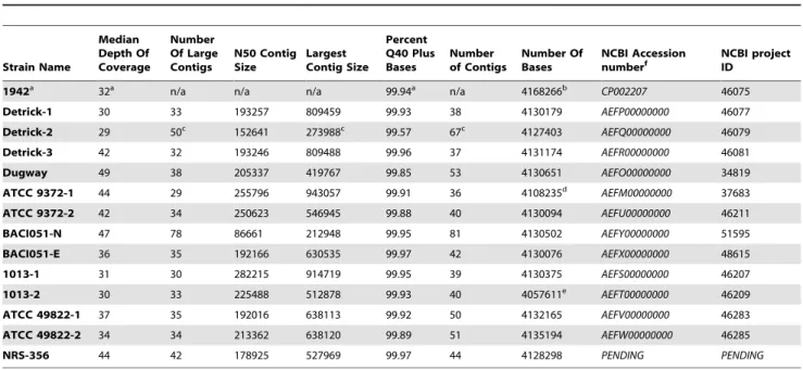
Table 3.Genome Sequencing andde novoAssembly Statistics.
Strain Name
Median Depth Of Coverage
Number Of Large Contigs
N50 Contig Size
Largest Contig Size
Percent Q40 Plus Bases
Number of Contigs
Number Of Bases
NCBI Accession
numberf NCBI projectID
1942a 32a n/a n/a n/a 99.94a n/a 4168266b
CP002207 46075
Detrick-1 30 33 193257 809459 99.93 38 4130179 AEFP00000000 46077
Detrick-2 29 50c 152641 273988c 99.57 67c 4127403 AEFQ00000000 46079
Detrick-3 42 32 193246 809488 99.96 37 4131174 AEFR00000000 46081
Dugway 49 38 205337 419767 99.85 53 4130651 AEFO00000000 34819
ATCC 9372-1 44 29 255796 943057 99.91 36 4108235d AEFM00000000 37683
ATCC 9372-2 42 34 250623 546945 99.88 40 4130094 AEFU00000000 46211
BACI051-N 47 78 86661 212948 99.95 81 4130502 AEFY00000000 51595
BACI051-E 36 35 192166 630535 99.97 42 4130076 AEFX00000000 48615
1013-1 31 30 282215 914719 99.95 39 4130375 AEFS00000000 46207
1013-2 30 33 225488 512878 99.93 40 4057611e
AEFT00000000 46209
ATCC 49822-1 37 35 192016 638113 99.92 50 4132165 AEFV00000000 46283
ATCC 49822-2 34 34 213362 638120 99.89 51 4135194 AEFW00000000 46285
NRS-356 44 42 178925 527969 99.97 44 4128298 PENDING PENDING
aGenome was finished and closed to a single contig. Depth of coverage and Q40
+metrics are forde novoassembly of 454 data only.
b454Draft sequences appear smaller than 1942 due to collapse of repeat regions. cThe average read length of this sample was considerably lower (239 versus
.300) for other genomes in this dataset, resulting in larger number of contigs and decreased assembly quality.
dDeletionof 23 kb with clear join points (positions 4,022,138–4,045,817) verified by optical mapping (Figure 4B). eDeletion of 73 kb with clear join points (positions 3,992,613 to 4,065,341) verified by optical mapping (Figure 4B).
Optical maps were generated by digestion with NcoI of DNA arrayed linearly on glass slides and the resulting maps were aligned and compared with the MapSolver software package (OpGen, Inc., Gaithersburg MD).
Information-based Genomic Distance (IBGD) analysis
Using an information-based method for genomic classification [23], the sequence contigs from BG isolates 1942, 1013-2 and 49822 were analyzed in order to map the phylogenetic relationships of these isolates to other Bacillus species. In this method, genomic content is characterized by the frequencies of occurrence of short n-mers contained within each sequence (n typically from 3 to 16). These n-mers are then rank ordered by genome. The pair-wise comparison of the rank of n-mers within two different genomes is then used to compute an information-based genetic distance (IBGD), where the sum of the differences in rank for all possible n-mers is weighted by an entropy factor that depends on the frequencies of occurrence of the respective n-mers in the two genomes. The pair-wise IBGD values are then used to construct a phylogenetic network [24]. Bacilli genomes were obtained from Genbank. This method for phylogenetic charac-terization enables computation even with the unassembled reads, and it can be applied to draft or partial genome sequence data, which was the case for the three B. atrophaeus genomes studied here.
Phenotype microarray (PM) analysis
The first seven BGstrains listed in Table 1 were streaked for single colonies on BHI plates and incubated at 33uC overnight, followed by subculturing a second time under the same conditions. Subsequently, cell suspensions were prepared according to Biolog specifications, with OD readings ranging between 0.35–0.45 at 600 nm. Biolog phenotypic microarray plates PM1 through PM20, were inoculated according to the manufacturer’s specifications, and incubated at 37uC for 72 hours. Readings were taken every 15 minutes, and data processed by OmniLog Phenotype Micro-array File Management/Kinetic Plot and Parametric modules. Two biological replicates of the experiment were conducted for each strain. PM1-10 contain single wells for each growth condition whereas PM11-20 contain quadruplicate wells for each condition.
Statistical analyses and heatmap of phenotype microarray data
The area under the curve (AUC) values were computed by adding all OmniLog values at all time points for each of the 1200 distinct phenotypes produced from the OmniLog software. The AUC values from the two different biological replicates for each unique phenotype were averaged. The ratio for each AUC was calculated between the 6 query strains (Detrick-1, Detrick-2, Detrick-3, 1013-1, 1013-2, and Dugway) and reference parent strain (1942). For the purpose of visualization, 1920 phenotypes were included in the heatmap (i.e. this better represents the locations of the phenotypes which correspond to different modes of action categories). The same ratios were used for the phenotypes that have replicates. The ratio values were formatted as PM1 to PM20 for each strain across the columns and wells Aito Hi, where
i= 1 to 12 for the rows. The results were plotted in a heatmap using R [25]. Positive growth wells are represented by greenblocks while negative growth wells are represented by red blocks.
Catalase assay
Catalase activitywas assayed by spotting drops of hydrogen peroxide (3%) onto isolated colonies on LB agar plates. Colonies
were monitored for bubble formation, signifying the release of water and oxygen. A colony was considered to be catalase positive by observation of bubbles.
Sporulation efficiency assays
Streaks of Detrick 1, Detrick 2, and 1013 strains were grown for two days on TSA plates containing SBA.Bacterial cell mass was scraped using an inoculating loop (1ml) from the streak and
resuspended in PBS. Sporulation was evaluated by bright field contrast microscopy. Phase-bright free sporesand phase-dark vegetative cellswere counted. Five representative viewing fields were counted from each strain for each experiment. This experiment was completed in triplicate by repeating once per day over the course of three consecutive days.
In order to compare the percent sporulation between Detrick 1 and Detrick 2, and Detrick 1 and 1013, a mixed analysis of variance (ANOVA) was used to complete the analysis. Strain and viewing field were evaluated as fixed factors, and replicate was included as a random factor. The natural log of the percent sporulation was taken to obtain a normal distribution of the residual error. Tukey’s method was applied to compare the difference between the mean log percent sporulation.
Results
Historical investigations of BG provenance
We traceda potential provenance of the commonly used BGstrains through an exhaustive search of the open literature and the archives of the University of Wisconsin,which suggested a possible lineage from which the ‘‘military’’ BGstrains were derived. The original source of the strains were the collections at the University of Wisconsin during the 1930s and 1940s, from which the strains were transferred to Camp Detrick at the initiation of the US Army’s BW program at the beginning of the Second World War [6,26]. At Camp Detrick, BG was used as a non-pathogenic surrogate in process development for spore-forming bacteria It is tempting to speculate that the University of Wisconsin supplied BG to Porton Down: A note found in the archive of Dr. Baldwin’s papers, dated February 19, 1943, contained an order from Dr. Fildes (presumably Sir Paul Fildes, a noted bacteriologist active in the British BW program at the time), for a batch ofB. subtilisspores. It is not clear whether BG or
B. subtilissubsp.subtiliswas supplied, or whether this material was actually delivered. Unfortunately, original records describing in detail the maintenance of the strains during the period 1942–1955 were destroyed as per US Army policy at the time (Dr. Mark Wolcott, USAMRIID; personal communication), and the person-nel who had first-hand knowledge of the strain passage histories and methods are deceased. Therefore, the actual source of the Camp Detrick isolates must be inferred from published work [6], limited available documentation (e.g. ATCC 9372) and the genome sequences presented hereFrom Camp Detrick the isolates were eventually transferred to ATCC asB. subtilisvar.niger‘‘red strain.’’ The desire to maintain a phenotypically and genotypically uniform simulant throughout the biodefense communityprompted us to elucidate whether significant phenotypic and/or genomic differences had accumulated in any of the commonly used isolates during the growth and transfer of strains to different institutions and to compare the isolates in broad use today to the so-called ‘‘Mil-Spec’’ strain (ATCC 9372).In contrast, the origin of ATCC 49822 prior to acquisition F. Young’s laboratory (the depositor) is unclear.
as far as 1942 (Figure 1; Table 1). These isolates included an archival stock dated 1942 that likely predated the transfer to Camp Detrick, as well as material that had been returned to the University of Wisconsin from Camp Detrick in 1952. A derivative of the 1942 strain that had been repeatedly passaged in vitro on agar slants over a period ofapproximately 30 years allowed us to compare the genomic signatures of deliberate selection with the effects of long-termin vitropassage. In addition, a sample of strain NRS-356 [13], which is mentioned as a possible parent strain in correspondence between various academic laboratories and Camp Detrick, was also obtained from the same source as the 1942 ‘‘Vogel’’ strain. These isolates were subsampled, germinated on LB plates, screened for colony morphology variation (see below). Genomic DNA was prepared from these isolates for sequencing.
BG strains exhibit distinct colony morphologies
Upon initial plating of the archival and modern-dayBG stocks, we noted distinct colony morphotypes for many of the strains, with some strains containing multiple variants (Figure 2, Table 2). Some of these morphotypes were consistent with those observed by Hayward et al.[6] whooriginally described the emergence of colony variants in ‘‘B. globigii.’’ As in the earlier report, individual morphotypes were stable and did not interconvert with high frequency (data not shown), suggesting that these morphotypes were the result of relatively rare chromosomal mutations, although 1013-1 occasionally threw off papillae in heavier streaks (not shown). Multiple morphotypes were noted for ATCC9372, ATCC 49822, Detrick, and 1013, while the archival 1942 isolate, the isolate obtained from Dugway Proving Ground (Dugway) and BACI051 appeared to be pure populations on LB. All strains tested positive for BGusing Real Time-PCR primers specific to the
recFgene (Methods S1) [27]. The appearance of multiple colony morphotypes even within single ‘‘strains’’ strongly suggested an as-yet undescribed level of genetic diversity within these samples that likely affected the expression of cell-surface components and/or sporulation. The intra-strain colony morphology variation was particularly dramatic in thein vitropassaged 1013 and ATCC9372 isolates, in which one variant of each lineage had lost the production of color on LB orSBAplates (Figure 2), suggesting more dramatic alterations to the genome.
Whole genome sequencing of BG isolates
Draft genome sequences were generated from several BGstrains in our collection. A summary of the results from the sequenced isolates is indicated in Table 3. All of the ‘‘military’’ isolates (Detrick clones1through 3, BACI051, Dugway) were extremely closely related to each other and to both ATCC9372 variants. The ATCC isolates possessed additional mutations that were absent in the ‘‘military’’ isolates. The size of the finished and closed genome ofB. atrophaeusvar.globigii1942 was 4,168,266 bp, and annotation using RAST [22] revealed 4433 features, including 4343 protein-coding sequencesand 90 RNA molecules [28]. The preliminary annotations derived from RAST are available as Genbank .gbk files in the supplementary material.
Bioinformatic analysis of sequence data
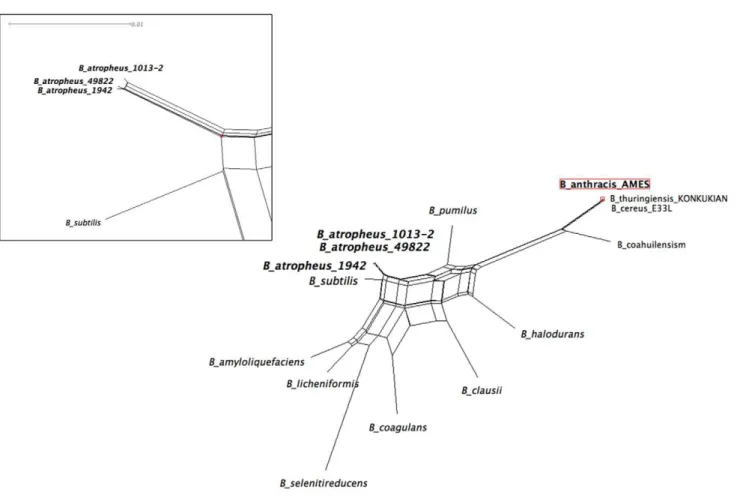
On average, the genome ofB. atrophaeusis approximately 86% identical to B. subtilis on the nucleotide level,supporting its delineation as a distinct species and agreeing well with previous estimates [29]. Analysis of the IBGD using whole-genome sequences (N-mer length .4) supported the identification of B. subtilis 168 as the closest relative among sequenced bacterial genomes (Figure 3). For this particular case, n = 5 (i.e., there were 45= 1024 total 5-mers used to compute the IBGD). The IBGD
values were relatively insensitive to the choice of n over the range of 4–8. Thethree BGgenomes analyzed grouped closely together, and our analysis of the Bacillus-wide phylogeny using IBGD revealed the phylogenetic distance of thatB. subtilis/B. atrophaeus
species from B. anthracis, supporting the inferences published elsewhere from rRNA sequence analysis (Figure 3) [30]. Primary amino acid sequences of RAST-annotated proteins are on average 72% (median 83%) identical betweenB. atrophaeusandB. subtilis. When only the proteins that yielded bidirectional BLAST hits in RAST are examined, the predicted proteome ofB. atrophaeusis, on average, 83% identical (86% median) toB. subtilis.
We utilized the finished sequence of the 1942 isolate as a reference strain for templated assembly of the remaining BG draft sequences. Two additional ATCC isolates ofB. atrophaeus(49337 and 6537) were distinguishable from var.globigii on the basis of very high SNP/indel counts, lower coverage ofand percentage of reads mapping to the 1942 reference, and unique genomic features which supported their proposed classification as var.atrophaeus[14]. The distinguishing genomic features of var.atrophaeus strains and the delineation of theB. atrophaeus clade from B. subtilis will be published elsewhere.
Scaffolding of ‘‘military’’ BG genomes using optical maps
Optical restriction mapping [31,32,33] was used to compare the overall genomic structure of selected isolates. No differencesin overall genome architecture between the ‘‘military’’ BG isolates, the archival 1942 isolate, or 1013-1 were observed (Figure 4, data not shown), suggesting that the global architecture of these isolates is relatively stable, even over 30 years of serial in vitro
passage.However, the optical maps and sequence coverage analysis of 1013-2 and 9372-1revealed substantial deletions of approximately 72,727and 23,678 bases, respectively, of genomic materialspanning from positions 3,992,613 to 4,065,341 (1013-2) or 4,022,138–4,045,817 (ATCC 9372-1) (Figure 4; Table S2). The genes within this deleted region are listed in Table S3 but notably contain genes encoding for nitrite reduction, germination (gerKABC), and biosynthesis of the lipopeptide surfactin (srfCAB) [34,35]. A defect in surfactin production is a particularly intriguing candidate for the morphology and pigmentation variations in 1013-2 and ATCC 9372-1, since disruption ofsrfAhas been shown to have dramatic effects on spreading motility on semisolid media, on biofilm formation [34,36], and low-grade hemolytic activity.
Mutation analysis of BG isolates
strain is in fact a mixed population of at least two genetically distinct substrains.
Effects of genotype on strain phenotypes
The 72 kb deletion in 1013-2 included the structural genes for biosynthesis of surfactin, a cyclic lipopeptide with a mild hemolytic activity [34]. To test whether the ‘‘military’’ andin vitropassaged strains possessed low-grade hemolytic activity, we streaked these variants on rich agar media containing 5% sheep’s blood and looked for hemolysis. To our surprise, all strains exhibited striking variation in their coloration (Figure 2A), with the 1942, 9372-1 and Detrick-1 isolates considerably darker on blood agar than the other ‘‘military’’ andin vitropassaged isolates. In addition, on LB the 1013-2 and 9372-1 isolates appeared white and off-white, respectively. Pigmentation ofB. subtiliscolonies is associated with production of a melanin-like pigment by the CotA protein, a major component of the spore coat [37]. In addition to the variations in pigmentation, streaks of the 1942 and Detrick-1 isolates were consistently translucent under transillumination (Figure 2B). These zones of translucency are suggestive of weak
b-hemolysis, which has previously been observed in B. subtilis
strains that produce high levels of surfactin [34,35,38]. The other strains exhibited either weaka-hemolysis or none at all, with the exception of the strongly hemolytic 49822-1 variant. At least in the ‘‘military’’ lineage, the quasi-hemolytic phenotype and dark-brown colony pigmentation correlated with the presence of a wild-typespo0Fallele, suggesting that the ability ofB. atrophaeusto lyse red blood cells may be regulated in part byspo0F. However this was not universally the case; the BACI051 strain had two discernible variants on SBA (not shown), one of which appeared to have recovered partial hemolytic activity (Figure 2B).
BG strains have distinct metabolic profiles
To gain insight into the effects of genetic divergence of the adapted isolates on their metabolic capacity, the Detrick isolates and the separate 1013 isolates were compared by multiphenotype analysis using the Omnilog system, which allows the high-throughput comparison of 96620 discrete growth conditions,
including carbon, nitrogen, phosphorus, sulfate, nutrient supple-ments, pH, osmolytes as well as a broad class of growth inhibitors. The growth of the 1942 strain was used as a reference for determining relative growth rates of the other strains. The results of
Figure 2. Appearance ofB. atrophaeusstrains on solid media. A) Appearance ofB. atrophaeusstrains on LB or blood agar plates after 24 hours at 37uC. Plates were illuminated directly.B)b-Hemolysis of someB. atrophaeusstrains. Transilluminated plates after 24 or 48 hours of growth on blood agar at 37uC.
Figure 3. Identification ofB. subtilisas nearest-neighbor toB. atrophaeusvar.globigiiby whole-genome phylogenetic analysis of
Bacillusgenomes.Information-based genomic distance (IBGD) was determined by comparing the relative distributions of n-mers within each genome to generate a pair-wise matrix of relative n-mer frequencies (see Materials and Methods). Variation of the n-mer length between 4 and 8 did not substantially affect the derived phylogeny. In this case n-mer length of 5 was utilized. For clarity, only three select species of theB. cereusgroup (of more than 30 that all cluster together) are labeled on the figure. The apparent divergence of isolate 1013-2 is due to alteration of the n-mer frequencies as a result of the deletion of 72 kb of genomic material.
doi:10.1371/journal.pone.0017836.g003
Figure 4. Optical mapping ofB. atrophaeusvar.globigiiand detection of a 72 kb deletion. A) Whole-genome consensus optical restriction maps (NcoI) ofB. atrophaeusATCC 9372-1 (Top), 1942 (middle) and 1013-2 (bottom) isolates.B) expanded view showingdetail of the deleted regions in ATCC 9372-1 and 1013-2.
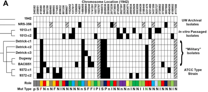
Figure 5. Whole-genome mutation analysis and evolutionary history of the ‘‘military’’ lineage ofB. atrophaeusvar.globigii.A) Whole-genome shotgun sequences of the other strains were mapped to thede novoassembled contigs of the 1942 strain using Newbler. Mutations exhibiting high quality scores in both reference and query sequences and with differences from the template exhibited in.85% of the individual sequencing reads are indicated as a blackened box. In one case (position 259001 in ATCC 9372-1) an initial false-negative due to the formation of a homopolymeric tract was found by direct inspection of the assemblies. The genes whose functions are altered by the given mutation are indicated in Table 4.B) Microevolutionary history ofB. atrophaeusvar.globigiistrains. ‘‘Enhancement’’ events are indicated in red.
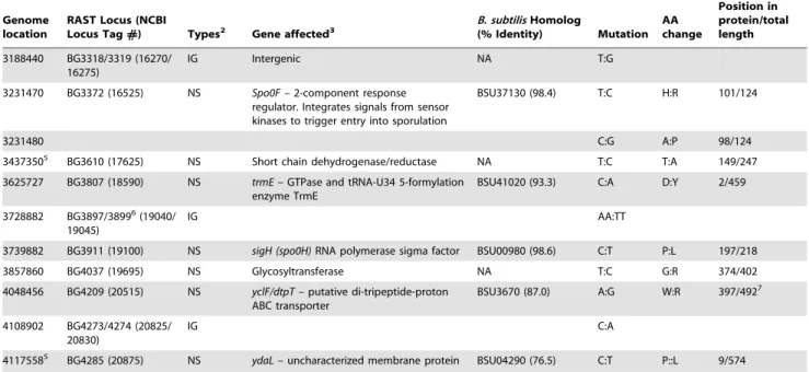
Table 4.SNP/Indel locations inB. atrophaeusgenomes1.
Genome location
RAST Locus (NCBI
Locus Tag#) Types2 Gene affected3
B. subtilisHomolog
(% Identity) Mutation
AA change
Position in protein/total length
246382 BG236 (01550) PS yetF– hypothetical protein BSU07140 (73.9) C:T * 217/231
259001 BG250 (12250) SYN yfnL– hypothetical protein BSU07230 (91.5) C:T
419580 BG405 (02000) 1 bp INS katA– vegetative catalase BSU08820 (94.4) +T 197/483
546358 BG536 (02620) NS hpr/scoC– MarR family regulator (extracellular protease expression and sporulation)
BSU1000 (85.7) C:G A:P 13/200
591250 BG584 (02840) NS hemAT– heme-based aerotactic transducer
BSU10380 (87.7) C:T R:K 215/432
686460 BG685 (03350) NS Oligopeptide ABC transporter, periplasmic oligopeptide-binding
protein OppA
BSU11430 T:G W:G 520/545
699955 BG706 (03425) PS yjbL– truncated hemoglobin BSU11590 (96.1) +C * 89/132
771405 BG795 (03855) NS rapA– response regulator (Dephosporylates Spo0F)
BSU12430 (91.2) T:G S:A 261/378
859632 BG904/905 (04400/ 04405)
IG T:C
881336 BG929 (04515) NS kinE– 2-component histidine kinase; regulates Spo0F activity
BSU13530 (84.6) C:T P:L 577/739
889607 BG939 (04560) NS Methylthioribulose-1-phosphate dehydratase (EC 4.2.1.109),mtnB
BSU13610 (87.6) A:G Y:C 86/209
1162042 BG1225 (05960) INS fliY– flagellar switch motor protein BSU16320 (87.2) +GTGGTC V3:V5 355/379
1317617 BG1320 (06415) NS pksR– Polyketide synthase BSU17720 (71.0) G:C C:S 1698/2574
1326096 BG1327 (06450) NS ebrB– multidrug resistance family protein BSU1729 (77.8) A:G S:P 24/115
1414651 BG1226 (07000) NS Chemotaxis regulator - transmits chemoreceptor signals to flagelllar motor components CheY
BSU17940 (86.0) C:T A:V 97/120
14864084 BG1487 (07220) SYN ppsA– Plipistatin
synthase/non-ribosomal peptide synthetase.
BSU18340 G:A 1872/2560
1668965 BG1681 (08240) 1 bp DEL yozB– putative inner membrane protein BSU19140 (93.8) 2T 55/178
1695248 BG1706 (08360) 5 bp INS yojO– hypothetical protein, potential nitricoxide synthase activation protein
BSU19380 (87.5) +GCTCT 140/638
1741391 BG1764/1765 (08650/ 08655)
IG A:G
1857762 BG1903 (09335) PS ypfA– possible pilZ homolog BSU22910 (60.1) G:A * 197/218 19237675 BG1978/1979 (09750/
09755)
IG C:T
1946572 BG1997 (09870) SYN Putative phage protein NA T:C 283/584
2029530 BG2096 (10375) PS 2-methylcitrate synthase BSU24140 (78.3) +GA * 197/218
2038809 BG2105 (10420) NS spo0A– 2-component response regulator, controls initiation of sporulation
BSU24220 (96.6) G:A A:V 225/265
2073239 BG2144/2145 (10615/ 10610)
IG Intergenic - possible promter region ofsinI
gene (repressor of SinR)
T:C
2073603 BG2146 (10620) NS SinR, regulator of post-exponential-phase responses genes (competence and sporulation)
BSU26100 (98.0%) A:G K:E
2105290 BG2186 (10820) NS yqgE– putative efflux transporter BSU25010 (92.5) G:C H:D 390/430
2827391 BG2944 (14450) NS gerAB– spore germination protein BSU33060 (67.7) G:A R:K 35/365 2828580 BG2945 (14455) NS gerAC– spore germination protein,
governs germination in response to alanine
BSU33070 (61.8) G:T D:Y 64/372
2843471 BG2960 (14530) NS yvrG– sensor histidine kinase involved in cell wall processes
BSU33210 (75.4) T:G I:L 18/580
2895934 BG3014/3015 (14785/ 14790)
IG A:G
3052929 BG3176 (15575) NS Flagellin NA A:G I:T 279/320
3053059 BG3176 (15575) NS Flagellin NA C:T A:T 236/320
3188439 BG3318/3319 (16270/ 16275)
IG Intergenic 10 bp
these experiments are summarized in Figure 6 and Table S4. In general, growth of the 1013 isolates was significantly diminished relative to the 1942 in many different growth conditions, most notably in the ability to use amino acids and peptides as carbon and nitrogen sources, to withstand osmotic stress, and to grow under reduced pH. In addition, the strains had developed sensitivity to beta-lactams, quinolones, and membrane-disrupting activities. These results suggested broad combined effects of several mutations on the phenotype of the strains. In addition to the spo0F(A98P)
allele, which is a likely candidate for highly pleiotropic effects on the decision to sporulate under many different conditions, both strains contain substitutions in the yetF and yqgE genes that may be contributing to the phenotypes observed. The more pronounced defect in 1013-2 may be attributable to defects in thegerABand
gerAC genes and the large 72 kb deletion which contains several genes involved in germination.
By contrast, the Detrick isolates in general grew more robustly than the 1942 strain under multiple growth conditions. Increased relative growth rates were particularly pronounced for acidic conditions and media containing osmolytes, but particularly for wells containing sodium lactate [6].
Another isolate in the ‘‘military’’ lineage, Dugway, is clearly derived from the Detrick lineage by SNP/indel profiling yet has a metabolic profile that is much closer to the parental strain. Like the Detrick isolates, the Dugway strain grows better at low pH, but many of the other conditions do not promote elevated growth relative to 1942. Only one mutation differentiates that isolate from the Detrick-2 isolate – a 2-bp insertion in theyojOgene encoding a
putative activator of nitric oxide (NO) synthesis. Again, the physiological role of this mutation is unclear, although nitric oxide synthesis plays a critical role in modulating antibiotic resistance in
Bacillusspp. [39]. In addition to its role in promoting resistance to antibacterial drugs, NO is known to modulate B. subtilis genes involved in nitrate respiration when oxygen is limited [40]; thus the lowered growth in this strain may reflect the inability to grow to higher densities and overcome the resulting lower oxygen tension. An additional isolate, BACI051 is clearly derived from Dugway, yet two variants have accumulated additional mutations insigH (spo0H),
hpr/scoC, andebrB. Notably, the phenotype of BACI051-E on plates more closely resembles the 1942 strain (Figure 2B).
Catalase activity of BG
sequencing of the ‘‘military’’ isolates revealed a frameshift mutation in thekatAgene encoding the major vegetative catalase [41]. The absence of catalase activity in ‘‘military’’ isolates was confirmed by adding a solution of 3% H2O2to smears of various
strains. In contrast to the 1942 strain, which exhibited immediate and robust catalase activity, the strains containing the frameshift lacked this activity. A small amount of bubbling could be seen, probably due to the presence of a second catalase normally packaged in spores [42].
Sporulation efficiency
To test whether the phenotype observed on blood agar was associated with differences in sporulation, selected strains were grown for two days as patches on blood agar, resuspended in PBS
Genome location
RAST Locus (NCBI
Locus Tag#) Types2 Gene affected3
B. subtilisHomolog
(% Identity) Mutation
AA change
Position in protein/total length
3188440 BG3318/3319 (16270/ 16275)
IG Intergenic NA T:G
3231470 BG3372 (16525) NS Spo0F– 2-component response regulator. Integrates signals from sensor kinases to trigger entry into sporulation
BSU37130 (98.4) T:C H:R 101/124
3231480 C:G A:P 98/124
34373505 BG3610 (17625) NS Short chain dehydrogenase/reductase NA T:C T:A 149/247
3625727 BG3807 (18590) NS trmE– GTPase and tRNA-U34 5-formylation enzyme TrmE
BSU41020 (93.3) C:A D:Y 2/459
3728882 BG3897/38996(19040/
19045)
IG AA:TT
3739882 BG3911 (19100) NS sigH (spo0H)RNA polymerase sigma factor BSU00980 (98.6) C:T P:L 197/218
3857860 BG4037 (19695) NS Glycosyltransferase NA T:C G:R 374/402
4048456 BG4209 (20515) NS yclF/dtpT– putative di-tripeptide-proton ABC transporter
BSU3670 (87.0) A:G W:R 397/4927
4108902 BG4273/4274 (20825/ 20830)
IG C:A
41175585 BG4285 (20875) NS
ydaL– uncharacterized membrane protein BSU04290 (76.5) C:T P::L 9/574
1In addition to the above mutations, putative SNPs at positions 39912, 45501, 61413, 611822, 1170114, 1251549, 2174033, 2771980, 3128882, 3269962, 3354410,
3528224, and 3880949 were common to all reference mapping experiments against the finished genome using 454 data, including the 1942 dataset. The commonality to all datasets suggest that these were errors in the finished sequence that are being verified.
2PS = Premature stop; SYN = Synonomous; INS = Insertion; NS = Non-synonomous; IG = Intergenic; DEL = Deletion. 3Annotations are a combination of RAST and directed tBLASTn and BLASTp searches vs
Bacillusdatabases.
4Forms part of a large polypeptide synthase containing highly homologous regions. 5Also shared with strain ATCC 49822.
6The conserved start codon of theradAgene (BG3899) ofB. atrophaeusandB. subtilisfalls within the BG3898 ORF. Therefore BG3898 as called by RAST is not deemed
likely to be a protein-coding gene.
7In putative transmembrane region.
doi:10.1371/journal.pone.0017836.t004
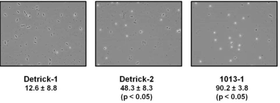
and counted directly. Strain Detrick-2exhibited significantly higher percentages of phase-bright spores than the Detrick-1 strain (Figure 7, Mean+/2standard error of the mean). Similar results were observed for the 1942 and Dugway strains (not shown). The 1013-1 strain exhibited an even higher degree of sporulation than the Detrick-1 strain under identical conditions (Figure 7).
Discussion
Bacillus atrophaeushas historically been grouped withB. subtilis, and is usually described as a black-pigmented variant (var. niger) because of its many phenotypic similarities to the better-characterized B. subtilis. Both organisms are soil-dwelling, non-pathogenic saprophytes, but have been differentiated by the ability
to produce pigment on nutrient media containing an organic nitrogen source [13]. The orange pigmentation of B. atrophaeus
var.globigii spores made it an attractive simulant for B. anthracis, facilitating the detection of dispersed spores in complex environ-mental samples. Recently, more sensitive phylogenetic approaches using AFLP have delineated B. atrophaeus as a separate species [13,14]. The taxonomic confusion has arisen due to inadequately sensitive typing methods, and has led to misattribution of pathogenic qualities associated with someB. licheniformisstrains to theB. atrophaeus
strains currently in use as simulants [12], for which no direct evidence of pathogenicity exists. This report defines the genomic composition ofB. atrophaeusvar.globigiiand clearly separates the species by whole-genome phylogenetic analysis.
In this study, we generated a high-quality, closed reference genome for the 1942 isolate using a combination of 454, Illumina, and directed Sanger sequencing. We expect the final genome to have an error rate of,1 in 50,000 basepairs. When we mapped
the 454 datasets for all of the isolates back to the finished sequence that was generated using the same DNA, we noted several putative SNPs that were common to all datasets (Table 4). We believe these represent errors introduced during generation of the final consensus sequence, as they did not appear when the isolates were mapped against draft sequence generated exclusively using the 454 platform; these are currently being verified and the final sequence will be updated.
Our sequences of multiple, closely related strains of this organism allow us to trace the derivation of the ‘‘military’’ BG isolates currently in use to a culture present at Camp Detrick during the 1940s and 1950s. The origin of ATCC 49822 is not as clear, but a publication from that era suggests a possible common origin at the University of Wisconsin [43]. While that strain is unlikely to be NRS-356 itself, given the presence of several strain-specific SNPs in our sequence, the SNPs common to both 49822 and the ‘‘military’’ lineage suggest a common ancestor that is not represented among the strains sequenced for this study. Given the lack of original records, it is unclear whether the NRS-356 variant in this study might have passed through Camp Detrick and been returned to the University of Wisconsin. However, given the date on the label and the general secrecy of operations at Camp Detrick during the Second World War [26] we consider this possibility unlikely.
During development of BGas a simulant forB. anthracis, strains were selected that exhibited the most desirable characteristics, those being rapid growth, high spore yield, and experimental reproducibility. Without being aware of the nature of the genetic alterations in their ‘‘optimized’’ strains, BW workers at Camp Detrick selected a mutant that provided dramatically higher total and relative spore yields, and generated consistent experimental results [6]. These strains were adopted into the inventories of numerous biodefense laboratories and have been used for many
Figure 7. Thespo0F(H101R)andspo0F(A98P)alleles are associ-ated with hypersporulation. Phase-contrast microscopy of BG strains after two days of growth on SBA. Vegetative cells appear as phase-dark rods, while spores appear as round, phase-bright globules. The mean percentage sporulation of each strain in a representative experiment is given 6SEM. The experiment was repeated on three consecutive days; representative results of a single experiment are shown. Statistical significance was determined by mixed ANOVA (Tukey’s method, p,0.05).
doi:10.1371/journal.pone.0017836.g007
Figure 6. Omnilog phenotypic arrays ofB. atrophaeussubsp.globigiistrains.Six strains were each inoculated into twenty 96-well Omnilog plates and grown at 37uC. Reduction of tetrazolium dye by respiring cells was measured every 15 minutes by optical density. Dye reduction relative to the 1942 strain is shown; the red ratio values indicate less respiration while the green ratio values indicate more respiration as compared to the 1942 strain. Individual arrays or strains are displayed in each of the six major columns labeled Detrick 1, Detrick 2, Detrick 3, 1013-1, 1013-2, and Dugway.A)Heat map of all conditions for each strain. Each of the twenty plates for each strain is represented by the notation PM01-PM20 (left-to-right for each strain) along the x-axis. The rows represent the well position, and are denoted as Aito Hi(i = 1 to 12) from the bottom to the top of the
plot in each array along the y-axis. Each cell ratio value represents the average of two biological replicates for each strain. Plates PM01-PM10 contains single wells for each growth condition, while plates PM11-PM20 contain quadruplicate wells for each growth condition. Solid circle indicates wells containing sodium lactate; dotted circle indicates well containing L-serine at pH 4.5. The details of the 1920 growth conditions can be found in the first worksheet labeled ‘‘All strain AUC data’’ in Table S4.B) Most significant phenotypes for each of the six test strains as compared to the 1942 strain. The phenotypes with statistically significant increases and/or the decreases in ratio values for each of the six strains are presented. For the 1013 isolates only the conditions giving the five largest changes are presented. The number in each color block indicates the ratio for the test strain relative to the parent strain for the phenotype specified. The details of all significant phenotypes for each test strain can be obtained in Table S4.Bold Italicfont indicates p,0.05.
decades in simulations of decontamination and dispersal [12]. By applying a combination of genomic and biochemical profiling techniques, our data demonstrate that the BG isolates were ‘‘enhanced’’ by researchers at Camp Detrick during the development of the organism as a simulant.
The selection of a strain with the desired properties appears to have occurred in at least two discrete steps, as shown by the genome sequences and metabolic profiles. The initial step appears to have been the adaptation of a strain to growth in corn steep liquor, an acidic medium rich in protein and lactate [44]. The robust growth of the Detrick strains relative to 1942 in low-pH medium containing high lactate levels is likely due to mutations in mmgD (2-methylcitrate synthase, position 2029530), or a short-chain 3-oxoacyl-[acyl-carrier-protein] reductase (position 3437350), or both. The most likely candidate for a mutation in the Detrick isolates that increases growth is the frameshift inmmgDthat occurred following the divergence from the 49822 lineage and results in an altered C-terminus (Figure S1). The
mmgDgeneencodes a 2-methylcitrate synthase that is expressed in the mother cell at the intermediate stages of sporulation [45]. A null mutation inmmgDhad no perceptible effect on sporulation, although other TCA-cycle enzymes when mutated led to a loss of sporulation [45]. The effects of the frameshift mutation on sporulation and cellular physiology on the function of the enzyme are not clear at this time. We speculate that the frameshift mutation alters the substrate specificity of MmgD in favor of citrate, thus increasing the flux of lactate-derived intermediates through the tricarboxylic acid cycle. Evidence for this possibility includes the observations that 2-methylcitrate synthases can have partial citrate synthase activity [45] and that theB. subtilis mmgD
gene can complement agltA(citrate synthase) mutant ofE. coli[46]. Alternatively, alteration of function ofmmgDmay have predisposed the lactate-adapted strain to acquisition of a hypersporulating phenotype, which is not readily isolated or stable inB. subtilis(see below); however the presence of a hypersporulating phenotype in an independently evolved lineage (1013) of BG indicates that the species may have an intrinsic predisposition to evolving such a phenotypein vitro.
The ‘‘military’’ strains also grow more readily on media containingD,L-diaminopimelic acid (meso-DAP), a major compo-nent of bacterial peptidoglycan. Corn steep liquor is derived from the incubation of corn in water at 42–55uC, during which a lactic fermentation by a community of wild organisms including numerous uncharacterized Bacillus spp. occurs. Total bacterial counts at the conclusion of CSL production can be quite high [44], thus the availability of such compounds for growth is not surprising. Another potential source of meso-DAP could be
bacterial autolysis during sporulation. The relative roles of each of the alleles in growth on lactate and/ormeso-DAP is the subject of current investigation in our laboratory.
The second step in the development of BG as a simulant appears to have been the deliberate selection of a hypersporulating variant [6,47]. Importantly, the selection of a strain optimized for spore yield resulted in the fixation of a newspo0Fallele that has no counterpart among the availablespo0F sequences (Figure 8). The sole Spo0Fsequence that differs at position 101 is that ofB. clausii, in which tyrosine replaces histidine. Notably, the spo0F(H101R) mutation is distinct from a separatespo0F(A98P)mutation present in thein vitropassaged 1013 isolates. Given that the amino acid sequence ofB. atrophaeusSpo0F is identical to that ofB. subtilisbut for two conservative substitutions, it is likely to have very similar if not identical biochemical properties. Detrick-1 and 1942 likely represent one of the two R colony morphotypes described by Hayward et al. [6], whereas the hypersporulating F morphotypes likely arose due to the emergence of thespo0F(H101R)mutation. However, the possibility that Detrick-1 represents a reversion mutant at this locus from Detrick-2 cannot formally be excluded, but since it represented the dominant morphotype in the 1952 Detrick vial we believe this is unlikely. The presence of thespo0F(H101R)allele in the ATCC 9372 strains suggests that these strains were acquired by ATCC after this mutation appeared within the Detrick lineage. Experiments to verify the roles of each allele in modulating sporulation are currently in progress. Preliminary results indicate that transformation ofB. subtilisDspo0FwithB. atrophaeusDNA and selection of spo+ cells dramatically alters colony morphology independently of thespo0Fallele introduced; additional studies to verify the effects of each allele are currently in progress (James Hoch, personal communication).
The H101R and A98P allelesare likely to alter the response to signals promoting sporulation. Aspo0F(H101A) allele results in a sporulation-proficient strain that throws off sporulation-deficient papillae [48], and the same mutation has been shown to suppress the spo2phenotype of a strain containing a defectivekinAallele. H101 has been proposed as a potential metal-binding site with particular affinity for Cu2+ [49]. Binding of Cu2+ (or another
divalent metal) at this site may modulate interaction with one or more sensor kinases that promote sporulation. Substitution of positively charged arginine at this position could potentially mimic the binding of a metal cation in the loop containing H101, resulting in altered sporulation of the strains due to a change in the interaction with the kinases governing sporulation. It is unclear why, given the proposed role of divalent Cu2+
in suppressing
Figure 8. Multiple alignment of Spo0F protein sequences.The predicted protein sequences of Spo0F from multipleBacillusspecies were aligned using ClustalW. Residues mutated in hypersporulating variants are indicated with grey (A98P) and black (H101R) arrows. Key: Batroph – Bacillus atrophaeus; Bsubtilis –B. subtilis168; B_amyloliq –B. amyloliquefaciens; B_NRRL -Bacillus. spp. NRRL; B_SG-1 –Bacillusspp. SG-1; B_thur –B. thuringiensis strains Al Hakam and var. Israelensis (Isr); B_coahuil – B. coahuilensis; B_weihenst – B. weihenstephanensis; B_pseudomyc – B. pseudomycoides.
sporulation, H101R would result in a hypersporulation phenotype. The mechanistic relationship between spo0F(H101R) and the hypersporulation phenotype will be tested in future experiments.
Both variants in the 1013 lineage possess an A98P allele in
spo0F. Although the presence of several other mutations within this lineage confounds the attribution of the hypersporulating phenotype to this allele at this time, the presence of a mutation in the same gene as another hypersporulating mutant is highly suggestive. The effect of proline substitution at position 98 on Spo0F functionis not immediately obvious, but the relatively inflexible proline residue can disrupt alpha-helices in protein structures. The 1013-1 lineage exhibits a hypersporulating phenotype even more pronounced thanspo0F(H101R) strains in the ‘‘military’’ lineage. The observation that hypersporulating phenotypes have emerged during cultivationof two independentB. atrophaeus lineages point to the possibility that certain in vitro
selection pressures may actually favor hypersporulating variants. The selection pressures acting on the sporulation pathwayare highlighted by the sheer number of mutations discovered within the entire data set that occur in proteins known to play roles in sporulation. Nine of the 38 mutations (23%) found in all lineages were in genes that directly or indirectly regulate either entry into stationary phase or sporulation; this number exceeds the number that would be expected if mutations were to occur by chance, since less than 5% of
B. subtilis genes are dedicated to regulatory processes of any kind [50,51]. In addition to the mutations found within the ‘‘military’’ lineage, the two variants of ATCC 49822 shown in Figure 2 differ by mutations in rpoB(Table S5) which also plays a role in entry into sporulation [52]. Null mutations inspo0Fresulting in asporogenous phenotypes contribute to colony morphology variation inB. anthracis,
B. thuringiensisandB. subtilis[53,54,55]. Enhancedin vitro‘‘fitness’’ is also a likely driver behind the recovery of asporogenic B. anthracis
mutants that were discovered during the investigation into the B. anthracisattacks of 2001 [56]. Because the process of sporulation is highly energy-intensive and irreversible once commenced, mutants that delay sporulation (or fail to sporulate altogether) to take advantage of remaining nutrients would out-compete wild-type cells during repeated passage in vitro in the absence of other selection pressures, as has been demonstrated in extended in vitroevolution studies withB. subtilisunder relaxed sporulation conditions [57]. This may not be universally the case, since gain-of-function mutations in sporulation such as those observed in this studymay compete favorably with wild-type cells if cannibalism of vegetative cells by sporulating bacteria is the dominant selective pressure [58]. Finally, horizontally transferred genetic elements can have dramatic effects on sporulation: for example, recent studies of phage lysogeny in B. anthracis have revealed the ability of several integrated phages to positively affect the kinetics of sporulation upon lysogeny of commonly usedB. anthracisstrains [59].
This study identifies thespo0F(H101R)allele as the signature of a deliberate selection during the development ofB. atrophaeusas a simulant. However, without the knowledge of the history and the analysis of the phenotypes of the strains originating from ‘‘Camp Detrick’’ as published in the open literature, attribution of this genotype to a deliberate selection event would not have been definitive, since a similar phenotype is observed in the 1013 lineage which to our knowledge was not deliberately selected for any specific trait. Any study designed to determine genomic ‘‘signatures’’ of deliberate enhancement or selection is likely to require an analysis of the baseline likelihood that mutations conferring a similar phenotype would emerge and become fixed by natural processes within an evolutionary timeframe consistent with a known time interval or number of passages.
Available evidence suggests that hypersporulation is not easily evolved in vitro. Maughan and coworkers attempted to evolve populations of a laboratory strain ofB. subtiliswith a hypersporulat-ing phenotype by repeatedly heat-shockhypersporulat-ing cultures. While their efforts to enrich for hypersporulators failed, other studies revealed that asporogenous mutants evolved readily [60,61], confirming many early studies ([62] and references therein). With the exception of the studies by Maughanet al., most ofthese investigators applied selections intended to inhibit sporulation rather than to enrich for strains with elevated sporulation rates. The 1013 lineage was never heat-shocked during its many transfers; thus the adaptations seen in this work are the result of balancing sporulation versus vegetative growth for prolonged periods on agar slants. However, because undomesticated isolates were observed to sporulate to 98–100% [60], we cannot formally exclude the possibility thatin vitroculture of the 1942 strain following its isolation for an unknown period by the University of Wisconsin might have selected for a hyposporulating variant. In this scenario, the H101Rand A98P mutations would represent suppressor mutations. We consider this possibility unlikely, given the phenotypic similarity of two environmental isolates in the UW collection (1942 and NRS-356). Furthermore, a progression toward darker pigmentation and greater hemolysisis evident in the ‘‘military’’ lineage (Figure 2B). These phenotypic changes are associated with the accumulation of additional mutations including a P145L substitution mutation in sigH, a positive regulator of sporulation [63,64] and an A13P mutation in
scoC, a negative regulator of sporulation [65]. Together, the strains analyzed in this study suggest strong selective pressures on the genes in the sporulation pathway, and more carefully controlled studies should be carried out to determine the dynamics ofin vitroevolution and adaptation of spore-forming organisms, as has been done extensively inE. coli[66,67,68,69,70].
Unexpectedly, the ‘‘military’’ lineages were also marked by the loss of catalase activity, whose presence is an identifying feature of bothB. subtilisandB. atrophaeus[13]. This activity was present in a separate lineage ofin vitropassaged organisms, so it is not immediately clear why ‘‘military’’ isolates, i.e. those subjected to selection within the early days of the development of BG as a simulant organism, would have lost the catalase activity characteristic of the parental isolate. Because the KatA gene product is not found in spores [41,71], we consider it unlikely that the absence of this activity would impact the resistance of spores to decontamination reagents, and thus any antioxidant resistance phenotype exhibited by spores of ‘‘military’’ isolates would likely have gone unnoticed. However, direct comparisons of the ‘‘military’’B. atrophaeuslineages to the progenitor strains have not been done, and pleiotropic effects of aspo0Fmutation on spore physiology cannot currently be excluded.
The genomic basis of interlaboratory strain variation is only beginning to become evident, with recent studies tracing the histories of commonly used lab strains of B. subtilis 168, E. coli,
Salmonella entericaserovar Typhimurium 14028s,Pseudomonas aerugi-nosaPA01 andMycobacterium tuberculosisH37Rv [76,77,78,79,80,81]. These have revealed significant divergence of putatively identical strains from one laboratory to another, largely arising from mutations that accumulate during serial passage. Like the earlier work, our study highlights the utility of approaches based on whole-genome sequencing for the discrimination of closely related strains, especially when investigating the provenance for a given isolate. Tragically, at least 13 institutions are known to have destroyed archival collections of Select Agents [82] following the implemen-tation of mandatory monitoring and reporting requirements, representing an incalculable loss of phenotypic and genomic diversity. This report underscores the importance of maintaining the genetic heritage preserved in the culture collections of individual investigators and institutions.
Supporting Information
Figure S1 Effect of frameshift mutation in themmgDgene on the C-terminus of the 2-methylcitrate synthase homolog of B. atrophaeus strain Detrick-1. Arrow indicates the location of the GA dinucleotide insertion. Multiple alignment was performed using the ClustalW algorithm in the MEGAlign module of LaserGene.
(TIF)
Table S1 Table S1 is a consolidated spreadsheet containing Newbler HCDiffs calls for each templated assembly to the finished sequence of the 1942 isolate.
(XLSX)
Table S2 Table S2 details the scaffolding of large contigs of the
de novoassembly of 454 data for the1942 strain based on optical maps.
(XLSX)
Table S3 Table S3 contains RAST annotation files (.gtf format) of the 1942 strain and indicates the location of the large deletions in ATCC 9372-1 and 1013-2.
(XLSX)
Table S4 Table S4 contains Omnilog phenotypic array data normalized to 1942 strain.
(XLSX)
Table S5 Table S5 contains HCDiffs calls from templated assembly of ATCC 49822 variants using the finished sequence of the 1942 isolate.
(XLSX)
Methods S1 Confirmation of BG Identity by RT-PCR. (DOCX)
Acknowledgements
We thank Dr. Kevin P. O’Connell for helpful discussions and insights into the manuscript and for facilitating the collaboration with the University of Wisconsin. We also thank Kristin Willner and Amy Butanifor assistance with sequencing. We thank Gary Ouellette for help with phylogenetic analysis and Drs. Mark Wolcott (USAMRIID) and James Hoch (Scripps) for helpful discussions. The opinions presented here are those of the authors and are not necessarily those of the U.S. Government or any of its agencies. Information in this report is unclassified and cleared for public release.
Author Contributions
Conceived and designed the experiments: HSG TDR EWS SS CD. Performed the experiments: SMB C. Hong CC AA HD DB RT SJ C. Han PAM LAM M. Karavis KAB-L SS. Analyzed the data: HSG LAM HD CC M. Karavis KHP AF LAM HD CC M. Krepps KHP AF JSL WEC BWH PD AL EF RT SJ KAB-L CNR. Contributed reagents/materials/ analysis tools: JL. Wrote the manuscript: HSG.
References
1. Turnbough CL, Jr. (2003) Discovery of phage display peptide ligands for species-specific detection ofBacillusspores. J Microbiol Methods 53: 263–271. 2. Stratis-Cullum DN, Griffin GD, Mobley J, Vass AA, Vo-Dinh T (2003) A
miniature biochip system for detection of aerosolizedBacillus globigiispores. Anal Chem 75: 275–280.
3. Kournikakis B, Ho J, Duncan S (2010) Anthrax letters: personal exposure, building contamination, and effectiveness of immediate mitigation measures. J Occup Environ Hyg 7: 71–79.
4. Phillips CR (1949) The sterilizing action of gaseous ethylene oxide; sterilization of contaminated objects with ethylene oxide and related compounds; time, concentration and temperature relationships. Am J Hyg 50: 280–288. 5. Sagripanti JL, Carrera M, Insalaco J, Ziemski M, Rogers J, et al. (2007) Virulent
spores of Bacillus anthracis and otherBacillusspecies deposited on solid surfaces have similar sensitivity to chemical decontaminants. J Appl Microbiol 102: 11–21.
6. Hayward AE, Marchetta JA, Hutton RS (1946) Strain Variation as a Factor in the Sporulating Properties of the So-calledBacillus globigii. J Bacteriol 52: 51–54. 7. Carrera M, Zandomeni RO, Fitzgibbon J, Sagripanti JL (2007) Difference between the spore sizes of Bacillus anthracis and otherBacillusspecies. J Appl Microbiol 102: 303–312.
8. Silva JM, Moreira AJ, Oliveira DC, Bonato CB, Mansano RD, et al. (2007) Comparative sterilization effectiveness of plasma in O2-H2O2mixtures and
ethylene oxide treatment. PDA J Pharm Sci Technol 61: 204–210. 9. United States Pharmacopeia (2000) Biological indicator for ethylene oxide
sterilization, paper strip. The United States Pharmacopeia/The National Formulary. RockvilleMD: US Pharmacopeia. pp 231–232.
10. Stein DC, Kopec LK, Yasbin RE, Young FE (1984) Characterization ofBacillus subtilis DSM704 and its production of 1-deoxynojirimycin. Appl Environ Microbiol 48: 280–284.
11. Harris-Warrick RM, Lederberg J (1978) Interspecies transformation inBacillus: sequence heterology as the major barrier. J Bacteriol 133: 1237–1245.
12. Page WF, Young HA, Crawford HM, Institute of Medicine (U.S.). Advisory Panel for the Study of Long-Term Health Effects of Participation in Project SHAD (2007) Long-term health effects of participation in Project SHAD (Shipboard Hazard and Defense). Washington, D.C.: National Academies Press xviii, 123 p.
13. Nakamura LK (1989) Taxonomic Relationship of Black-Pigmented Bacillus subtilisStrains and a Proposal forBacillus atrophaeussp. nov. Int J Syst Bacteriol 39: 295–300.
14. Burke SA, Wright JD, Robinson MK, Bronk BV, Warren RL (2004) Detection of molecular diversity in Bacillus atrophaeus by amplified fragment length polymorphism analysis. Appl Environ Microbiol 70: 2786–2790.
15. Margulies M, Egholm M, Altman WE, Attiya S, Bader JS, et al. (2005) Genome sequencing in microfabricated high-density picolitre reactors. Nature 437: 376–380. 16. Bennett S (2004) Solexa Ltd. Pharmacogenomics 5: 433–438.
17. Zerbino DR, Birney E (2008) Velvet: algorithms for de novo short read assembly using de Bruijn graphs. Genome Res 18: 821–829.
18. Ewing B, Green P (1998) Base-calling of automated sequencer traces using phred. II. Error probabilities. Genome Res 8: 186–194.
19. Ewing B, Hillier L, Wendl MC, Green P (1998) Base-calling of automated sequencer traces using phred. I. Accuracy assessment. Genome Res 8: 175–185. 20. Gordon D, Abajian C, Green P (1998) Consed: a graphical tool for sequence
finishing. Genome Res 8: 195–202.
21. Han CS, Chain P. Finishing repeat regions automatically with DupFinisher. In: Arabnia HR, Valafar H, eds. 2006. CSREA Press. pp 141–146.
22. Aziz RK, Bartels D, Best AA, DeJongh M, Disz T, et al. (2008) The RAST Server: rapid annotations using subsystems technology. BMC Genomics 9: 75. 23. Yang AC, Goldberger AL, Peng CK (2005) Genomic classification using an
information-based similarity index: application to the SARS coronavirus. J Comput Biol 12: 1103–1116.