Microstructural analysis of demineralized primary enamel after
in
vitro
toothbrushing
Análise microestrutural do esmalte decíduo desmineralizado após
escovação dentária
in vitro
Aline de Almeida Neves* Rodolfo de Almeida Castro* Eduardo Tavares Coutinho** Laura Guimarães Primo***
ABSTRACT:The aim of this study was to investigate,in vitro, the morphological characteristics of demineralized pri-mary enamel subjected to brushing with a dentifrice with or without fluoride. In order to do so, 32 enamel blocks were divided in 4 different groups containing 8 blocks each. They were separately immersed in artificial saliva for 15 days. The experimental groups were: C - control; E - submitted to etching with 37% phosphoric acid gel (30 s); EB - submit-ted to etching and brushing 3 times a day with a non-fluoridasubmit-ted dentifrice; EBF = submitsubmit-ted to etching and brushing 3 times a day with a fluoridated dentifrice. The toothbrushing force was standardized at 0.2 kgf and 15 double strokes were performed on each block. After the experimental period, the samples were prepared and examined under SEM. The control group (C) showed a smooth surface, presenting scratches caused by habitual toothbrushing. The etched samples (E) exhibited different degrees of surface disintegration, but the pattern of acid etching was predominantly the type II dissolution. The brushed surfaces were smooth, with elevations which corresponded to the exposure of Tomes’ process pits and depressions which corresponded to interrod enamel. Particles resembling calcium carbonate were found in the most protected parts of the grooves. No morphological differences were observed between brushing with fluoridated (EBF) and non-fluoridated (EB) dentifrice. The results suggest that the mechanical abrasion caused by brushing demineralized enamel with dentifrice smoothes the rough etched surface, and the presence of fluoride does not cause morphological modifications in this pattern.
UNITERMS:Tooth, deciduous; Dental enamel/ultrastructure; Tooth demineralization.
RESUMO:O objetivo do estudo foi investigar as características morfológicas do esmalte decíduo desmineralizado sub-metido à escovação com dentifrício fluoretado ou não fluoretado. Para isto, 32 blocos de esmalte foram separados em 4 grupos diferentes, contendo 8 elementos cada que foram imersos em saliva artificial por 15 dias: C = controle; E = ata-cado com gel de ácido fosfórico a 37% por 30 segundos; EB = ataata-cado e escovado 3 vezes ao dia com dentifrício não fluoretado; EBF = atacado e escovado 3 vezes ao dia com dentifrício fluoretado. A escovação foi padronizada em 0,2 kgf e 15 movimentos de vai-e-vem foram executados em cada bloco. Após o período experimental, as amostras foram pre-paradas e examinadas no MEV. O grupo controle (C) apresentou lisura superficial e riscos causados pela escovação habitual; as amostras atacadas (E) apresentaram diferentes graus de desintegração superficial, porém o padrão de ataque ácido foi predominantemente a dissolução do tipo II. As superfícies escovadas apresentaram-se alisadas, com exposição das elevações correspondentes aos processos de Tomes e as depressões de esmalte interprismático. Partícu-las semelhantes a carbonato de cálcio foram encontradas nas partes mais protegidas das depressões. Não houve dife-rença quando os grupos foram escovados com dentifrício fluoretado (EBF) e dentifrício sem fluoreto (EB). Os resulta-dos sugerem que a abrasão mecânica da escovação com dentifrício sobre o esmalte desmineralizado alisa a superfície rugosa causada pelo condicionamento ácido e que a presença do fluoreto não altera morfologicamente este padrão.
UNITERMOS:Dente decíduo; Esmalte dentário/ultra-estrutura; Desmineralização do dente.
INTRODUCTION
The tooth enamel surface is constantly exposed to great acidic challenges, such as those caused by undisturbed plaque accumulations in stagnation
areas or by etching techniques in restorative or orthodontic procedures. The morphology of the enamel affected by each of these factors is charac-terized by direct dissolution and roughness of the outer surfaces3,14
.
*Graduate students (MSD); ***MSD, PhD, Assistant Professor – Department of Pediatric Dentistry and Orthodontics, School of Dentistry, Federal University of Rio de Janeiro, Brazil.
Active incipient enamel caries lesions are one of the frequent clinical situations that require non-operative treatment; comparatively, the fate of et-ched enamel surfaces that are left uncovered, ex-posed to the oral environment, is still a matter of discussion in the literature. The effect of reminera-lizing agents19
, professional prophylaxis8
and of the abrasion caused by brushing with dentifrice18
over these demineralized surfaces has been extensively studied. Fluoride is said to enhance remineraliza-tion, which, in turn, means recovery of deminerali-zed enamel20
. Although in the presence of mecha-nical cleaning procedures fluoride is known to promote the arrest and regression of carious lesi-ons13,23
, the effects of fluoride on the morphology of demineralized enamel has not yet been verified.
The aim of this study was to observe, using the scanning electron microscope (SEM), the morpho-logy of demineralized primary enamel after a con-trolled period of toothbrushing with fluoridated and non-fluoridated dentifrices,in vitro.
MATERIALS AND METHODS
Eight second primary lower molars were selec-ted from a bulk of extracselec-ted primary teeth collecselec-ted at the Pediatric Dental Clinic, Federal University of Rio de Janeiro. The teeth were caries-free and had no other gross surface defects. They were kept in 4% aqueous formalin solution (pH 7.0) until the beginning of the experiment. The teeth were was-hed in distilled water and cut into six smooth-sur-face sections, in buccolingual direction (n = 48 blocks), with a carborundum disc.
The enamel blocks were subsequently positio-ned over a vinyl polysiloxane (3M Express STD Fir-mer Set) standardized matrix with the aid of a
5-centemeter-long orthodontic wire. The matrix was filled with epoxy resin (XGY 1109 and HY 850) and stored at room temperature for 24 hours to al-low for resin polymerization (Figure 1). Each block was mounted over the resin, with the outer enamel surface exposed.
Each block was pumiced with a rotating rubber cup for 30 s and washed in distilled water for 10 s. After compressed air drying, the enamel blocks were examined under a stereomicroscope (10 X magnification) for surface defects. Thirty-two sam-ples were selected and randomly assigned to 4 dif-ferent groups: C - control (not etched); E - etched with 37% phosphoric acid gel (Dentsply - Caulk) for 30 seconds, EB - etched as described in Group E and brushed with a non-fluoridated dentifrice (Phillips - The Sydney Ross Co.); EBF - etched as described in Group E and brushed with a fluorida-ted dentifrice (Philips 2 - Sydney Ross Co., 1,200 ppm MFP). The abrasive component of the dentifrices was calcium carbonate.
The groups were separately immersed in 60 ml of artificial saliva (KCl - 0.96 mM; NaCl - 0.674 mM; MgCl2.H2O - 0.0408 mM; CaCl2.H2O - 0.1168 mM;
K2H2(PO4)2- 0.274 mM; 0.8% carboxymetylcellulose,
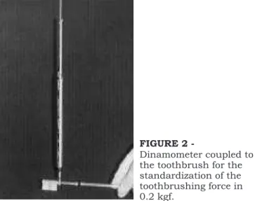
pH 7.0 – School of Pharmacy, Federal University of Rio de Janeiro) for 15 days. The samples from groups EB and EBF were removed from the saliva three times a day, with a minimum 3-hour interval, and brushed with a toothbrush (Oral B Indicator Soft 35). The toothbrushing force was standardized at 0.2 kgf by means of a dynamometer coupled to a small hole carved on the toothbrush (Figure 2). At each brushing period, 15 double strokes were per-formed on each enamel block previously painted with a thin pellicle of dentifrice. After brushing, the blocks were rinsed in water, dried with
high-com-FIGURE 1 -Vinyl polysiloxane standardized base with enamel blocks positioned, by means of an orthodontic wire, in a resin-filled spot.
FIGURE 2
pressed air and put back in the artificial saliva. A toothbrush was used for each brushed group du-ring the experimental phase. The enamel blocks of groups C and E remained immersed in the artificial saliva throughout the 15 days.
At the end of the experimental phase, the sam-ples were washed in distilled water for 2 minutes, dried at room temperature, sputter-coated with gold, and visualized by means of SEM at 20 kV (Zeiss DSM 960, Germany). Electronmicrographs were obtained, and a descriptive analysis was car-ried out. The typical features observed are descri-bed and illustrated in Figures 3 to 6.
RESULTS
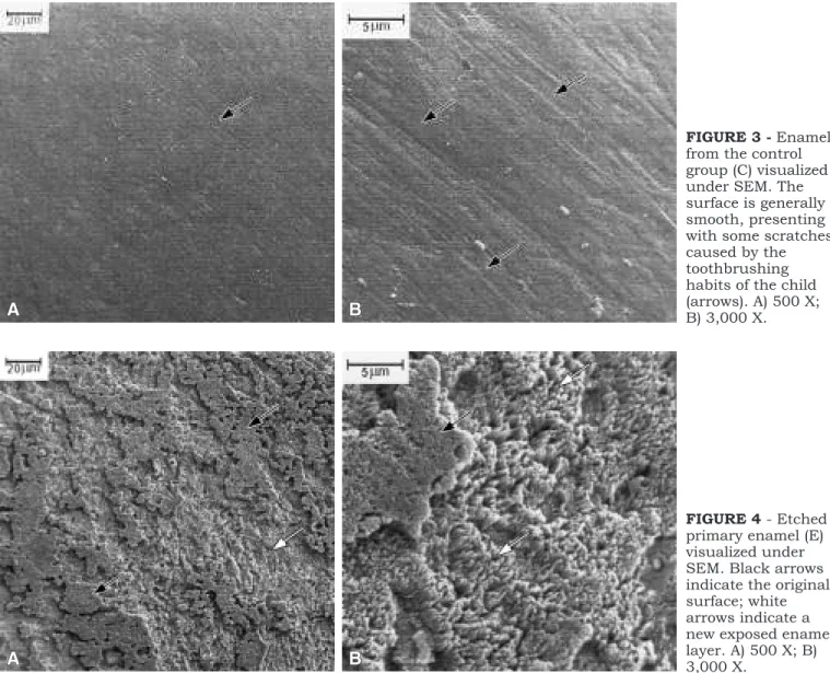
Figure 3 shows the results of the control Group. The surface is generally smooth, while various scratches can be observed in a specific direction.
The samples subjected to acid etching presen-ted direct dissolution of the surface. The lower magnification (Figure 4A) revealed an extensively eroded enamel surface. The higher magnification showed enhanced porosity and prism irregulariti-es caused by the etching procedurirregulariti-es. The pattern of dissolution, in the majority of the surfaces, was classified21
as type II. Figures 5 and 6 show typical features found in the groups brushed with non-fluoridated (EB) and fluoridated dentifrices (EBF), respectively. It is possible to note a general leveling and smoothening of the samples in lower magnification, with exposure of Tomes’ process pits (Figures 5B and 6B) separated by the smoot-hened interrod enamel of the underlying enamel layer (Figures 5B and 6B). Small particles, resem-bling insoluble remnants of the components of dentifrices were found, especially in the most
pro-FIGURE 3 -Enamel from the control group (C) visualized under SEM. The surface is generally smooth, presenting with some scratches caused by the toothbrushing habits of the child (arrows). A) 500 X; B) 3,000 X.
tected parts of the surface, such as in the grooves between Tomes’ process pits (Figures 5B and 6B). No differences were found between groups EB and EBF, as to surface morphology.
DISCUSSION
The smooth and scratched appearance of clini-cally sound enamel of erupted teeth, observed in the samples of the control group, was a common finding in other studies with permanent teeth car-ried out with SEM. Mannenberg17
(1960), using the replica technique, reported the appearance of new scratches,in vivo, after 4 weeks of toothbrushing
with a dentifrice containing abrasive. Thylstrup, Fredebo24
(1982) concluded that the scratched pat-tern and the smoothening of developmental irregu-larities are the gradual reaction of the enamel
sur-face to the abrasion caused by toothbrushing. The anatomical structure of the tooth surface at the time of eruption, with the typical perikymata pat-tern, is gradually lost and replaced by a scratched pattern.
In this study, the acid etching technique was used to simulate the morphologic alterations that occur on the enamel surface when initial caries le-sions are taking place or when enamel is etched but not covered with composites during restorative procedures. Although enamel caries is considered a subsurfacial lesion, as observed in polarized light microscopic studies6,15
, correlated SEM studi-es show that together with subsurfacial porosity there is an intense dissolution of the outer enamel surface11,14
. In fact, according to Thylstrup et al.23
(1994), the chalky appearance of demineralized
FIGURE 5 -Enamel brushed with a non-fluoridated dentifrice (EB) visualized under SEM. A) 500 X. The surface is smoothed down. B) 3,000 X. Black arrows indicate Tomes’ process pits; white arrows, interrod enamel; dotted arrows, remnants of insoluble components of the dentifrice abrasive.
enamel is enhanced by the irregular surface gene-rated by direct erosion of the enamel surface, gi-ving rise to a diffuse back-scattering of light. The method of demineralization chosen in this study intended to simulate the morphological alterations that take place on the enamel surface in those two clinical situations.
The pattern of demineralization in primary ena-mel is considered to be greatly altered by the pre-sence of aprismatic enamel21. In fact, Costaet al.4
(1998) reported a random distribution of demine-ralization patterns in unerupted deciduous teeth etched with 35% phosphoric acid. However, Fava
et al.7
(1997), using the same technique, though with a larger number of samples, found a slightly greater prevalence of type II demineralization, which is in agreement with our findings.
As most studies report variations on the tooth-brushing force applied by patients1,17
, the stan-dardization employed in this study intended to minimize possible differences in the results. Man-nemberg17(1960) found values ranging from 0.2 to
1 kgf in the toothbrushing force applied by 48 indi-viduals, while Björn, Lindhe1
(1966), studying 73 patients, found a mean maximum force of 0.45 kgf during horizontal brushing. In this study, a force of 0.2 kgf was chosen for being the most comforta-ble for the operator in the position of brushing the enamel blocks, while still in the range of the previ-ously found values.
It is usually accepted that the etched enamel re-mineralizes few moments after being exposed to the oral environment9. Nevertheless, Garberoglio,
Cozzani8(1979) observed that etched enamel
sur-faces do not show, microscopically, complete resti-tution of their structure after being exposed to the oral environment. After 90 days, the etched ena-mel cleaned off from the organic debris maintained its surface irregularities. The action of pumicing appeared to smooth the irregularities caused by the etching agent8
. Also, Hachiya et al.10
(1985) etched and stored extracted permanent teeth in artificial saliva for 1 month. During that period, a group was submit-ted to 15 brushing sessions (60 hand strokes in each session) with a dentifrice containing calcium carbonate. The enamel that was not brushed retai-ned the irregularities produced by etching, while the brushed group presented rounded surface de-fects, although depressions were not completely eliminated. In our study, a smoother enamel sur-face was also present after brushing, and the
ob-served depressions correspond to the interrod ena-mel of the brushed surfaces presented in Figures 5 and 6.
Holmenet al.13
(1987) observed, in high magnifi-cations under SEM, that the carious enamel re-exposed to oral mechanical forces showed removal of the external loosely packed crystals, correspon-ding, in broader terms, to the removal of the outer and demineralized surface. In the present study, the morphology of the brushed surfaces was cha-racterized by a general leveling of the enamel sur-face and by the visualization of Tomes’ process pits separated by smooth interrod enamel (Figures 5 and 6).
Although Head12
(1912) had previously stated that sound enamel shows insignificant wear to the dentifrice abrasive and its friction, while softened enamel may loose material from its surface, a diffe-rent approach to the removal of the outer dissolved and porous demineralized enamel surface by toothbrushing was reported by Kuroiwa et al.16
(1994). The authors observed,in vivo, the gradual
abrasion of demineralized enamel after toothbrus-hing with dentifrice. Samples brushed without dentifrice showed organic and mineral deposi-tion, which was assumed to originate from sali-vary components and remnants of loose enamel crystallites smeared over the surface. The authors concluded that toothbrushing without dentifrice would remineralize etched enamel, while paste brushing would merely abrade the weakened su-perficial enamel layers, without remineralization.
While the subsuperficial characteristic of caries lesion is observed by means of polarized light, the remineralization of enamel is generally investiga-ted by means of microhardness measurements20
. The formation of an organic-mineral coating is a frequent observation8,16
that may contribute to the increasing hardness of the remineralized etched enamel. However, the clinical presence of such an organic coating over the enamel surfaces fre-quently leads to staining of tooth surfaces. Man-nenberg17
(1960) observed the appearance of a brownish film over tooth surfaces in patients using only a mouthwash during toothcleaning procedu-res. This fact supports the importance of some de-gree of abrasivity during toothcleaning procedu-res.
Although the fluoridated dentifrice used in this study contained MFP, which is supposed to de-pend on intra-oral hydrolysis to be effective, Bru-unet al.2(1987) found out that enamel apatite
study, the contact of the fluoridated dentifrice with enamelin vitrowas considered as a catalyst to the
breakdown of MFP.
Calcium fluoride-like material was not found. Such finding is in disagreement with those of Cruz
et al.5
(1994). However, those authors had enamel surfaces treated for 24 hours with dentifrice su-pernatants, while no mechanical procedures was carried out. In the present study, mechanical abrasion or dissolution in artificial saliva probably impaired the observation of deposited calcium fluoride.
In spite of being generally acquainted that remi-neralization in the presence of fluoride restores the structure of the enamel surface, in this study, the appearances of surfaces brushed with either a fluoridated or non-fluoridated dentifrice were quite similar. However, using a morphological approach, it is not possible to preclude differences as to the chemical structures of samples brushed in the presence of fluoride or not. Corroborating this, Thylstrup, Bruun22
(1992) stated that the success of the individual caries control therapy based on the use of fluoridated dentifrices derives from me-chanical plaque control combined to the mainte-nance of optimal levels of fluoride in the oral ca-vity.
In a morphological context, the leveling of the eroded surface of initial caries lesions or
uncove-red etched enamel, produced by mechanical treat-ment of the enamel (toothbrushing, professional toothcleaning), may have a special role in the fate of demineralized enamel since it renders a smoot-her surface, making it more difficult for bacteria to adhere and contributing to the disappearance of the chalky white appearance caused by erosion. In clinical situations, this fact may be misinterpreted as remineralization or re-incorporation of minerals into the enamel.
CONCLUSION
Brushing demineralized enamel surfaces with dentifrice led to smoothening of the outer dissolved surface by removing the irregularities caused by acid etching. There was no difference, as to the morphology of the enamel surface, between sam-ples brushed with fluoridated and non-fluoridated dentifrices.
ACKNOWLEDGMENTS
The authors would like to thank the technical assistance of Noemia Rodrigues Gonçalves (De-partment of Biophysics, Federal University of Rio de Janeiro) and Maria de Fátima Lopes (Depart-ment of Material Sciences and Metallurgy, Pontifi-cal Catholic University - Rio de Janeiro).
REFERENCES
1. Björn H, Lindhe J. On the mechanisms of toothbrushing. Odont Rev 1966;17:9-16.
2. Bruun C, Giskov H, Thylstrup A. Intraoral hydrolysis of monofluorophosphate. Scand J Dent Res 1987;95:202-4. 3. Buonocore MG. A simple method of increasing the
adhe-sion of acrylic filling materials to enamel surfaces. J Dent Res 1955;34:849-53.
4. Costa LRRS, Watanabe L, Fava M. Three-dimensional as-pects of etched enamel in non-erupted deciduous teeth. Braz Dent J 1998;9:95-100.
5. Cruz R, Rolla G, Ogaard B. Alkali-soluble fluoride deposi-tion on human enamel exposed to monofluorophospha-te-containing toothpastes in vitro. Acta Odont Scand 1994;52:72-6.
6. Darling AI. Studies of the early lesion of enamel caries. Its nature, mode of spread, and points of entry. Brit Dent J 1958;105:119-36.
7. Fava M, Watanabe L, Moraes FF, Costa LRRS. Observa-tions on etched enamel in non-erupted deciduous molars. Rev Odont Univ São Paulo 1997;11:157-60.
8. Garberoglio R, Cozzani G.In vivoeffect of oral environment on etched enamel: a scanning electron microscopic study. J Dent Res 1979;58:1859-65.
9. Gwinnett AJ, Buonocore MG. Adhesives and caries preven-tion. Brit Dent J 1965;119:77-80.
10. Hachiya Y, Takatsu T, Hosoda H, Fusayama T. A varnish to prevent etching unrestored enamel. J Prosth Dent 1985;53:46-50.
11. Haikell Y, Frank RM, Voegel JC. Scanning electron micros-copy of the human enamel surface layer of incipient cari-ous lesions. Caries Res 1983;17:1-13.
12. Head J. A study of saliva and its action on tooth enamel in reference to its hardening and softening. J Am Med Assoc 1912;59:2118-22.
13. Holmen L, Thylstrup A, Artun J. Surface changes during the arrest of active enamel carious lesionsin vivo: a scan-ning electron microscopic study. Acta Odont Scand 1987;45:383-90.
14. Holmen L, Thylstrup A, Ogaard B, Kragh F. A scanning electron microscopic study of progressive stages of enamel cariesin vivo. Caries Res 1985;19:355-67.
15. Holmen L, Thylstrup A, Ogaard B, Kragh F. Polarized light microscopic study of progressive stages of enamel cariesin vivo. Caries Res 1985;19:348-54.
16. Kuroiwa M, Kodaka T, Kuroiwa M, Abe M. Brushing-indu-ced effects with and without a non-fluoride abrasive denti-frice on remineralization of enamel surfaces etched with phosphoric acid. Caries Res 1994;28:309-14.
18. Miura F, Nakagawa K, Masuhara E. New direct bonding system for plastic brackets. Am J Orthod 1971;59:350-61. 19. Monteiro Junior S, Andrada MAC, Baratieri LN.
Reminera-lização de lesões cariosas incipientes. RGO 1985;33:185-9. 20. Silvestone LM. Remineralization phenomena. Caries Res
1977; 11 suppl1:59-84.
21. Silverstone LM, Dogon IL. The effect of phosphoric acid on human deciduous enamel surfaces in vitro. J Int Assoc Dent Child 1976;7:11-5.
22. Thylstrup A, Bruun C. The use of dentifrices in the treat-ment of dental caries.In:Embery G, Rolla G. Clinical and
biological aspects of dentifrices. Oxford: IRL Press; 1992. p.131-143.
23. Thylstrup A, Bruun C, Holmen L.In vivocaries models: me-chanisms for caries initiation and arrestment. Adv Dent Res 1994;8:144-57.
24. Thylstrup A, Fredebo L. A method for studying surface coa-tings and the underlying enamel features in the scanning electron microscope.In:Frank RM, Leach SA. Surface and colloid phenomena in the oral cavity: methodological as-pects. St. Louis: IRL Press; 1982. p.169-184.