vol. 38, n. 3, jul./set., 2002
Human platelet nitric oxide synthase activity: an optimized method
Elisa Mitiko Kawamato
1, Isaias Glezer
1, Carolina Demarchi Munhoz
1, Cristiane Bernardes
1,
Cristoforo Scavone
1*, Tania Marcourakis
1,21Departamento de Farmacologia, Instituto de Ciências Biomédicas, 2Centro de Investigações em Neurologia,
Hospital das Clínicas/FMUSP (LIM-15), Universidade de São Paulo
We investigated the kinetic analysis of human platelet Nitric Oxide Synthase (NOS) activity by the rate of conversion of [3H] arginine
to [3H]-citrulline in unstimulated fresh platelets. NOS activity was
present in the membrane fraction and cytosol, and was Ca2+- and
calmodulin dependent which is a characteristic of endothelial NOS. NOS activity was also dependent of NADPH since the omission of this cofactor induced an important decrease (85,2%) in the enzyme activity. The kinetic varied with protein and arginine concentration but optimum concentrations were found up to 60 minutes, and up to 80 μg of protein at 120 nM of arginine and 0.5 μCi of 3H-arginine.
NOS activity in the absence of FAD (flavin adenine dinucleotide), FMN (flavin mononucleotide) and BH4 (tetrahydrobiopterin) was only 2.8% of the activity measured in the presence of these three cofactors. The enzyme activity was completely inhibited by L-NAME (1 mM) (98.1 %) and EGTA (5 mM) (98.8 %). Trifluoperazine (TFP) caused 73.2% inhibition of the enzyme activity at 200 μM and 83.8 % at 500 μM. Under basal conditions, NOS Km for L-arginine was 0.84 ± 0.08 μM and mean Vmax values were 0.122 ± 0.025 pmol.mg-1.min-1. Mean human NOS platelet activity was 0.020
± 0.010 pmol.mg-1.min-1. Results indicate that the eNOS in human
platelet can be evaluated by conversion of [3H]-arginine to [3
H]-citrulline in an optimized method, which provide reproducible and accurate results with good sensitivity to clinical experiments involving neurological and psychiatric diseases.
*Correspondence:
C. Scavone
Avenida Professor Lineu Prestes, 1524 Departamento de Farmacologia Instituto de Ciências Biomédicas Universidade de São Paulo CEP 05508-900 – Cidade Universitária São Paulo – Brazil
E-mail: [email protected]
Uniterms:
• Nitric Oxide Synthase • Platelets
• Arginine • Citrulline
INTRODUCTION
Platelets, skin fibroblasts, lymphocytes, plasma and cerebrospinal fluid were identified in the search for peripheral tissues or body fluids suitable for exploring
pathophysiology of neurodegenerative diseases such as Alzheimer ’s disease and psychiatric disorders, as depression, bipolar disorder and panic disorder (Gasparini
1955 (Pletscher, et al., 1956). A variety of biochemical perspectives is offered by platelets since they can synthesize a vast number of compounds, such as second messengers, have metabolizing enzymes (MAO), express different kinds of receptors, including adrenoceptors (α2 and β2), serotonin (5-HT2) and benzodiazepine receptors (Pletscher, 1988, Camacho, Dimsdale, 2000). Moreover, platelets share some similarities with neurons mainly in respect to the uptake system of serotonin (Pletscher, 1988). In addition, platelets can be used as model for the study of stimulus-response coupling mechanism since they yield homogeneous cell suspension that respond reliably and quantifiably to many pharmacological agonists (Hourani, Cusack, 1991).
The involvement of free radical in neuro-degeneration is well established and the use of platelets to assess the role of these agents is of great interest. Peroxynitrite is formed from the interaction between nitric oxide (NO) and superoxide anions. Interestingly, NO has been suggested to protect against superoxide anion-mediated injury under certain conditions (Miles et al., 1996). NO is an important physiological messenger with several biological functions such as regulation of vascular tone, blood pressure, vasodilation, sodium excretion, neurotransmission, macrophage cytotoxicity (Rees et al., 1989; Aisaka et al., 1989; Garthwaite, Boulton, 1995; Bredt, Snyder, 1990; Hibbs et al., 1990; Mckee et al., 1994). Moreover, NO also inhibits platelet aggregation through the activation of soluble guanylyl cyclase with the production of cGMP (Radomski et al., 1990a; Radomski
et al., 1990b). NO synthase (NOS) is the enzyme responsible for the oxidation of L-arginine to generate NO and L-citrulline in a reaction that needs molecular oxygen, NADPH as a co-substrate and BH4, FAD, FMN and Ca2+
-calmodulin as cofactors (Lincoln, Messersmith, 1995). In humans, three distinct forms of NOS have been identified. The neuronal NOS (nNOS) is found in neuronal and epithelial cells and acts in neurotransmission. The inducible NOS (iNOS) is found in macrophages and many other cells (such as glia) after stimulation by inflammatory signals such as cytokines or endotoxin. The endothelial NOS (eNOS) is found basically in endothelial cells. The
activity of both eNOS and nNOS is Ca2+
-calmodulin-dependent, whereas that of iNOS is Ca2+-insensitive
(Bredt, Snyder, 1990; Mckee, et al., 1994).
Different methodological approaches have also been used to characterize NOS isoforms in platelets. Thus, studies using Western blots of cytosolic and membrane fractions of human platelets as well as sequential affinity chromatography were able to confirm the presence of a Ca2+/CAM dependent isoform similar to the eNOS (Sase,
Michel, 1995; Muruganandam, Mutus, 1994). These
results are also supported by spectroscopic determination (Radomski et al., 1990b), eletrochemical NO detection (Lantoine et al., 1995) and porphyrinic microsensor assay (Malinski et al., 1993), after platelet stimulation with collagen. In addition, there is evidence for an inducible isoform in human platelet NOS using reverse transcription polymerase chain reaction (Mehta et al., 1995; Berkels et al., 1997).
However, the problem among all these methods is the lack of sensitivity because of the small amount of NO production by platelets. Thus, the radiochemical assay based on the biochemical conversion of L-arginine with the production of stoechiometric quantities of L-citrulline and NO is considered as a simple, sensitive and specific method to measure the production of NO. When optimized, the conversion assay can provide reproducible and accurate results with good sensitivity.
We present here a method to the determination of NOS activity in human platelets by the conversion of tritium labeled L-arginine to tritium labeled L-citrulline. NOS activity was assessed under a variety of different conditions in order to optimize conditions for NOS activity in platelet homogenate to permit using small volume of whole blood and protein. Such information is important to validate NOS studies in human and rat platelet as well as in other peripheral tissues where eNOS has shown to act as an intracellular messenger. Furthermore, the optimized study will permit a simultaneous determination of the enzyme activity, cyclic GMP levels and cyclic GMP-dependent protein kinase activity in patients with several diseases.
MATERIALS AND METHODS
Chemicals
3H- Arginine (specific activity 45.2 Ci/mmol) was
obtained from Dupont – New England Nuclear (Natick, MA) and scintillation cocktail was purchased from Ulti-ma Gold TM (Packard BioScience BV, Groningen). 3
H-arginine from New England Nuclear is not 100% pure and decomposition or bacterial contamination can bring unspecific high radioactivity into the blank sample. Since the levels of radioactivity in the blanks largely determine the sensitivity of the assay, the 3H-arginine was purified
before NOS assay in human platelets. This was performed by passing 3H-arginine through a Dowex ion-exchange
column. The column was washed four times with 1 mL of distillated water, and the 3H-arginine was eluted with
0.02 M NaOH (2 mL). 3H-arginine was stored in aliquots
was supplied by Sigma-Aldrich Chemicals (St. Louis, MO). The resin was converted to the sodium form by washing in 1 N NaOH for approximately five minutes and after in de-ionized water until pH 9-12, followed by washing in HEPES buffer (pH=5,5) until pH < 7. HEPES, sucrose, EDTA, dithiothreitol, PMSF, leupeptin, pepstatin, calcium chloride, NADPH, calmodulin, FAD, FMN, BH4, L-NAME and arginine were obtained from Sigma-Aldrich Chemicals.
Preparation of washed platelets
The study was approved by the hospital’s ethic committee and all subjects signed informed consent forms. Blood from normal healthy volunteers (20 mL), who were not taking any drug in the previous days, was drawn from an antecubital vein into citrate acid dextrose anticoagulant (2.73% citric acid, 3.68% sodium citrate, 2% glucose). Platelet rich plasma was prepared within 30 min after blood collection by centrifuging the blood at 394 x g. The plasma was then centrifuged at 1500 x g to obtain the platelet pellet. The pellet was washed twice in Krebs buffer (pH 6) containing [mM] 140 NaCl, 5 KCl, 12 sodium citrate, 10 glucose, 12.5 sucrose and centrifuged at 1500 x g. All centrifugations were performed for 15 min at room temperature.
The platelets were ressuspended in a buffer (pH 7.4) with 20 mM HEPES, 0.32 mM sucrose, 1 mM
dithio-threitol (DTT), 10 μg/mL leupeptin, 0.1 mM EDTA,
1 mM pepstatin, 1 mM PMFS, and sonicated at 4 °C. The sample was then treated with the ion-exchange resin (Dowex 50WX8-400, sodium form) to remove endo-genous arginine. NOS activity was measured in the homogenate with an assay adapted from methods published previously (Bredt, Snyder, 1990; Mckee et al., 1994).
Determination of Nitric Oxide Synthase Activity (NOS) in platelets
The incubation medium (pH=7.4) contained 4 μM
FAD, 4 μM FMN, 10 μg/mL calmodulin, 1.25 mM Ca2+,
1 mM NADPH, 25 μM BH4 and one of the three different L-arginine concentrations (10 μM, 1 μM, 120 nM). The reaction was started by adding the platelet samples in the desired protein concentration. Total platelet protein was determined in the homogenate using Biorad (Melville, NY) assay (Bradford, 1976).
After 30 and 60 minutes of incubation at 37 °C, the reaction was stopped by the addition of 1 mL of cold Hepes 20 mM (pH=5.5) and keeping the tubes in ice. 3
H-citrulline was separated from 3H-arginine through an ion
exchange resin column (Dowex 50WX8-400, sodium form) and the addition of 1 mL of distilled water. Total eluate was collected in a scintillation vial containing 10 mL of Ultima Gold scintillation fluid. The radioactivity was determined in a Packard Liquid Scintillation Counter with 54% efficiency. Blanks consisted of homogenizing medium, without platelet samples, which was carried out through the identical process. All measurements were made in triplicate and corrected for the appropriate blanks. Total count refers to a vial containing [3H]-arginine and
L-arginine without passing by the resin. The data were converted from cpm to dpm and specific activity (pmol.mg-1.min-1) was calculated by using the following
formula: [arginine] x [sample count]/[total count] x [protein] x time. Determination of Km and Vmax was done based on Lineweaver-Burk regression (Conrad, Davis, 1995).
RESULTS
Linearity with time, protein and substrate concentration
It might be assumed that NOS assay is highly dependent on the enzyme concentration and the incubation time. However, the activity of the enzyme can rapidly reach a plateau with time and with protein concentration. Thus, the first step was to determine the optimum conditions of time incubation and protein concentration to ensure that this plateau has not been reach as suggested by Hevel and Marletta (1994). For kinetic analysis of platelet NOS activity, it was necessary to carry out the NOS assay using different concentrations of the L-arginine substrate. Therefore, three different L-L-arginine concentrations were tested: 10 μM, 1 μM, 120 nM with a fixed concentration of 3H-arginine (0.5 μCi) (Figure 1).
The increase of 3H-arginine concentration was not useful
as NOS activity did not increase with higher concen-trations of that (data not shown). Preliminary experiments demonstrated that under standard conditions the assay was linear when protein concentration was up to 80 μg and the time of reaction was up to 60 minutes at 120 nM of arginine (Figure 1). Under basal conditions, NOS Km for L-arginine was 0.84 ± 0.08 mM and mean Vmax values were 0.122 ± 0.025 pmol.mg-1.min-1. Mean human NOS
platelet activity was 0.020 ± 0.010 pmol.mg-1.min-1.
Cofactor requirement of platelet NOS activity
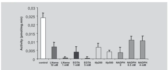
Inhibition of human NOS platelet by L-NAME
L-NAME (10 μM and 1 mM), a inhibitor of the all isoforms of NOS was used to characterize the enzyme. L-NAME (10 μM) inhibited approximately 70% of platelet NOS and 1 mM caused a complete inhibition of this enzyme activity (Figure 3). The total NOS activity can be expressed as the difference in activity in the presence and absence of L-NAME.
FIGURE 2 - Effects of the co-factors, FAD, BH4 and FMN, on NOS activity in human platelet. The clear bar represents control activity under standard conditions. Cofactors were investigated either singly (single hatching) or in combination (cross hatching). The concentration (M) used has been indicated under each bar. The composition of all the other components of the incubating medium was kept constant. Results were expressed as pmol. mg-1. min-1 and have been
given as the mean ± SEM of 3-5 experiments each consisting of triplicate determination.
FIGURE 1 - Effect of arginine, protein concentration, and incubation time on NOS activity in a preparation from fresh human platelet. Three different L-arginine concentrations (10 μM, 1 μM, 120 nM) were tested with different protein concentration (40 , 80 S and 160 ug T) a fixed concentration of 3H-arginine (0.5 μCi) at 30 and 60 minutes of
incubation. Results have been expressed as cpm, which was corrected for its own blank (homogenate omitted). Data consist of a representative experiment, which was repeated, and have been given as the mean of triplicate determination.
(approximately 2.8%) was detected when none of the cofactors were added to the incubation medium. The
addition of FAD, FMN or BH4 individually, enhances
platelet NOS activity, but not to the level observed when they were present all together. The combination of two of
the cofactors (FAD+FMN, FAD+BH4, FMN+BH4)
Ca2+/calmodulindependence on platelet NOS activity
To further characterize human NOS platelet activity, it is important to test Ca2+-calmodulin dependence of this
enzyme. To this purpose, a range of Ca2+ concentration
was added to the incubation medium: 0, 0.625, 1.25 (reference concentration) (Figure 4). In addition, EGTA
was used as a calcium chelator (1 mM and 5 mM) and two different concentration (200 and 500 mM) of TFP were used to determine calmodulin dependence in NOS platelet (Figure 3)
EGTA caused 80% inhibition of NOS activity at 1 mM and total inhibition at 5mM (Figure 5). The optimum concentration of NADPH was 1.0 mM and of FIGURE 3 - Effects of the co-substrate, NADPH, and of the inhibitors, L-NAME , EGTA and TFP, on NOS activity in human platelet. The clear bar represents control activity measured under standard conditions. The incubation media were altered as indicated under each of the hatched bars. Results are expressed as pmol. mg-1. min-1 and have been given as
the mean ± SEM of 3-5 experiments each consisting of triplicate determination.
FIGURE 4 - Effects of Ca2+ concentration on NOS activity in human platelet. The clear bar represents control activity
measured under standard conditions (Ca2+ concentration, 1.25 mM). Ca2+ concentrations were altered as indicated under
the hatched bars. Note that these concentrations represent those added exogenously and not the final concentration in the incubation medium which contains a non-specific chelator, EDTA. Results are expressed as pmol. mg-1. min-1 and
Ca2+ was 1.25 mM (Figure 3). Increases of NADPH
concentration and Ca2+ induced a reduction of 140% and
60 % of the NOS activity, respectively (Figure 3). TFP induced 73.2% inhibition of the enzyme activity at 200 mM and 83.8 % at 500 mM (Figure. 3)
DISCUSSION
In the present study, an optimized NOS assay was characterized from a preparation of homogenate of human platelet. Preliminary results suggest that although eNOS activity in platelets is membrane-bound and is thus localized in the particulate fraction, we could detect NOS enzyme activity in platelet cytosol, as well as in homogenate of human platelets disrupted by sonication. The profile of higher NOS activity in human platelet homogenate when compared to either membrane or cytosol correlates well with results reported by Sase and Michel (1995), and suggests that this fraction should be used for studying the basic biochemical features of eNOS in platelet.
Evidences emerging from various laboratories shows that there are several different conditions in the assay of NOS activity in platelet, like different time of incubation, different concentrations of cofactors, NADPH, buffer, protein, L-arginine, [3H]-arginine (Chen,
Mehta, 1996; Mehta et al., 1995; Riddell et al., 1997; Sase, Michel, 1995). In addition, it has been reported that only large amount of whole blood (from ~ 200 mL of whole blood) and protein (~ 10 mg/mL) has to be used in order to obtain a good NOS activity in human platelet (Sase, Michel, 1995). However, the kinetic analysis of NOS activity by using different concentrations of the L-arginine, and other requirements for eNOS activity has not been evaluated in human NOS platelet study. It is very important to establish that the concentration of arginine used is not rate limiting since it might be hard to demonstrate changes in the enzyme activity in experimen-tal study where NOS activity increases (Lincoln, Messersmith, 1995). Our results confirm previous findings that the sensitivity of NOS assay on platelets is very low when compared with NOS activity on cerebellum (Sase, Michel, 1995; Mckee et al., 1994). Since the basis of the NOS assay is to measure the conversion of radioactive arginine to citrulline, the higher the levels of endogenous, non-radioactive arginine, the lower is the sensitivity of the assay. Therefore, it was necessary to remove endogenous arginine from crude supernatants by passing the sample through a Dowex ion-exchange column. In addition, it was important to run control experiments in which NOS activity was measured
after different incubation times and using different concentrations of tissue. Our experiments showed that the linear portion of the curve could be obtained with 80 µg of protein at 30 and 60 minutes of incubation.
The kinetic analysis of platelet NOS activity was based on Garvey et al. (1994) and Wolf et al. (1994) by using three different L-arginine concentrations with a fixed concentration of 3H-arginine (0.5 μCi). In order to
have high enough arginine concentration in the assay, 3
H-arginine can be mixed with non-radioactive H-arginine. Thus, it was necessary to determine the relative proportions of radioactive and non-radioactive arginine in the working solution so that the change in specific activity of 3H-arginine can be calculated. Experiments in platelets
using 1 and 10 mM concentration of L-arginine have obtained high NOS activity but there is a reduction in specific activity, which reduce the sensitivity of the assay. In addition, the dose-response curve was not linear when high concentrations of arginine were used in NOS assay. Thus, the working solution with the best specific activity was obtained in the presence of 120 nM of L-arginine with a fixed concentration of 3H-arginine (0.5 μCi). Under
basal conditions, NOS Km for L-arginine was 0.84 ±
0.08 μM and mean Vmax values were 0.122 ±
0.025 pmol.mg-1.min-1. Mean human NOS platelet activity
was 0.020 ± 0.010 pmol.mg-1.min-1 .
The dependence of the presence of co-substrate and cofactors in NOS activity have been reported in different tissue preparation, such as kidney, myenteric plexus, dorsal brain stem, neutrophils, dorsal periaquedutal gray, spinal cord and LLC-PK1 cells (Lincoln, Messersmith, 1995; Mckee et al., 1994; Barjavel, Bhargava, 1995; Pontieri et al., 1998; Cerchiaro et al., 2001; Chiavegatto
concentration of BH4 from 0.25 to 25 µM and the maximal activity was achieved with 25 µM.
The enzyme activity was completely inhibited by L-NAME (1 mM) (98.1 %) and EGTA (5 mM) (98.8 %). The results also showed that human platelet NOS activity is highly dependent on NADPH and Ca2+ in the incubation
medium since the absence of both induced a tremendous decrease in the enzyme activity. Interestingly, the increase
of Ca2+ concentration above 1.25 mM resulted in
inhibition of NOS activity. It should be noted that Ca2+
concentration added in the medium does not represent the final free Ca2+ concentration since the non-specific
divalent cation chelator EDTA is present in the homo-genizing buffer. It should be noted that the omission of calmodulin from the incubating media was not sufficient to induce any decrease in the enzyme activity (data not shown) probably due to the binding of this compound to the enzyme during tissue preparation as it has been suggested to iNOS (Cho et al., 1992). However, TFP, a calmodulin antagonist, caused73.2% inhibition of the enzyme activity at 200 µM and 83.8 % at 500 μM.
Therefore, the present studies indicate that NOS activity in human platelet are highly dependent on the appropriate substrate and Ca2+ concentration, as well as on
exogenous co-fators for maximal activity. Given the extensive involvement of NOS in disease states, the procedure described here should be able to provide good indications of NOS activity in human platelet when investigating changes in the enzyme activity in patho-logical conditions.
ACKNOWLEDGMENTS
This work was supported by research grants from Fundação de Amparo à Pesquisa do Estado de São Paulo-FAPESP and Bunka Fund - Sumitomo Bank for E.M.K, I.G., E.M.K. and C.D.M. are supported by student research fellowship from FAPESP and C.S.B. is supported by University of São Paulo (Projeto IV). C.S. are research fellow of Conselho Nacional de Desenvolvimento Cien-tífico e Tecnológico.
RESUMO
Atividade da Óxido Nítrico Sintase em plaquetas hu-manas: um método otimizado
A análise cinética da atividade da óxido nítrico sintase (NOS) plaquetária foi avaliada pela conversão de [3H]-arginina em [3H]-citrulina em plaquetas humanas
frescas não estimuladas. A atividade da NOS foi detecta-da na fração citosólica e na membrana, além de ser de-pendente de Ca2+-calmodulina, que é uma característica
da NOS endotelial (eNOS). A omissão de NADPH levou à diminuição da atividade da NOS dependente da dose causando redução de 85,2% da atividade enzimática. A cinética variou de acordo com as concentrações de pro-teína e de arginina, sendo que as melhores leituras foram obtidas com 80 μg de proteína, 120 nM de arginina em 0,5 μCi de 3H arginina, em 60 minutos de incubação. A
atividade da NOS na ausência de FAD (flavina adenina dinucleotídeo), FMN (flavina mononucleotídeo) e BH4 (tetrahidrobiopterina) foi de apenas 2,8% da atividade medida na presença destes três cofatores. A atividade da enzima foi completamente inibida pelo L-NAME (1 mM; 98,1 %), EGTA (5 mM; 98,8 %) e adição de trifluo-perazina (TFP), nas concentrações de 200 μM e 500 μM, inibiu a atividade da enzima em 73,2% e 83,8 %, respec-tivamente. Em condições basais, o Km da NOS para L-arginina foi de 0,84 ± 0,08 μM e o valor de Vmax foi de 0,122 ± 0,025 pmol.mg-1.min-1. A atividade média da NOS
plaquetária humana foi de 0,020 ± 0,010 pmol.mg-1.min-1.
Os resultados indicam que a eNOS em plaquetas humanas pode ser avaliada pelo método da conversão de [3H]-arginina em [3H]-citrulina, que em condições
otimizadas, fornece resultados reprodutíveis e precisos com ótima sensibilidade para experimentos clínicos en-volvendo doenças neurológicas e psiquiátricas.
UNITERMOS: NOS. Plaquetas. Humano. Arginina. Citrulina.
REFERENCES
AISAKA, K., GROSS, S. S., GRIFFITH, O. W., LEVI, R. Nγ-methylarginine, an inhibitor of endothelium derives nitric oxide synthesis, is a potent pressor agent in the guinea-pig: does nitric oxide synthesis regulate blood pressure in vivo ? Biochem. Biophys. Res. Commun., v. 160, p. 881-886, 1989.
BARJAVEL, M. J., BHARGAVA, H. N. Nitric oxide synthase activity in brain regions and spinal cord of mice and rats: kinetic analysis. Pharmacology, v. 50, p. 168-174, 1995.
BRADFORD, M. M. A rapid and sensitive methods for the quantification of microgram quantities of protein utilizing the principle of protein-dye binding. Anal. Biochem., v. 72, p. 278, 1976.
BREDT, D. S., SNYDER, S. H. Isolation of nitric oxide synthase, a calmodulin-requiring enzyme. Proc. Natl. Acad. Sci. USA, v. 87, p. 682-685, 1990.
CAMACHO, A., DIMSDALE, J. E. Platelets and psychiatry: lessons learned from old and new studies. Psychosomatic Med., v. 62, p. 326-336, 2000.
CERCHIARO, G. A., SCAVONE, C., TEIXEIRA, S., SANNOMIYA, P. Inducible nitric oxide synthase in rat polymorphonuclear leukocytes: role of insulin. Biochem. Pharmacol., v. 62, p. 357-362, 2001.
CHEN, L. Y., MEHTA, J. L. Variable effects of L-arginine analogs on L-arginine- nitric oxide pathway in human neutrophils and platelets may relate to different nitric oxide synthase idoforms. J. Pharmacol. Exp. Ther., v. 276, p. 253-257, 1996.
CHIAVEGATTO, S., SCAVONE, C., CANTERAS, N. S. Nitric oxide synthase activity in the dorsal periaqueductal gray of rats expressing innate fear response.
NeuroReport, v. 9, p. 571-576, 1998.
CHO, H. J., XIE, Q. W., CALAYCAY, J., MUMFORD, R. A., SWIDEREK, K. M., LEE, T. D., NATHAN, C. Calmodulin is a subunit of nitric oxide synthase from macrophages. J. Exp. Med., v. 176, p. 599-604, 1992.
CONRAD, K. P., DAVIS, A. K. Nitric oxide synthase activity in placentae from women with pre-eclampsia. Placenta, v.16, p. 691-699, 1995.
GARTHWAITE, J., BOULTON, C. L. Nitric oxide signaling in the central nervous system. Ann. Rev. Physiol., v. 57, p. 683-706, 1995.
GARVEY, P., TUTTLE, J. V., COVINGTON, K., MERRILL, B. M., WOOD, E. R., BAYLIS, S. A., CHARLES, I. G. Purification and characterization of the constitutive nitric oxide synthase from human placenta.
Arch. Biochem. Biophys., v. 311, p. 235-241, 1994.
GASPARINI, L., RACCHI, M., BINETTI, C., TRABUCCHI, M., SOLERTE, S. B., ALKON, D., ETCHEBERRIGARAY, R., GIBSON, G., PAOLETTI, R., GOVONI, S. Peripheral markers in testing pathophysiological hypothesis and diagnosing Alzheimer’s disease. FASEB J.,v. 12, p. 17-34, 1998.
HIBBS, J. B., TAINTOR, R. R., VAVRIN, Z., GRANGER, D. L., DRAPIER, J. C., AMBER, I. J., LANCASTER, J. R. Synthesis of nitric oxide from a terminal guanidino atom of L-arginine: a molecular mechanism regulating cellular proliferation that targets intracellular iron. In: MONCADA, S., HIGGS, E. A., eds. Nitric oxide from L-arginine: a bioregulatory system. Amsterdam: Elsevier, 1990. p. 189-223.
HEVEL, J. M., MARLETTA, M. A. Nitric-oxide synthase assays. Methods Enzymol., v. 233, p. 250-258, 1994.
HOURANI, S. M., CUSACK, N. J. Pharmacological receptors on blood platelets. Pharmacol. Rev., v. 43, p. 243-298, 1991.
LANTOINE, F., BRUNET, A., BEDIOUI, F., DEVYNCK, J., DEVYNCK, M. A. Direct measurement of nitric oxide production in platelets: relationship with cytosolic Ca2+ concentration. Biochem. Biophys. Res. Commun., v.
215, p. 842-848, 1995.
LINCOLN, J., MESSERSMITH, W. A. Conditions required for the measurement of nitric oxide synthase activity in a myenteric plexus/ smooth muscle preparation from the rat ileum. J. Neurosci. Methods, v. 59, p. 191-197,1995.
MALINSKI, T., RADOMSKI, M. W., TAHA, Z., MONCADA, S. Direct eletrochemical measurement of nitric oxide released from human platelets.Biochem. Biophys. Res. Commun., v. 194, p. 960-965, 1993.
MCKEE, M., SCAVONE, C., NATHANSON, J. A. Nitric oxide, cGMP, and hormone regulation of active sodium transport. Proc. Natl. Acad. Sci., v.91, p. 12056-12060, 1994.
MILES, A. M., BOHLE, D. S., GLASSBRENNER, B., HANSERT, D. A., WINK, G., GRISHAM, M. B. Modulation of superoxide-dependent oxidation and hydroxylation reactions by nitric oxide. J. Biol. Chem., v. 271, p. 40-47, 1996.
MURUGANANDAM, A., MUTUS, B. Isolation of nitric oxide synthase from human platelets. Biochim. Biophys. Acta, v. 1200, p. 1-6, 1994.
PLETSCHER, A., SHORE, P. A., BRODIE, B. B. Serotonin as a mediator of reserpine action in brain. J. Pharmacol. Exp. Ther., v. 116, p. 84-89, 1956.
PLETSCHER, A. Platelets as models: use and limitations.
Experientia, v. 44, p. 152-155, 1988.
PONTIERI, V., VENEZUELA, M. K., SCAVONE, C., MICHELINI, L. C. Role of endogenous nitric oxide in the nucleus tractus solitarii on baroreflex control of heart rate in spontaneously hypertensive rats. J. Hypertens., v. 16, p. 1993-1999, 1998.
RADOMSKI, M. W., PALMER, R. M. J., MONCADA, S. Characterization of the L-arginine: nitric oxide pathway in human platelets. Brit. J. Pharmacol., v.101, p. 325-328,1990a.
RADOMSKI, M. W., PALMER, R. M. J., MONCADA, S. An L-arginine/ nitric oxide pathway present in human platelets regulates aggregation. Proc. Natl. Acad. Sci., v.87, p. 5193-5197, 1990b.
REES, D. D., PALMER, R. M. J., HODSON, H. F., MONCADA, S. A specific inhibitor of nitric oxide formation from L-arginine attenuates endothelium dependent relaxation. Brit. J. Pharmacol., v.96, p. 418-424, 1989.
RIDDELL, D. R., GRAHAM, A., OWEN, J. S. Apolipoprotein E inhibits platelet aggregation through the L-arginine:nitric oxide pathway. J. Biol. Chem., v. 272, p 89-95, 1997.
SASE, K., MICHEL, T. Expression of constitutive endothelial nitric oxide synthase in human blood platelets. Life Sci., v. 57, p. 2049-2055, 1995.
WOLF, D. J., LUBESKIE, A., UMANSKY, S. The inhibition of the constitutive bovine endothelial nitric oxide synthase by imidazole and indazole agents. Arch. Biochem. Biophys., v. 314, p. 360-366, 1994.