Article
Printed in Brazil - ©2015 Sociedade Brasileira de Química0103 - 5053 $6.00+0.00
A
*e-mail: [email protected]
Main Degradation Products of Dabigatran Etexilate Evaluated by LC-UV and
LC-ESI-MS, Degradation Kinetics and
in vitro
Cytotoxicity Studies
Raquel M. Bernardi,a Felipe B. D’Avila,*,a Vítor Todeschini,b Juliana M. M. Andrade,a
Pedro E. Fröehlicha and Ana M. Bergolda
aPrograma de Pós-Graduação em Ciências Farmacêuticas, Universidade Federal do Rio Grande do Sul,
Av. Ipiranga 2752, 90610-000 Porto Alegre-RS, Brazil
bFaculdade de Farmácia, Universidade Federal do Rio de Janeiro,
Av. Aluísio da Silva Gomes 50, 27930-560 Macaé-RJ, Brazil
The present study reports the stability profile of an antithrombotic drug: dabigatran etexilate (DAB). The drug was subjected to thermal degradation at 60 °C and products formed were investigated by liquid chromatography-UV (LC-UV) and liquid chromatography-mass spectrometry (LC-ESI-MS). Chromatographic separation of the degradation products was performed on a GL Sciences Inc. Inertsil ODS-2 column (250 mm × 4.6 mm i.d., with a particle size of 5 µm and pore size of 110 Å) with mobile phase consisting of acetonitrile and ammonium acetate buffer (pH 5.5; 10 mmol L−1) (65:35, v/v) pumped at 1.0 mL min−1 flow rate. Column temperature was set at 30 °C
and detection at 225 nm using a UV detector. LC-UVmethod previously validated was extended to LC-ESI-MS for the characterization of the degradation products (DP-01 and DP-02) formed, without complicated isolation or purification processes, based on retention times and confirmation of molecular weight. Degradation kinetics of DAB was also evaluated and could be described as a first-order process (R2 = 0.9900). Furthermore, no evidence of cytotoxicity in human mononuclear
cells was observed for DAB degraded samples.
Keywords: cytotoxity, dabigatran etexilate, degradation kinetics, degradation products, LC-ESI-MS
Introduction
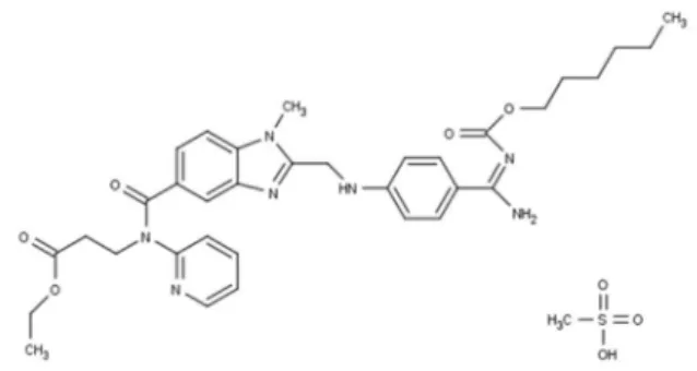
Dabigatran is a non-peptidic, synthetic, oral, potent and specific agent, a direct inhibitor of free thrombin, fibrin-bound thrombin and thrombin-induced platelet aggregation with rapid onset of action, predictable pharmacodynamic effects and pharmacokinetic characteristics.1-3 Dabigatran
is available as a pro-drug dabigatran etexilate (ethyl N
-{[2-({[4-((E)-amino{[hexyloxy)carbonyl]imino} methyl)
phenyl]amino} methyl)-1-methyl-1H-benzimidazol-5-yl]
carbonyl}-N-pyridin-2-yl-β-alaninate methanesulfonate (DAB) (Figure 1), converted into the active metabolite by non-specific plasma and liver esterases.
DAB is the first new oral anticoagulant to become available for the prevention of stroke and systemic embolism in patients with atrial fibrillation in over 50 years.4 It has
been approved for the prevention of stroke and systemic embolism in patients with non-valvular atrial fibrillation by
US Food and Drug Administration since October 2010 and by European Medicines Agency (EMA) since August 2011. EMA also approved it in March 2008 for the prevention of venous thromboembolic events after elective total hip replacement and/or total knee replacement.5,6
a key role in fibrin formation and is very important for blood coagulation and platelet activation, it is a prime target for the development of anticoagulant agents for the prevention and treatment of thromboembolic disorders.9,10
It has been well documented that drug products will undergo physicochemical degradation during manufacturing processes and storage.11 According to the International
Conference on Harmonization (ICH), stress studies of pharmaceutical drugs can support the identification of probable degradation products. Therefore it is necessary to adopt stability-indicating methods.12 The identification
of degradation products in dosage formulations plays an important role in the drug development process.11,13
Understanding drug degradation in formulated product is critical in pharmaceutical development because drug stability and degradation products could have a significant impact on formulation development, analytical method development, package development, storage conditions and shelf-life determination, safety and toxicology concerns. In formulation development, pharmaceutical stress testing has been widely used to predict drug stability problems, to develop stability-indicating analytical methods, and to identify degradation products and their reaction pathways.14
Traditional methods involving process scale-up, isolation, and purification of trace components such as degraded products and impurities are expensive and time-consuming. Besides, it is not possible to identify the chemical structure of the compounds by liquid chromatography (LC) alone.15 Therefore, the use of
hyphenated techniques, like the liquid chromatography-mass spectrometry (LC-ESI-MS) method is a versatile and attractive alternative to rapidly characterize degradation products and impurities.16-19 Moreover, the chromatographic
resolution of unresolved or co-eluting peaks detected by UV is not required to obtain product ion data for structural analysis, due to the high selectivity of MS.15
Most of the analytical aspects of the new anticoagulant remain unpublished. There are works describing the determination of DAB and metabolites in biological fluids by LC-MS-MS and radioactivity, but no information exists on its decomposition behavior.4,20-23
Previously, stress testing was performed to provide stability-indicating properties for analytical methods, and thus used to determine the analytes in the presence of degradation products by our group. The instability of DAB under thermal conditions was observed.24 However, these
components were not characterized.
The aim of the present study is to characterize the degradation products observed. Additionally, degradation kinetics was studied and a preliminary in vitro toxicity study
was performed using mononuclear cells.
Experimental
Chemicals
Dabigatran etexilate (purity 99.5%) was purchased from Sequoia Research Products (Oxford, United Kingdom). The commercial Pradaxa® Boehringer Ingelheim do Brasil (São
Paulo, Brazil) capsules containing 150 mg of dabigatran etexilate were obtained from commercial sources within their shelf life period. LC-grade acetonitrile was obtained from Tedia (Fairfield, USA). Ammonium acetate was purchased from Merck (Frankfurt, Germany). All chemicals used were of pharmaceutical grade. For all the analyses, ultrapure water was obtained using a Milli-Q Plus system Millipore (Bedford, USA).
Preparation of reference solution
Stock solution of DAB was prepared by weighing 10 mg of reference substance, transferred to individual 10 mL volumetric flasks and diluted to volume with methanol, obtaining a concentration of 1 mg mL−1. The stock solution
was stored at −20 °C, protected from light, and daily diluted to an appropriate concentration in mobile phase to produce the final concentrations of 30 and 60 µg mL−1.
LC-UV method
LC-UV method was developed on an Agilent® Infinity 1260 (Waldbronn, Germany) equipped with a 1260 quaternary pump, Standard degasser, 1260 TCC column oven, ALS 1260 autosampler and 1260 VWD detector. Peak areas were integrated automatically by computer using a ChemStation software program. UV detector was set at 225 nm. The experiments were performed on a reversed-phase GL Sciences Inc. Inertsil ODS-2 column (250 mm × 4.6 mm i.d., with a particle size of 5 µm and pore size of 110 Å) (Tokyo, Japan). LC system was operated isocratically at 30 °C, using a mobile phase consisting of acetonitrile and ammonium acetate buffer (pH 5.5; 10 mmol L−1) adjusted with acetic acid 2 mol L−1
(65:35, v/v); filtered through a 0.45 µm Millipore membrane filter (Bedford, USA) with a flow rate of 1.0 mL min−1.
Injection volume was 20 µL for both standard and samples. LC-UV method was previously validated, following the ICH guidelines.24-26
LC-ESI-MS method
method. Agilent quadrupole mass spectrometer (Waldbronn, Germany), model 6120, was equipped with an electrospray (ESI) source in positive mode. Studies were performed with capillary voltage, flow nebulizer, gas nebulizer pressure and dry temperature of 3000 V, 12 L min−1, 55 psig and
300 °C, respectively. Degradation samples were injected and full scan spectra were acquired, then the selected ions were monitored in single-ion recording (SIR) mode by the LC-ESI-MS system. Data acquisition and analysis were performed using the ChemStation software.
Forced thermal degradation studies
Forced degradation protocol was performed in order to provide suitable analytical conditions for a stability study of the drug. Thermal evaluation of DAB was performed with solutions (60 µg mL−1) exposed to 60 °C for 4 h (protected
from light). Each time interval (0.25, 0.5, 0.75, 1, 1.5, 2, 3, and 4 h) was evaluated separately.
After thermal degradation treatment, samples were diluted with mobile phase to final theoretical concentrations of 30 µg mL−1 for LC-UV analysis. Subsequently, samples
were diluted to 2 µg mL−1 with mobile phase and then
analyzed by LC-ESI-MS. All samples were analyzed against a freshly prepared control sample (without degradation treatment).
Thermal degradation kinetics study
Degradation kinetics of DAB was analyzed by validated stability-indicating LC-UV26 method and established using
same experimental stress conditions described previously in section forced degradation studies.
DAB thermal degradation rate was determined by plotting the drug concentration (zero-order process), log (first-order process) and reciprocal (second-order process) concentration versus time. Determination coefficient (R2)
was obtained and the best observed fit indicated the reaction order. Kinetic parameter like apparent order degradation rate constant (k) and time at which 90% of original
concentration of the drug is left(t90) was also calculated.
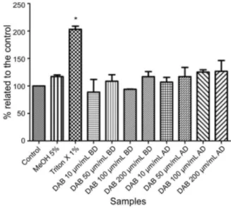
Cytotoxicity assay
For cytotoxicity experiments, the peripheral blood of three healthy volunteers was collected and subsequently diluted with Hank’s balanced salt solution. The human mononuclear cells were obtained by separation on Ficoll-Paque® gradient centrifuge, Sigma Aldrich (St. Louis, USA), washed and resuspended in Hank’s medium to a concentration of 1.0 × 10⁷ viable cells in
1.0 mL. The monitoring of total cells was performed using an Alphaphot-2 YS2 microscope from Nikon Instrument Inc. (New York, EUA).
All reagents, including cell suspension, commercial kit solutions Doles (Goiânia, Brazil) and samples before and after thermal degradation treatment (previously described) were distributed in a 96-well plate and maintained at 37 °C, for 30 min. Final analyzed concentrations were 10, 50, 100 and 200 µg mL−1 for DAB. Concurrently, a blank
test (without sample addition) and positive control, which shows 100% cell death (Triton X-100 1%) were performed. Interference of methanol (final concentration of 5%) used to prepare the samples was also evaluated. All samples were performed in triplicate. Cellular injury (cytotoxicity) was determined by measuring lactate dehydrogenase (LDH) at 492 nm using a plate reader Envision, Perkin Elmer Inc. (Waltham, USA).
A one-way analysis of variance (ANOVA) followed by Dunnet’s test was used for comparison of the means. Differences were considered statistically significant, if
p < 0.05. Data analyses were performed using Prism 5.0,
GraphPad Software Inc. (La Jolla, USA).
Results and Discussion
DAB degradation behavior
Stability testing is an essential part of any drug development because the quality of a drug product changes with time under the influence of environmental factors such as temperature, humidity and light.12 The purpose
of stability testing is to investigate those changes during the storage that are likely to influence quality, safety and efficacy, and to establish a shelf life for the drug product as well as recommend storage conditions. With ever increasing regulatory and compendial stringency on the control of degradation products in drug substances and finished pharmaceutical formulations, great emphasis is being laid on their characterization and analysis at trace levels.27
containers. In addition, the information on drug degradation products could be crucial for understanding potential toxicity or side effects associated with degradation. Therefore, a rapid and accurate elucidation of the drug degradation process has become essential to reduce drug development timelines.13,27
Following degradation behavior of DAB was investigated by the validated stability-indicating LC-UV26 method and
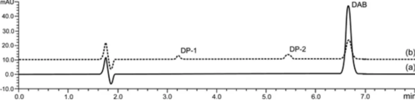
LC-ESI-MS studies. The overlay chromatograms of a DAB reference substance and DAB after thermal degradation are shown in Figure 2.
As previously mentioned, DAB was found to be labile under thermal conditions. After 4 h of exposure, approximately 75% reduction of concentration was observed (sufficient degradation to allow assessment of kinetics degradation with reliability). LC-UV chromatogram showed, besides significant decrease of peak area of DAB, rise of two signals detected by UV at 3.2 min and 5.5 min.
Based on their retention times, LC-ESI-MS studies indicated two major degradation peaks with m/z 500.2 and m/z 264.1, which were nominated DAB degradation products
(DP). LC-ESI-MS chromatograms are shown in Figure 3.
Figure 2. Overlapping LC-UV chromatograms of DAB (60 µg mL−1): (a) DAB without exposure to stress; (b) DAB after thermal stress condition.
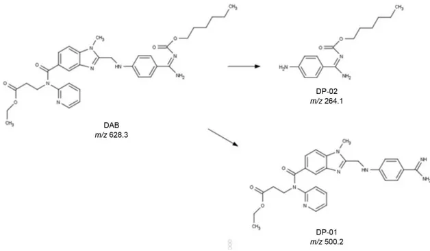
Figure 4. Proposed structures of DP-01 and DP-02.
The product ion at m/z 500.2 (DP-01) is a
carbamimidoylphenyl derivate formed by cleavage of carbamate, and was chemically characterized as ethyl 3-(2(((4-carbamimidoylphenyl)amino)methyl-N
-(pyridine-2-yl)-1H-benzol[d]imidazole-5-carboxamide) propanoate.
DP-01 is also identified as a DAB metabolite.4
The product ion at m/z 264.1 (DP-02) was chemically
characterized as (E)-hexyl(amino(4-aminophenyl)
methylene)carbamate and was formed by thermochemical cleavage of phenyl aminomethyl benzimidazolyl linkage to phenylamine.
Proposed structures of degradation products are shown in Figure 4.
Degradation kinetics studies
Degradation studies provide information on the quality of a drug under influence of different conditions and the degradation kinetics allows measuring speed of these processes.28 In this study, thermal decomposition of DAB
was carried out during different periods of time used for kinetic evaluation by LC-UV method.
The purpose of kinetics testing is to provide evidence on how quantity of a drug varies with time under the influence of stress factor. Furthermore, the information learned may be used to extrapolate rates of degradation and help to predict long-term stability under normal storage conditions.29,30
Thermal degradation kinetics was calculated through the decreasing concentration values of the remaining
drug versus time. The remaining DAB concentration
was calculated at each time interval for three replicates, comparing the mean drug concentration of the standard solution.
Through analysis of R2 obtained by plotting drug
concentration, log and the reciprocal concentration versus
time, degradation kinetics of the drug could be better described as first-order. These findings indicate that DAB degradation speed is proportional to one component concentration.29
In this experimental protocol, the obtained degradation rate constants (k) and t90 were: 0.0028 min−1 and 37.49 min.
Concentration, log and reciprocal concentration plots of remaining drug concentration versus time obtained during
kinetics are shown in Figure 5. Additionally, ambient temperature sample was evaluated and no considerable variation was observed.
Cytotoxicity studies
Cytotoxicity assays are among the most used in vitro
methods to predict potential toxicity of a substance in a cell culture.31
In this experimental protocol, non-significant differences (p > 0.05) were observed when DAB samples, before and
Conclusion
Evaluation of degradation products is essential for future biological and toxicological investigations, aiming at maintenance of safety and efficacy of pharmaceutical products. In this study, DAB stability study was performed using thermal stress conditions. The main degradation products formed were analyzed by LC-UV and LC-ESI-MS methods and structures were proposed. Additionally, degradation kinetics of the drug and cytotoxicity of the degraded samples were also studied, contributing to the improvement of the quality control of this drug.
Acknowledgements
The authors wish to thank Coordenação de Aperfeiçoamento de Pessoal de Nível Superior (CAPES) for the financial support.
References
1. van Ryn, J.; Stangier, J.; Haertter, S.; Liesenfeld, K.-H.; Wienen, W.; Feuring, M.; Clemens, A.; Thromb. Haemost.2010, 103, 1116.
2. Eriksson, B. I.; Dahl, O. E.; Ahnfelt, L.; Kälebo, P.; Stangier, J.; Nehmiz, G.; Hermansson, K.; Kohlbrenner, V.; J. Thromb. Haemost.2004, 2, 1573.
3. Fuji, T.; Fuijita, S.; Ujihira, T.; Sato, T.; J. Arthroplasty2010,
25, 1267.
4. Hu, Z.-Y.; Parker, R. B.; Herring, V. L.; Laizure, S. C.; Anal. Bioanal. Chem.2013, 405, 1695.
5. European Medicines Agency (EMA) http://www.ema.europa. eu/docs/en_GB/document_library/EPAR_-_Summary_for_the_ public/human/000829/WC500041060.pdf accessed in January 2015.
6. US Food and Drug Administration (FDA) http://www.fda.gov/ downloads/AdvisoryCommittees/CommitteesMeetingMaterials/ Drugs/CardiovascularandRenalDrugsAdvisoryCommittee/ UCM247244.pdf accessed in January 2015.
7. Franchini, M.; Mannucci, P. M.; Eur. J. Intern. Med.2009, 20, 562.
8. Borris, L. C.; Arch. Orthop. Trauma Surg.2010, 130, 583. 9. Stangier, J.; Stähle, H.; Rathgen, K.; Fuhr, R.; Clin.
Pharmacokinet.2008, 47, 47.
10. Mavrakanas, T.; Bounameaux, H.; Pharmacol. Ther.2011, 130, 46.
11. Liu, Z.-Y.; Zhang, H.-H.; Chen, X.-J.; Zhou, X.-N.; Wan, L.; Sun, Z.-L.; Int. J. Mass Spectrom.2011, 303, 90.
12. International Conference Harmonization (ICH); Stability Testing of New Drug Substances and Products Q1A (R2);ICH: Geneva,
2003.
Figure 5. Degradation kinetics assay and plots obtained for first-order reaction of DAB after thermal degradation.
13. Wu, Y.; Biomed. Chromatogr.2000, 14, 384.
14. Pan, C.; Jiang, Y.; Wen, H.; Motto, M.; J. Pharm. Biomed. Anal.
2012, 57, 99.
15. Volk, K. J.; Hill, S. E.; Kerns, E. H.; Lee, M. S.; J. Chromatogr. B: Biomed. Sci. Appl.1997, 696, 99.
16. Modhave, D. T.; Handa, T.; Shah, R. P.; Singh, S.; J. Pharm. Biomed. Anal.2011, 56, 538.
17. Ardrey, R. E.; Liquid Chromatography-Mass Spectrometry: An Introdution, John Wiley & Sons: Chichester, 2003.
18. Fukutsu, N.; Kawasaki, T.; Saito, K.; Nakazawa, H.; J. Chromatogr. A2006, 1129, 153.
19. Lee, H.; J. Liq. Chromatogr. Relat. Technol.2005, 28, 1161. 20. Stangier, J.; Rathgen, K.; Stähle, H.; Gansser, D.; Roth, W.;
Br. J. Clin. Pharmacol.2007, 64, 292.
21. Stangier, J.; Eriksson, B. I.; Dahl, O. E.; Ahnfelt, L.; Nehmiz, G.; Stähle, H.; Rathgen, K.; Svärd, R.; J. Clin. Pharmacol.2005,
45, 555.
22. Delavenne, X.; Moracchini, J.; Laporte, S.; Mismetti, P.; Basset, T.; J. Pharm. Biomed. Anal.2012, 58, 152.
23. Blech, S.; Ebner, T.; Ludwig-Schwellinger, E.; Stangier, J.; Roth, W.; Drug Metab. Dispos.2008, 36, 386.
24. Bernardi, R. M.; Fröehlich, P. E.; Bergold, A. M.; J. AOAC Int.
2013, 96, 37.
25. International Conference Harmonization (ICH); Validation of Analytical Procedures: Text and Methodology Q2(R1); ICH: London, 2005.
26. Bernardi, R. M.; D’Avila, F. B.; Todeschini, V.; Fröehlich, P. E.; Bergold, A. M.; Anal. Methods2013, 5, 4777.
27. Singh, S.; Handa, T.; Narayanam, M.; Sahu, A.; Junwal, M.; Shah, R. P.; J. Pharm. Biomed. Anal.2012, 69, 148.
28. Marques, L. A.; Maada, I.; de Kanter, F. J. J.; Lingeman, H.; Irth, H.; Niessen, W. M.; Giera, M.; J. Pharm. Biomed. Anal.
2011, 55, 85.
29. Waterman, K. C.; Handbook of Stability Testing in Pharmaceutical Development; Huynh-Ba, K., ed.; Springer New York: New York, 2009.
30. Nudelman, N. E. S.; Estabilidad de Medicamentos, Librería El Ateneo: Buenos Aires, 1975.
31. Martinez, V.; Corsini, E.; Mitjans, M.; Pinazo, A.; Vinardell, M. P.; Toxicol. Lett.2006, 164, 259.
Submitted: August 27, 2014
Published online: February 6, 2015