Article
Printed in Brazil - ©2013 Sociedade Brasileira de Química0103 - 5053 $6.00+0.00
A
*e-mail: [email protected]
New Hybrid Material Based on Layered Double Hydroxides and Biogenic Silver
Nanoparticles: Antimicrobial Activity and Cytotoxic Effect
Priscyla D. Marcato,a,b Natália V. Parizotto,a Diego Stéfani T. Martinez,a Amauri J. Paula,a
Iasmin R. Ferreira,c,d Patrícia S. Melo,c,d Nelson Duráne,f and Oswaldo L. Alves*,a
aLaboratório de Química do Estado Sólido (LQES), Instituto de Química,
Universidade de Campinas, 13083-970 Campinas-SP, Brazil
bFaculdade de Ciências Farmacêuticas de Ribeirão Preto, Universidade de São Paulo,
14040-903 Ribeirão Preto-SP, Brazil
cFaculdade Integrada Metropolitana de Campinas (METROCAMP), 13035-270 Campinas-SP, Brazil
dFaculdade de Ciências Aplicadas, Universidade de Campinas, 13484-350 Limeira-SP, Brazil
eLaboratório de Química Biológica, Instituto de Química, Universidade de Campinas,
13083-970 Campinas-SP, Brazil
fCentro de Ciências Naturais e Humanas, Universidade Federal do ABC, 09210-170 Santo André-SP, Brazil
Hidróxidos duplos lamelares (HDLs) têm sido amplamente investigados devido às suas diversas aplicações nas indústrias de materiais e biotecnologia. A combinação de nanopartículas de prata com o material biocompatível HDL pode criar um novo material híbrido com novas propriedades sinergísticas. Neste trabalho, nanopartículas de prata biogênicas (AgNPbio) foram associadas com Mg-Al HDL para obter o material híbrido HDL-AgNPbio. O novo material híbrido obtido foi caracterizado por difratometria de raios X (XRD), microscopias eletrônica de transmissão (TEM) e de varredura com espectroscopia de energia dispersiva por raios-X (MEV-EDS), espectrometria de emissão atômica por plasma acoplado indutivamente (ICP-OES) e espectroscopia na região do infravermelho com transformada de Fourier (FTIR). O HDL foi eficiente em adsorver nanopartículas de prata devido à carga superficial oposta entre as AgNPbio (ζ = −13,2 mV) e o HDL (ζ = +3,2 mV). Além disso, as AgNPbio não foram lixiviadas do material híbrido, mesmo após cinco ciclos de lavagem, indicando uma forte interação. Uma importante propriedade deste material híbrido foi a sua atividade antimicrobiana contra Staphylococcus aureus e Escherichia coli e ausência de efeito
citotóxico em células de fibroblastos (V79). Este material híbrido é um interessante e promissor nanobiocompósito para aplicações biomédicas e cosméticas.
Layered double hydroxides (LDHs) have been widely investigated due to their several applications in the material and biotechnology industries. The combination of silver nanoparticles with biocompatible LDH material can create a new hybrid material with new properties. In this work, biogenic silver nanoparticles (AgNPbio) were associated withMg-Al LDH to obtain the hybrid material LDH-AgNPbio. The new hybrid material obtained was characterized by X-ray diffractometry (XRD), scanning electron microscopy with energy-dispersive X-ray spectroscopy (SEM-EDS), transmission electron microscopy (TEM), inductively coupled plasma optical emission spectrometry (ICP-OES) and Fourier transform infrared spectroscopy (FTIR). LDH was efficient to absorb silver nanoparticles due to an opposite surface charge between AgNPbio (ζ = −13.2 mV) and LDH (ζ = +3.2 mV). Furthermore, AgNPbio was not lixiviated from LDH-AgNPbio, even after five washes, indicating a strong interaction. An important property of this hybrid material was its antimicrobial activity against Staphylococcus aureus and Escherichia coli and
absence of cytotoxic effect to fibroblast cell (V79). This hybrid material is an interesting and promising nanobiocomposite for biomedical and cosmetic applications.
Introduction
Layered double hydroxides (LDHs) have been widely studied due to their potential applications in different areas, such as, catalysts, pharmaceuticals, UV stabilizers, adsorbents and others. These compounds are clays represented by the empirical formula [M2+
1-xM3+x(OH)2] (An−)x/n mH2O, in which M2+ and M3+ are divalent and trivalent cations and An− is an anion, thus
having positive or negative charge layers.1,2
LDHs show interesting properties such as the ability to incorporate and to transport several molecules in the porous clay matrix, low cytotoxicity, anti-fire properties, ion-exchange ability, high surface area and others.3-5 Another interesting property of LDHs is their capacity to adsorb molecules on surface. Due to this property, LDHs have been used in effluent treatments as showed by Alves
et al.6 In this study, carbon nanotube effluent was efficiently treated with LDH, showing to be an efficient material towards a safe nanotechnology. This characteristic led to the association of LDH with metallic nanoparticles, such as, gold and silver nanoparticles, mainly to improve the catalytic effect of these nanomaterials.7 LDHs supported with gold nanoparticles have shown to be highly efficient to catalyze the amidation reaction of alcohols with amines in the absence of hydrogen receptor and organic ligands.8 LDH associated with metallic nanoparticles can be obtained by different methods such as the addition of reducing agent in a LDH/metallic ions dispersion followed by heating at high temperature, by deposition- precipitation (DP) and successive calcinations or by simple mixtures of LDH and metallic nanoparticle dispersions.4,7
LDH supported with metallic nanoparticles can exhibit antimicrobial activity, in special, when combined with silver nanoparticles. These particles show high antimicrobial activity against several microorganisms as bacteria, fungi, virus and yeast.4,9 One of this metallic material could be silver nanoparticles. Silver nanoparticles (AgNP) can be produced by physical, chemical or biological methods. Biological synthesis is an eco-friendly method that produces stable AgNPs due to a protein coating from microorganisms used in the biosynthesis.10 Different microorganisms are able to reduce silver ions to metallic silver (silver nanoparticles) such as bacteria, fungi and yeast.11 Due to the AgNP antimicrobial activity, these particles have been applied in several areas, such as, biomedical, pharmaceutics and cosmetic. For instance, the association of antibiotics and AgNP showed a synergistic effect enhancing the antibiotic effect,12 nanocomposites of polymer and AgNP were developed for water filtration or as food packaging,13 and some cosmetic products with
AgNP are already on the market (deodorant, toothpaste, shampoo and others). However, although there are great properties associated to AgNP, these particles exhibit toxic effects.14,15 Due to this effect, a concern of the lixiviation of AgNP from the products and of its penetration in skin has increase in the last years. Thus, the association of AgNPs with other materials that could prevent or reduce these undesirable effects is highly promising. In this way, this work reports a simple method to produce a hybrid antimicrobial material based on LDH and biogenic silver nanoparticles that present a high antibacterial activity against Gram-positive and Gram-negative bacteria, absence of in vitro cytotoxicity to a fibroblast cell line and great
stability against AgNPs lixiviation.
Experimental
Preparation of the biogenic silver nanoparticles
Fusarium oxysporum strain (551) from the Genetics and
Molecular Biology Laboratory at the Escola Superior de Agricultura “Luiz de Queiroz” (ESALQ, Universidade de São Paulo, Piracicaba-SP, Brazil) was used. The fungal inoculums were prepared in 2% malt extract (Acumedia®) and 0.5% yeast extract (Difco®) at 28 oC in Petri dishes. The liquid fungal growth was performed in the presence of 0.5% yeast extract (Difco®) at 28 oC for 7 days. Afterwards, the biomass was filtrated, resuspended in sterile water (0.1 g of biomass per mL of water) and kept for
72 h at 28 oC. Then, the biomass was removed by filtration thus resulting in the fungal filtrated. AgNO3 (Nuclear®) (10-3 mol L-1)was added in the fungal filtrated and the system was kept for several hours at 28 oC. Periodically, aliquots of the reaction solution were removed and the absorption was measured in a UV-Vis spectrophotometer (Agilent 8453, diode array) at 450 nm.
Synthesis of the layered double hydroxides
Preparation of the hybrid material LDH-AgNPbio
The hybrid material was obtained by mixing the AgNPbio with LDH gel. Briefly, 5 mL of AgNPbio dispersion (1400 mg L-1) was mixed with LDH gel at different weights (25-250 mg of gel). The system was magnetically stirred (300 rpm) for 2 h at room temperature (25 oC). Afterwards, the hybrid material was washed thrice with deionized water and centrifuged at 4000 rpm for 15 min. The supernatant was analyzed by ICP-OES (inductively coupled plasma optical emission spectrometry, Perkin-Elmer spectrometer, model Optima 3000DV) to determine AgNPbio that did not adsorb on LDHs or that was lixiviated.
Transmission electron microscopy (TEM)
LDHs and the hybrid material were characterized by transmission electron microscopy (TEM). Bright field images and diffraction patterns were obtained using a Carl Zeiss (Libra) transmission electron microscope (120 keV). One drop of the AgNPbio or LDH-AgNPbio dispersions diluted in water was deposited on carbon-coated parlodion films supported in 300 mesh copper grids (Ted Pella).
Fourier transform infrared spectroscopy (FTIR)
The Fourier transform infrared spectra (FTIR) were recorded using a Bomem MB spectrometer in the 4000-400 cm-1 frequency range using an attenuated total reflectance (ATR) mode. A total of 250 scans and a resolution of 4 cm-1 were employed in obtaining the spectra.
X-ray powder diffraction (XRD)
X-ray powder diffraction (XRD) patterns were obtained in a Shimadzu XRD 6000 diffractometer, using Cu Kα (1.5406 Å) radiation, operating with 30 mA and 40 kV. The scan speed was 0.02 degree min-1 and the time constant was 2 s.
Antibacterial assay
The antibacterial activity was carried out by the microdilution assay against Staphylococcus aureus
(ATCC 6538) and Escherichia coli (ATCC 25923).16 In a
plate with 96 wells, it was added different concentration of AgNPbio, LDH or LDH-AgNPbio (0-250 µg mL-1). The nanomaterials dispersion was diluted in Müller Hinton medium. In each well, it was added 10-5 CFU mL-1 of bacteria. The plates were incubated at 37 oC for 24 h. Afterwards, the bacterial growth was evaluated. Each
concentration was tested in 3 different experiments (n = 12).
Cytotoxicity assay
The cytotoxicity of the nanomaterials (AgNPbio, LDH and LDH-AgNPbio) was assessed in a permanent lung fibroblast cell line (V79) culture derived from Chinese hamster. These are cells commonly used for cytotoxicity studies.17 V79 fibroblasts were grown as monolayers in Dulbecco’s modified Eagle’s medium (DMEM) supplemented with 10% of fetal bovine serum, 100 IU of penicillin per mL and
100 µg mL-1 of streptomycin in a humidified incubator with 5% of CO2 in the air at 37 oC. The cells were plated at a density of 3 × 10⁴ cells mL-1 in 96-well plates. After 48 h of cell seeding, semiconfluent cultures were exposed to different nanomaterials at different concentrations (0-50 µmol L-1). The cells were exposed for 24 h to the test medium with or without the compounds studied (control). Each concentration was tested in six replicates in each of three separate experiments. At the end of the incubation, the classical MTT (3-(4,5-dimethylthiazol-2-yl)-2,5-diphenyl tetrazolium bromide) reduction cytotoxicity assay was performed as described by Denizot and Lang.18 Briefly, cells were washed once with PBS (phosphate buffered saline) before adding 0.1 mL of serum-free medium containing 0.05% of MTT salt to each well. After incubation for 5 h, the culture medium was removed and 0.1 mL of ethanol was added to each well to solubilize the formazan formed. The plates were shaken gently for 10 min and the absorbance was measured at 570 nm (VersaMaxk, Tunable Microplate Reader, Molecular Devices, Co., Sunnyvale, CA, USA).
Results and Discussion
Hybrid material LDH-AgNPbio
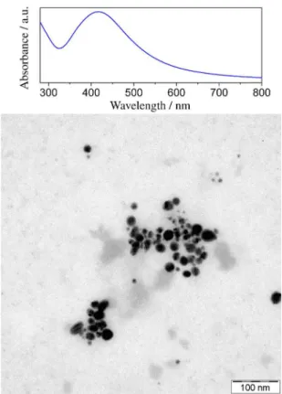
Biological synthesis (or “green chemistry”) of metallic nanoparticles is an interesting method to obtain silver nanoparticles in large scale and low costs, and to obtain particles with high stability.9 In this work, biogenic silver nanoparticles (AgNPbio) with average size of 20 nm were efficiently produced by fungi Fusarium oxyporum
(Figure 1). The AgNPbio production was evidenced by the presence of the plasmon band at 450 nm, as shown in Figure 1. Durán et al.11 described a possible mechanism
When AgNPbio was associated with layered double hydroxides (LDHs), it was verified a high capacity of LDHs to adsorb AgNPbio, as shown in Figure 2 (>80% of adsorption with 250 mg of the LDH gel, which correspond to 153 mg of dry LDH). The insert in Figure 2 presents the LDH-AgNPbio hybrid before and after the centrifugation, showing a colorless supernatant without AgNPs, as confirmed by ICP-OES. This capacity is due to the high surface area of LDHs.3
The interaction between LDH and AgNPbio was energetically very stable, preventing the lixiviation
of AgNPbio even after washing the hybrid suspension for 5 times. This stability manifests from electrostatic interactions between the negatively charged AgNPbio (ζ =−13.2 mV) and positively charged LDHs (ζ = +3.2 mV), resulting in a negative net surface charge on the hybrid LDH-AgNPbio (ζ = – 12 mV). Furthermore, AgNPbio interacted with the LDH surface and was not intercalated among the inorganic layers, since it was observed no significant difference in the interlamellar distance of LDHs without (8.48 Å) and with AgNPbio (8.50 Å), as verified by XRD analysis (Figure 3). This result was expected due to the size of AgNPbio (ca. 20 nm). The SEM image (insert in Figure 4) of LDH-AgNPbio hybrid (obtained with backscattered electrons) shows that LDH surface is covered by AgNPbio (gray regions), confirmed by EDS analysis (Figure 4).
Comparing the FTIR spectra for LDH, AgNPbio and the new hybrid material LDH-AgNPbio, it can be observed
Figure 1. TEM image and UV-Vis spectrum of AgNPbio.
Figure 2. Adsorption profile of the biogenic silver nanoparticles (AgNPbio)
on LDH (insert figure: LDH-AgNPbio dispersion (1) before and (2) after centrifugation).
Figure 4. EDS spectrum of the new hydrid material LDH-AgNPbio
(backscattering image of LDH-AgNPbio).
Figure 3. XRD patterns of LDH and of the new hydrid material
characteristic bands of fungal proteins and characteristic bands of LDH, despite the subtle differences between the spectra of LDH and LDH-AgNPbio. In the AgNPbio and LDH-AgNPbio spectra, it was observed the bands related with fungal proteins around at 955 cm-1, attributed to the wagging mode of a trans-ethylenic moiety, and bands at 1060 and 1031 cm-1, attributed to the N–H stretching mode of the free amino groups of biomacromolecules (Figure 5).19 Further, in the LDH and LDH-AgNP
bio spectra, characteristic bands of LDHs were identified at 1369 cm-1, attributed to the ν
3 vibration mode of CO32−, and the band at 1097 cm-1 is assigned to the ν
3 vibration mode of SO4−3. By combining XRD, FTIR and SEM images data it is possible to confirm that interaction of AgNPbio with LDH occurs by adsorption rather than intercalation of AgNPs in LDHs.
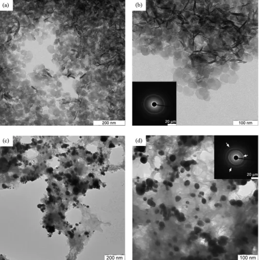
LDH morphology was assessed through transmission electron microscopy (TEM), which indicated the presence of crystalline nanoparticles around 20 nm (Figures 6a and 6b). When adsorbed on LDHs, protein-coated biogenic silver
nanoparticle tend to agglomerate the LDH nanoparticles after the sample drying, as seen in TEM images (Figure 6c and 6d). The diffraction pattern (insert Figure 6d) suggests that AgNPbio in the hybrid material is as monocrystal, exhibiting spots of diffraction relative to a face-centered cubic lattice.19
Figure 5. FTIR spectra of LDH, AgNPbio and LDH-AgNPbio.
Figure 6. TEM images of (a-b) LDH (insert in (b) is the SAED pattern recorded on the LDH), and (c-d) LDH-AgNPbio (insert in (d) is the SAED pattern
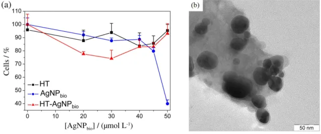
Figure 7. (a) MTT cytotoxicity assay of LDH, AgNPbio and LDH-AgNPbio on fibroblast V79 cells and (b) TEM image of new hybrid material LDH-AgNPbio.
Antibacterial activity and toxicity of the new hybrid material LDH-AgNPbio
The antibacterial activity of LDH, AgNPbio and the hybrid material LDH-AgNPbio was investigated against Gram-positive (S. aureus) and Gram-negative (E. coli)
bacteria. LDH did not exhibit antibacterial activity against both strains tested even at the highest concentration studied (250 µg mL-1). However, LDH-AgNP
bio showed antimicrobial activity with a minimum inhibitory concentration (MIC) of 6.6 and 12 µg mL-1 against S. aures and E. coli, respectively. This MIC values were the same values verified for AgNPbio, demonstrating that the association of AgNPbio with LDH did not modify the antibacterial activity of AgNPbio (Table 1). In this direction, this new hybrid material allows for an efficient interaction between AgNPbio and bacteria, providing the same antibacterial effect compared to free AgNPbio.
About the in vitro toxicity, the LDH-AgNPbio did not exhibit cytotoxic effect on fibroblast cell lines at the concentration range studied (0-50 µmol L-1 of AgNP
bio) as well as pure LDH (Figure 7). However, it was observed approximately 50% of cell mortality for AgNPbio at 45 µmol L-1 (Figure 7). This difference can be attributed to the immobilization of AgNPbio on the LDH surface, as
shown in Figure 7b, suggesting that LDHs can be used as a vehicle for the compatibilization of AgNPbio in medical and cosmetic applications. Some authors described in the literature the dependence of capping stabilizers on AgNP cytotoxicity. Suresh et al.20 studied the cytotoxicity
of AgNP with different surface coatings, and verified that the cytotoxic effect was dependent on the coating molecules. Uncoated AgNP exhibited a lower toxic effect compared to biogenic AgNPs (with a protein coating) and AgNPs covered with oleate, against macrophage and lung epithelial cells. In this way, the lower cytotoxic effect observed for the hybrid material LDH-AgNPbio tested may be explained, by the capacity of LDH to adsorb and neutralize toxins from the biomolecular coating. This effect was observed when antacid hydrotalcite LDH adsorbed and neutralized proteins and toxins secreted by Helicobacter pylori. Possibly, this is an ulcer-healing
action of this inorganic material.21 In this context, the new hybrid material LDH-AgNPbio produced here can decrease the cytotoxic effect of silver nanoparticles, improving the safety of AgNPbio towards a great potential in medical or cosmetic applications.
Conclusions
The present study reports the development of a new hybrid material based on layered double hydroxides (LDHs) and biogenic silver nanoparticles which presents antimicrobial activity with a great potential for application in cosmetics and medicine. Considering its characteristics, the hybrid is suitable for the production of nanocomposites with several combined properties, such as antimicrobial (AgNPbio) and anti-fire (LDH). Furthermore, in contrast to the known toxic effect of AgNPsbio, this material did not show
in vitro cytotoxic effect against fibroblast cells, a result of the Table 1. Minimum inhibitory concentration (MIC) values for AgNPbio,
LDH and for the new hybrid material LDH-AgNPbio (n = 12)
Sample MIC / (µg mL
-1)
S. aureus E. coli
AgNPbio 6.6 12
LDH a a
LDH-AgNPbio 6.6 12
compatibilization capacity of LDHs towards the obtainment of friendly and safe nanomaterials.
Acknowledgments
The authors are grateful to Conselho Nacional de Desenvolvimento Científico e Tecnológico (CNPq) and Fundação de Amparo à Pesquisa do Estado de São Paulo (FAPESP) for financial support. This is a contribution of the National Institute of Science, Technology and Innovation on Complex Functional Materials (Inomat).
References
1. Del Hoyo, C.; Appl. Clay Sci.2007, 36, 103.
2. Gouveia, D. X.; Ferreira, O. P.; Filho, A. G. S.; da Silva, M. G.; de Paiva, J. A. C.; Alves, O. L.; Filho, J. M.; J. Mater. Sci. 2007, 42, 534.
3. Cross, H. E.; Parkes, G.; Brown, D. R.; Appl. Catal., A2012, 24, 429.
4. Carja, G.; Kameshima, Y.; Nakajima, A.; Dranca, C.; Okada, K.; Int. J. Antimicrob. Agents2009, 34, 534.
5. Ferreira, O. P.; Alves, O. L.; Gouveia, D. X.; Filho, A. G. S.; de Paiva, J. A. C.; Filho, J. M.; J. Solid State Chem.2004, 177, 3058.
6. Alves, O. L.; Stéfani, D.; Parizotto, N. V.; Filho, A. G. S.; J. Phys. Conf. Ser.2011, 304, 012024.
7. Takagaki, A. A.; Tsuji, A.; Nishimura, S.; Ebitani, K.; Chem. Lett.2011, 40, 150.
8. Zhu, J.; Zhang, Y.; Shi, F.; Deng, Y.; Tetrahedron Lett.2012, 53, 3178.
9. Durán, N.; Marcato, P. D.; Durán, M.; Yadav, A.; Gade, A.; Rai, M.; Appl. Microbiol. Biotechnol. 2011, 90, 1609.
10. Durán, N.; Marcato, P. D.; Alves, O. L.; De Souza, G. I. H.; Esposito, E.; J. Biomed. Nanotechnol.2007, 3, 203.
11. Durán, N.; Marcato, P. D.; Alves, O. L.; De Souza, G. I. H.; Esposito, E.; J. Nanobiotechnol.2005, 3, 1.
12. Gajbhiye, M.; Kesharwani, J.; Ingle, A.; Gade, A.; Rai, M.; Nanomed. Nanotechnol. Biol. Med. 2009, 5, 382.
13. Ducan, T. V.; J. Colloid Interface Sci.2011, 363, 1.
14. Durán, N.; Marcato, P. D.; De Conti, R.; Alves, O. L.; Costa, F. T. M.; Brocchi, M.; J.Braz. Chem. Soc.2010, 21, 949. 15. De Lima, R.; Seabra, A. B.; Durán, N.; J. Appl. Toxicol.2012,
32, 867.
16. Brazilian National Health Surveillance Agency (Anvisa) with permission to translate from Clinical and Laboratory Standards Institute (CLSI); Metodologia dos Testes de Sensibilidade a Agentes Antimicrobianos por Diluição para
Bactéria de Crescimento Aeróbico, 6a. ed.; Norma M7-A6 Anvisa; Clinical and Laboratory Standards Institute (CLSI): Pennsylvania, EUA, 2003, p. 81.
17. Corrêa, D. H. A.; Melo, P. S.; Carvalho, C. A. A.; Azevedo, M. B. M.; Durán, N.; Haun, M.; Eur. J. Pharmacol.2005, 510, 17.
18. Denizot, F.; Lang, R.; J. Immunol. Methods1986, 89, 271. 19. Mukherjee, P.; Roy, M.; Mandal, B. P.; Dey, J. K.; Mukherjee,
P. K.; Ghatak, J.; Tyagi, A. K.; Kale, S. P.; Nanotechnology 2008, 19, 1.
20. Suresh, A. K.; Pelletier, D. A.; Wang, W.; Morrell-Falvey, J. L.; Gu, B.; Doktyc, M. J.; Langmuir2012, 28, 2727.
21. Tarnawski, A.; Pai, R.; Itani, R.; Wyle, F. A.; Digestion1999, 60, 449.
Submitted: October 16, 2012
Published online: February 14, 2013