Article
Printed in Brazil - ©2013 Sociedade Brasileira de Química0103 - 5053 $6.00+0.00
A
*e-mail: [email protected]
A Fast Sonochemical Method to Prepare 1D and 3D Nanostructures of Bismuth Sulfide
Paulo R. R. Mesquita,a Jorge S. Almeida,a Leonardo S. G. Teixeira,a,b Antônio F. da Silvac and Luciana A. Silva*,a,b
aInstituto de Química, Universidade Federal da Bahia, Campus Ondina,
40170-290 Salvador-BA, Brazil
bInstituto Nacional de Ciência e Tecnologia, INCT de Energia e Ambiente,
Universidade Federal da Bahia, Campus Ondina, 40170-290 Salvador-BA, Brazil
cInstituto de Física, Universidade Federal da Bahia, Campus Ondina,
40170-290 Salvador-BA, Brazil
Nesse trabalho, um método sonoquímico de síntese de nanoestruturas de sulfeto de bismuto em 1D e 3D foi desenvolvido e comparado com uma rota sintética empregando aquecimento sob refluxo. O método sonoquímico monstrou ser mais rápido e eficiente na obtenção de nanoestruturas com alta homogeneidade morfológica. A forma e qualidade dos nanocristais foram dependentes do tipo de solvente empregado na síntese. Superestruturas em 3D semelhantes a flores foram obtidas quando etileno glicol puro foi utilizado como solvente, enquanto estruturas em 1D na forma de nanobastões foram obtidas quando utilizada uma mistura de dimetilsulfóxido e etileno como solvente.
In this work, a sonochemical method to synthesize nanostructures of bismuth sulfide in 1D and 3D framework was developed and compared with a synthetic route with heating under reflux. The sonochemical method showed to be faster and more efficient than refluxing method to obtain nanostructures with high morphological homogeneity. Form and quality of the nanocrystals were dependent on the type of solvent employed in the synthesis procedure. 3D flower-like superstructures were obtained when ethylene glycol was used as solvent, while 1D nanorods were obtained when a mixture of dimethyl sulfoxide and ethylene glycol was used as solvent.
Keywords: bismuth sulfide, sonochemical method, nanomaterials
Introduction
During the last decades, a lot of attention was devoted to study nanocrystalline materials due to their unusual
properties and potential applications.1-5 However, the
control over particle size and its morphology is still a challenge to science. In semiconductor area, bismuth
sulfide (Bi2S3) has been considered a promising material
to be applied in the field of photoelectricity, sensors and thermoelectricity since its direct band gap (in range of 1.3-1.7 eV) can be tuned by different particle size and
shape, resulting in different properties.6,7
Traditionally, Bi2S3 is synthesized by reaction of
bismuth and sulfur vapor in a quartz flask under high
temperature.8 Several methods have been developed
to prepare Bi2S3 with or presenting different size and
morphology, such as thermal decomposition,9 hydrothermal or
solvothermal methods,10,11 biomolecule-assisted pathways,12
microwave irradiation,13,14 evaporation method15 and ionic
liquid-assisted.16 Nevertheless, these methods usually spend
too much time and use high temperature and pressure, besides most procedures are complex. In this direction, the development of fast and simple methods to prepare bismuth sulfide nanoparticles is still required. Ultrasonic waves have been successfully used in the synthesis of new materials since they provide smaller particle size and higher surface
area than reported by other methods.17
Sonochemistry uses high power ultrasonic waves
(20 kHz-10 MHz) to promote chemical reactions.18,19 The
high purity. Despite the potential of sonochemical method in the synthesis of nanomaterials, there is a lack of studies
concerning the preparation of Bi2S3 nanostructures using
this method. Wang et al.20 were the only ones who reported
the application of sonochemical method in the synthesis
of Bi2S3 nanostructures. In that work, Bi2S3 nanorods were
prepared from bismuth nitrate aqueous solution and sodium thiosulfate in the presence of a complexing agent, with ultrasonic irradiation for 2 h.
Structures with specific morphology are of great interests due to their physical and chemical properties and potential applications in designing new materials and devices. Thus, it is reported herein a simple sonochemical
method to synthesize 1D and 3D Bi2S3 nanostructures,
using different solvents and 15 min of reaction time, and the method is compared with a refluxing route. All products were characterized by X-ray powder diffractometry (XRD), scanning electron microscopy (SEM) and transmission electron microscopy (TEM).
Experimental
Materials
All the reagents used in our experiments were of analytical purity and were used without further
purification. Bi(NO3)3.5H2O, sodium thiosulfate and
cetyltrimethylammonium bromide (CTAB) were purchased from Vetec. Ethylene glycol (EG) and dimethyl sulfoxide (DMSO) were purchased from Synth. Acetylacetone was purchased from Aldrich.
Synthesis of nano-sized Bi2S3 by sonochemical method
(S1-S3)
In a typical experiment, 1.89 g (3.9 mmol) of
Bi(NO3)3.5H2O, 3.70 g (15.5 mmol) of Na2S2O3 and 0.18 g
(0.5 mmol) of CTAB were added to 50 mL of different solvents. This mixture was stirred at 50 ºC until all chemicals were dissolved. Then, the solution was exposed to high-intensity ultrasound irradiation using a ultrasonic waves source UP400S, Hielscher. The parameters were
the following: 24 kHz, 400 W cm-2, 4 cm2, titanium direct
immersion horn, at 20% of amplitude and 80% of pulse of duty cycle wave under ambient air for 15 min. When the reaction was finished, black precipitates were obtained. After being cooled to room temperature, the precipitates were centrifuged, washed with distilled water, absolute ethanol, and acetone in sequence, and dried in air at room temperature.
Synthesis of nano-sized Bi2S3 by refluxing method (R1)
In a typical experiment, 1.89 g (3.9 mmol) of
Bi(NO3)3.5H2O, 3.70 g (15.5 mmol) of Na2S2O3 and 0.18 g
(0.5 mmol) of CTAB were added to a round-bottom flask which contained 50 mL of ethylene glycol. The mixture was stirred until all the chemicals were well-dispersed. This mixture was heated up to 125 ºC under constant stirring and was kept under reflux in oil bath for 90 min. When the reaction was finished, black precipitates were obtained. After being cooled to room temperature, the precipitates were centrifuged, washed with distilled water, absolute ethanol, and acetone in sequence, and dried in air at room temperature.
Table 1 summarizes the experimental conditions for preparation of the nanomaterials by refluxing and sonochemical routes.
Characterization
Nano-sized Bi2S3 obtained were characterized by
X-ray powder diffraction (XRD), scanning electron microscopy (SEM) and transmission electron microscopy (TEM). X-ray powder diffraction analyses were carried
out on Shimadzu XRD 6000 diffractometer, using Cu Kα
(λ = 1.5418 Å). The scanning rate is 1 degree min-1
in the 2θ range of 10-80º. SEM images were taken in
JSM-6610LV scanning electron microscopy (JEOL) operated at 20 kV. TEM images were recorded on a FEI Tecnai transmission electron microscope, operating at 200 kV. The samples used for TEM observations were prepared by dispersing some products in ethanol followed by ultrasonic treatment for 5 min and then placing a drop of dispersion onto copper grid coated with a layer of amorphous carbon.
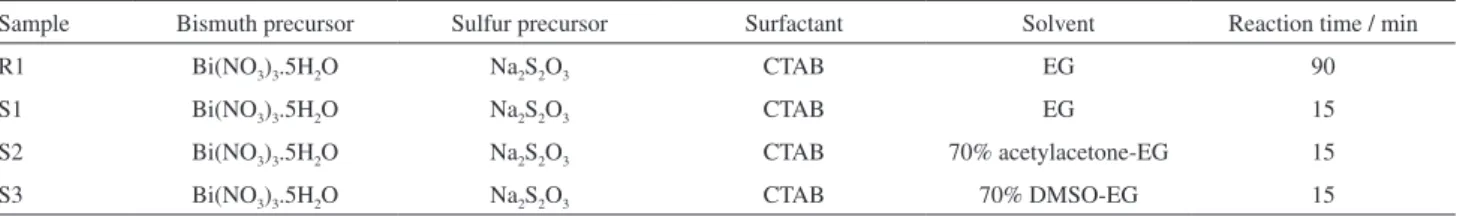
Table 1. Experimental conditions for the preparation of nano-sized Bi2S3
Sample Bismuth precursor Sulfur precursor Surfactant Solvent Reaction time / min
R1 Bi(NO3)3.5H2O Na2S2O3 CTAB EG 90
S1 Bi(NO3)3.5H2O Na2S2O3 CTAB EG 15
S2 Bi(NO3)3.5H2O Na2S2O3 CTAB 70% acetylacetone-EG 15
Results and Discussion
Figure 1 shows the X-ray diffraction patterns of the samples prepared from refluxing (R1) and sonochemical (S1) methods. All the diffraction peaks in the X-ray diffraction patterns for both samples can be indexed to
the orthorhombic phase of Bi2S3 (JCPDS 17-0320). The
diffraction peaks in XRD pattern from the R1 sample (Figure 1b) are sharper than those in XRD pattern from the S1 sample (Figure 1a), suggesting higher crystallinity of the sample obtained by refluxing method. On the other hand, broader peaks in X-ray diffraction patterns of the sample obtained from sonochemical method could be an evidence of smaller particle sizes. The crystallite size can be estimated from XRD data by the Scherrer formula:
, where λ is the X-ray wavelength of 1.5418 Å,
θ is the Bragg diffraction angle and B is the full width at half
maximum (FWHM) of 2θ.21,22 Crystallite sizes calculated
using the widths of Bi2S3 (130) reflecting plane are very
similar for both samples, 11.8 nm for S1 sample and 13.9 nm for R1 sample, suggesting that the difference in XRD patterns profile is due to an effect of crystallinity.
The morphology of Bi2S3 prepared by refluxing and
sonochemical methods, R1 and S1, respectively, was investigated by SEM imaging. Figure 2 shows that in both cases flower-like superstructures are formed. However, sonochemical method yields flower-like superstructures with better morphological homogeneity (Figures 2c and 2d) when compared with refluxing method (Figures 2a and 2b). Figure 3 shows TEM images of the products obtained from sonochemical and refluxing methods. TEM images
showed in Figure 3a corroborate the flower-like Bi2S3
superstructures formation with diameter around of 500 nm.
Figure 3b indicates that flower-like Bi2S3 superstructures
are formed by rods with diameter of 11-15 nm. Analyzing
Figure 3c, which shows TEM image of Bi2S3 obtained
by refluxing method, it is possible to see an aggregation of particles with predominance of rods larger than those
obtained from sonochemical method. Lu et al.23 reported a
similar morphology of Bi2S3 prepared using Bi(NO3)3 and
thiourea in ethylene glycol by microwave irradiation method.
To understand the role of solvents play in the synthesis
of flower-like Bi2S3 superstructures, nano-sized Bi2S3
was prepared by sonochemical method in the presence of Figure 1. Powder XRD patterns of nano-sized Bi2S3 obtained by
(a) refluxing method at 90 min (R1) and (b) sonochemical method at 15 min (S1).
Figure 2. SEM images of the products obtained for (a) and (b) refluxing method (R1); (c) and (d) sonochemical method (S1).
different solvents, such as EG (S1), 70% acetylacetone-EG (S2) and 70% DMSO-EG (S3), keeping the same experimental conditions.
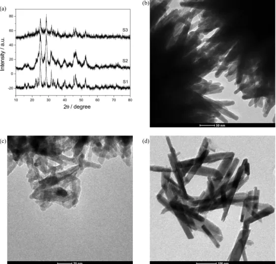
Figure 4a displays the XRD patterns of nano-sized Bi2S3
prepared using different solvents (S1-S3). In each case,
Bi2S3 formation can be confirmed by comparison with the
pattern JCPDS 17-0320. No peaks of any other phases are detected, indicating the high purity of the products.
The morphologies of the samples S1-S3 were depicted by TEM images. As shown in Figure 4b, a large quantity of
Bi2S3 nanorod aggregates in 3D flower-like superstructures
was obtained when EG was used as a solvent (S1). However, smaller rod-like nanoparticles were obtained in acetylacetone-EG (S2) showed in Figure 4c, while 1D nanorods were obtained in DMSO-EG (S3), as can be seen in Figure 4d. The above results showed that the blend of
DMSO-EG induces the formation of Bi2S3 nanorods (1D),
without formation of flower-like superstructures (3D). With the procedure proposed in this work, it is possible to obtain nanomaterials in only 15 min with morphological
homogeneity similar to nanostructures obtained by
Wang et al.20 that spends 2 h in the synthesis procedure.
Conclusions
A fast and simple sonochemical method for the
preparation of nano-sized Bi2S3 was developed. 3D
flower-like Bi2S3 superstructures can be successfully
prepared via a sonochemical route from an EG solution of bismuth nitrate and sodium thiosulfate in the presence of CTAB, while the use of DMSO-EG mixture leads to the
formation of 1D Bi2S3 nanorods. The developed method is
a fast, simple, convenient and efficient route for preparing
Bi2S3 nanomaterials and it can be a good alternative to
control the morphology.
Acknowledgements
The authors acknowledge the Brazilian research funding agencies Conselho Nacional de Desenvolvimento Científico Figure 4. Powder XRD patterns (a) and TEM images of nano-sized Bi2S3 prepared by sonochemical method in different solvents: (b) EG (S1),
e Tecnológico (CNPq) and Fundação de Amparo à Pesquisa do Estado da Bahia (FAPESB) for financial support. The authors are also thankful to Centro de Tecnologias Estratégicas do Nordeste (CETENE) for the TEM analyses and Laboratório Multi-Usuário de Microscopia Eletrônica da UFBA (LAMUME) for the SEM analyses.
References
1. Feldheim, D. L.; Keating, C. D.; Chem. Soc. Rev.1998, 27, 1.
2. Rincón, M. E.; Suárez, R.; Nair, P. K.; J. Phys. Chem. Solids 1996, 57, 1947.
3. Colvin, V. L.; Schlamp, M. C.; Alivisatos, A. P.; Nature1994,
370, 354.
4. Klein, D. L.; Roth, R.; Lim, A. K. L.; Alivisatos, A. P.; McEuen, P. L.; Nature1997, 389, 699.
5. Shiang, J. J.; Kadavanich, A. V.; Grubbs, R. K.; J. Phys. Chem. 1995, 99, 17417.
6. Nayak, B. B.; Acharya, H. N.; Mitra, G. B.; Mathur, B. K.; Thin Solid Films1983, 105, 77.
7. Farrugia, L. J.; Lawlot, F. J.; Norman, N. C.; Polyhedron 1995,
14, 311.
8. Kaito, C.; Saito, Y.; Fujita, K.; J. Cryst. Growth1989, 94, 967. 9. Shen, X. P.; Yin, G.; Zhang, W. L.; Xu, Z.; Solid State Commun.
2006, 140, 116.
10. Li, L.; Cao, R.; Wang, Z.; Li, J.; Qi, L.; J. Phys. Chem. C2009, 113, 18075.
11. Wei, F.; Zhang, J.; Wang, L.; Zhang, Z. K.; Cryst. Growth Des. 2006, 6, 1942.
12. Sigman, M. B.; Korgel, B. A.; Chem. Mater.2005, 17, 1655. 13. Liao, X. H.; Wang, H.; Zhu, J. J.; Chen, H. Y.; Mater. Res. Bull.
2001, 36, 2339.
14 Wu, J.; Qin, F.; Cheng, G.; Li, H.; Zhang, J.; Xie, Y.; Yang, H. J.; Lu, Z.; Yu, X.; Chen, R.; J. Alloys Compd.2011, 509, 2116.
15. Ye, C.; Meng, G.; Jiang, Z.; Wang, Y.; Wang, G.; Zhang, L.;
J. Am. Chem. Soc.2002, 124, 15180.
16. Jiang, J.; Yu, S. H.; Yao, W. T.; Ge, H.; Zhang, G. Z.; Chem. Mater.2005, 17, 6094.
17. Gedanken, A.; Ultrason. Sonochem.2004, 11, 47.
18. Suslick, K. S.; Hyeon, T. W.; Fang, M. W.; Chem. Mater.1996, 8, 2172.
19. Suslick, K. S.; Choe, S. B.; Cichowlas, A. A.; Grinstaff, M. W.;
Nature1991, 353, 414.
20. Wang, H.; Zhu, J. J.; Zhu, J. M.; Chen, H. Y.; J. Phys. Chem. B 2002, 106, 3848.
21. Almeida, C. G.; Andrade, H. M. C.; Mascarenhas, A. J. S.; Silva, L. A.; Mater. Lett.2010, 64, 1088.
22. Guimarães, T. B. F.; Pepe, I.; Ferreira da Silva, A.; Mangrich, A. S.; de Andrade, J. B.; Silva, L. A. J. Alloys Compd.2009,
481, 654.
23. Lu, J.; Han, Q.; Yang, X.; Lu, L.; Wang, X.; Mater. Lett.2007,
61, 2883.
Submitted: October 16, 2012