Article
Printed in Brazil - ©2013 Sociedade Brasileira de Química0103 - 5053 $6.00+0.00A
*e-mail: [email protected], [email protected]
Tautomerism in Quinoxalines Derived from the 1,4-Naphthoquinone Nucleus:
Acid Mediated Synthesis, X-ray Molecular Structure of
5-Chlorobenzo[
f
]quinoxalin-6-ol and Density Functional Theory Calculations
Javier A. G. Gomez, Mateus R. Lage, José Walkimar de M. Carneiro,* Jackson A. L. C. Resende and Maria D. Vargas*
Instituto de Química, Universidade Federal Fluminense, Campus do Valonguinho, Centro, 24020-141 Niterói-RJ, Brazil
A reação de tert-butil 2-(3-cloro-1,4-dioxo-1,4-di-hidronaftalen-2-ilamino)etilcarbamato
com CF3COOH/CH2Cl2 fornece 5-cloro-3,4-di-hidrobenzo[f]quinoxalin-6(2H)-ona. Este composto sofre desidrogenação promovida por ácido na presença de água para dar a 5-clorobenzo[f]quinoxalin-6-ol, inédita. A estrutura molecular no estado sólido, determinada por
um estudo de difração de raios X (XRD), e os dados em solução confirmam que a benzoquinoxalina existe na forma do tautômero enol-imina, tanto no estado sólido, quanto em solução, diferentemente de 5-cloro-3,4-di-hidrobenzo[f]quinoxalin-6(2H)-ona que exibe o arranjo ceto-amino. Cálculos de teoria do funcional da densidade (DFT) confirmam a preferência da benzoquinoxalina e dos compostos análogos contendo grupos H ou CH3 no lugar do Cl pela forma enol-imina. Sugere-se que a preferência da benzoquinoxalina pela estrutura enol-imina se deva ao maior caráter aromático desta estrutura em comparação com a forma ceto-amina. Os cálculos DFT dos dois tautômeros das benzo[a]fenazin-5(7H)-onas análogas às benzo[f]quinoxalin-6(4H)-onas indicaram que as estabilidades relativas são dominadas por efeitos de solvatação, no primeiro caso, e pelo grau de aromaticidade no segundo.
The reaction of tert-butyl 2-(3-chloro-1,4-dioxo-1,4-dihydronaphthalen-2-ylamino)
ethylcarbamate with CF3COOH/CH2Cl2 yields 5-chloro-3,4-dihydrobenzo[f ]quinoxalin-6(2H)-one which undergoes acid-promoted dehydrogenation in the presence of water to give novel 5-chlorobenzo[f]quinoxalin-6-ol. The molecular structure of 5-chlorobenzo[f
]quinoxalin-6-ol in the solid state, determined by an X-ray diffraction (XRD) study, and the solution data confirm that it exists as the enol-imine tautomer, both in the solid state and in solution, differently from 5-chloro-3,4-dihydrobenzo[f]quinoxalin-6(2H)-one, which exhibits the keto-amine
arrangement. Density functional theory (DFT) calculations confirmed the preference of 5-chlorobenzo[f]quinoxalin-6-ol and of the derivatives containing H and CH3 groups in place of the Cl atom for the enol-imine tautomer. It is suggested that the enol-imine structure is preferred for 5-chlorobenzo[f]quinoxalin-6-ol as a consequence of the higher aromatic character of this
structure in comparison with the keto-amine form. DFT calculations carried out on the two tautomers of the benzo[a]phenazin-5(7H)-ones analogous to the benzo[f]quinoxalin-6(4H)-ones
showed that the relative stabilities are dominated by solvation effects in the first case and the degree of aromaticity, in the latter.
Keywords: quinoxalines, 1,4-naphthoquinones, tautomerism, X-ray structure, DFT calculations
Introduction
Quinoxalin derivatives have been the subject of intensive industrial and academic investigation due to their importance as intermediates for the synthesis of pharmaceuticals and new materials. This nucleus is a
privileged molecular scaffold, present in a large number of molecules with a wide spectrum of biological activities,1
e.g., trypanocidal,2,3 antimalarial,4,5 antitubercular,6,7
antimicrobial,8 antitumoral,9 anti-proliferative,10,11
antithrombotic,12 anti-HIV,13 among others. Quinoxalines
have been investigated as luminescent materials,14,15
fluorescent probes16 and as constituents of donor-acceptor
ability has been used for the preparation of molecular sensors18,19 and hybrid coordination polymers.20
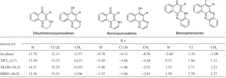
Of interest to this paper are the quinoxalines derived from the 1,4-naphthoquinone nucleus which include
the benzophenazines reported by Silva Junior et al.
(Figure 1a, a21 and the dihydrobenzoquinoxalines
(Figure 1b), studied extensively by Kallmayer and Seyfang.25-34 The benzophenazines shown in Figure 1a
have been obtained from the reactions of phenylenediamine with 2-hydroxy-3-R-1,4-naphthoquinones in AcOH, under heating, in the presence (a-f)21,22 or absence of
AcONa (g).23 Unusual dimeric phenazines h have been
synthesized in high yields by reacting lapachol (2-hydroxy-3-(3-methyl-2-butenyl)-1,4-naphthoquinone) with neat alkylamines.24 The reactions of ethylene diamines or
1,2-cyclohexyldiamine34 with 1,4-naphthoquinone
(R = H), 2,3-dibromo or dichloro-1,4-naphthoquinone (R = Cl or Br), 2,3-phthalimido-1,4-naphthoquinone (R = NHAc, NH2)29 and 2-alkyl or
2-aryl,3-halogen-1,4-naphthoquinones, in CH2Cl2/ ethanol,26 have yielded
the dihydrobenzoquinoxalines shown in Figure 1b. Following our continuing interest in aminonaphtho-quinones35-38 and their metal complexes39-41 as potential
antimicrobial and anticancer agents, our group has investigated the possibility of synthesizing molecular hybrids from 2,3-dichloro-1,4-naphthoquinone and N-Boc-monoprotected diamines (NH2(CH2)nNHBoc), whose deprotection would be followed by further extension of the diamine chain with nitrogen or oxygen containing fragments for coordination with metal ions. This tactic has proven successful for the diamines with a linear carbon chain longer than n = 3,42 and the results will be
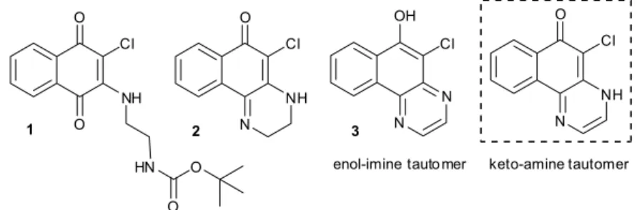
reported elsewhere. Under the conditions employed for the deprotection of the NH2(CH2)2NHBoc derivative 1, however,
5-chloro-3,4-dihydrobenzo[f]quinoxalin-6(2H)-one 226
was obtained instead, together with the dehydrogenated compound 5-chlorobenzo[f]quinoxalin-6-ol 3 (Figure 2).
5-R-3,4-Dihydrobenzo[f]quinoxalin-6(2H)-ones (R = CH3, OCH3, CN and NHAc, Figure 1b) were reported previously
to undergo base and acid promoted oxidation to the respective benzoquinoxalin-6-ol derivatives analogous to 3, in good yields and trace amounts, respectively.43
The benzophenazines and dihydrobenzoquinoxalines illustrated in Figure 1 in their keto-amine form could, in principle, exist as the enol-imine tautomer observed for 3
(Figure 2) and analogous benzoquinoxalines.44
Herein we describe a novel route to the acid mediated oxidation of 2 and the solution and solid state characterization
of 5-chlorobenzo[f]quinoxalin-6-ol 3, including an X -ray
diffraction (XRD) analysis. We also report the results of density functional theory (DFT) calculations to determine the relative stabilities of the two tautomers of quinoxalines related to 2 and 3 containing different substituents in place
of Cl, and of benzophenazines b, d and g (Figure 1a).
Experimental
General methods
2,3-dichloro-1,4-naphthoquinone (Aldrich), 1,2-ethano-diamine (Aldrich), di-tert-butyl-dicarbonate (Aldrich), trifluoroacetic acid (Aldrich), MeCN, EtOAc, hexane, EtOH (Vetec), CH2Cl2, Na2SO4 and NaHCO3 (Vetec) were
used as received. tert-Butyl N-(2-aminoethyl)carbamate
was prepared as described in the literature for tert-butyl N-(3-aminopropyl)carbamate.45 All solvents were removed
under reduced pressure. The reactions were monitored by thin layer chromatography (TLC) analysis on silicagel 60 F254 TLC plates with detection by UV absorption (254 nm).
Column chromatography (CC) was performed using Acros Organics silica gel (35-70 µm) as the stationary phase and the solvent systems indicated in each experiment. Melting points (mp) were obtained on a ThermoFisher Scientific Digital Melting Point IA9100 apparatus. Infrared spectra were recorded as thin films using a Varian 660 FTIR spectrometer equipped with an attenuated total reflectance (ATR) sampling accessory, and the spectral data are reported in wavenumbers (cm-1). Nuclear magnetic resonance (NMR) spectra were
acquired in CDCl3, DMSO-d6 or CD3OD as indicated
using a Varian VNMRS 300 MHz or Varian VNMRS 500 MHz spectrometer, where 1H and 13C were measured
at 500/300 and 125 MHz, respectively. 1H NMR spectra are
referenced to the CDCl3 (d 7.26 ppm), DMSO-d6 (d 2.50
ppm) and CD3OD (d 3.31 ppm) residual solvent peaks. The
hydrogen signals were attributed through coupling constant values and 1H × 1H COSY (correlation spectroscopy)
experiments. 13C NMR spectra were proton decoupled and
referenced to CDCl3 (d 77.0 ppm) and DMSO-d6 (d 39.4
ppm) residual solvents peaks.
Synthesis of tert-butyl
2-(3-chloro-1,4-dioxo-1,4-dihydro-naphthalen-2-ylamino)ethylcarbamate (1)
Compound 1 was prepared from
2,3-dichloro-1,4-naphtho quinone and tert-butyl N-(2-aminoethyl) carbamate according to literature procedures42,46 to give a
red solid, 70%, mp 131 °C; anal. calcd. for C17H19N2O4Cl:
C, 58.21; H, 5.46; N, 7.99; found: C, 58.15; H, 5.70; N,
7.97%; 1H NMR (500 MHz, CDCl
3) d 1.44 (s, 9H, H13),
3.45 (br q, 2H, J 5.0 Hz, H12), 3.97 (br q, 2H, J 5.0 Hz, H11), 4.86 (br, 1H, NH), 6.50 (br, 1H, NH), 7.61 (td, 1H, J 1.3, 7.6 Hz, H6/H7), 7.71 (td, 1H, J 1.3, 7.6 Hz, H6/H7), 8.01 (dd, 1H, J 0.9, 7.7 Hz, H5/H8), 8.13 (dd, 1H, J 0.9, 7.7 Hz, H5/H8); IR (thin film) ν/cm-1 3373 (N-H, amide),
3338 (N-H, amine), 1669 (C=O) and 1648 (C=O).
Synthesis of 5-chloro-3,4-dihydrobenzo[f
]quinoxalin-6(2H)-one(2)26
To a stirred solution of tert-butyl
2-(3-chloro-1,4-dioxo-1,4-dihydronaphthalen-2-ylamino)ethylcarbamate (1,
0.70 g, 2.0 mmol) in 5 mL of CH2Cl2 cooled in an ice
bath, was added trifluoroacetic acid (0.77 mL, 1.14 g, 10.0 mmol), and the resulting wine red solution was stirred at room temperature in a closed system for 24 h. After evaporation to dryness a saturated Na2CO3 solution was
added and the mixture was washed with EtOAc (40 mL). The organic layer was separated and the aqueous layer
was extracted with EtOAc (3 × 40 mL). The combined
organic extracts were dried (anhydrous Na2SO4) and the
solvent, evaporated. The residue was purified by CC (hexane/EtOAc, 10/1 to 2/1, v/v) to give an orange solid, 0.40 g, 86%, ; anal. calcd. for C12H9N2OCl.0.25C4H8O2:
C, 61.31; H, 4.35; N, 11.00; found: C, 61.79; H, 4.42; N, 11.02%, mp 128 °C (lit. 135-138 oC, C
12H9N2OCl);26
1H NMR (500 MHz, CDCl
3) d 3.50 (td, 2H, J 2.6, 6.5 Hz,
H11), 4.19 (t, 2H, J 6.5 Hz, H12), 7.57 (m, 2H, H5 e H8),
8.15 (m, 1H, H6/H7), 8.18 (m, 1H, H6/H7); 13C NMR
(125 MHz, CDCl3) d 37.2, 48.4, 109.0, 123.8, 126.1,
131.1, 131.2, 139.5, 152.2, 176.3; IR (thin film) ν/cm-1
3324 (N-H), 1737 (C=O), 1575 (C=C). Solubility: DMSO, acetone, methanol, ethanol, CH2Cl2, CHCl3 and hexane;
partially soluble in water and EtOAc.
Synthesis of 5-chlorobenzo[f]quinoxalin-6-ol (3)
Prepared from tert-butyl
2-(3-chloro-1,4-dioxo-1,4-dihydronaphthalen-2-ylamino)-ethylcarbamate (1,
1.50 g, 4.3 mmol) in 10 mL of CHCl2 and trifluoroacetic
acid (1.64 mL, 2.43 g, 21.5 mmol) according to the procedure described above. Evaporation to dryness was
followed by addition of EtOAc (2 × 20 mL) and the
insoluble residue discarded. The solution was then washed with water (50 mL), the organic layer was separated and the aqueous layer was extracted with EtOAc (4 × 40 mL).
purified by CC (hexane/EtOAc from 10:1 to 2:1) to give a pale yellow solid, 0.49 g, 46%, mp 216 °C; anal. calcd. for C12H7N2OCl.0.25H2O: C, 61.31; H, 4.35; N, 11.00; found:
C, 61.79; H, 4.42; N, 11.02%; 1H NMR (300 MHz, CDCl 3)
d 7.82 (m, 2H, H11-H12), 8.37 (m, 1H, H6-H7), 8.83 (d, 1H, J 2.1 Hz, H5-H8), 8.93 (d, 1H, J 2.1 Hz, H5-H8), 9.18
(m, 1H, H6-H7); 13C NMR (125 MHz, CDCl
3) d 109.7,
122.5, 127.1, 128.3, 129.2, 129.3, 137.1, 139.9, 141.1, 144.7, 151.0; IR (thin film) ν/cm-1 3199 (O-H), 1596 (C=C).
Solubility: DMSO, acetone, methanol, EtOAc, hexane and CH2Cl2, partially soluble in H2O and CHCl3.
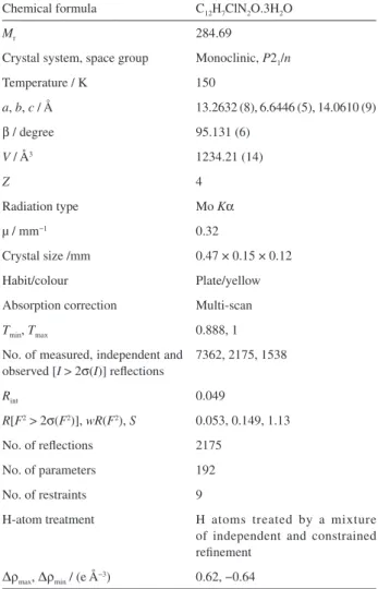
Crystallography: molecular and crystal structure of 5-chlorobenzo[f]quinoxalin-6-ol (3)
XRD data were collected at 150 K on an Agilent Xcalibur Atlas Gemini Ultra diffractometer with Mo Kα radiation (see Table 1). Data processing (including integration, scaling and absorption correction) was performed using CrysAlisPro software.47 The structure was solved using SHELXS-97 and
refined on F2 with SHELXL 97.48 All the non-hydrogen
atoms were refined anisotropically and the hydrogen atoms were refined using a riding model. The aqua hydrogens were located in Fourier difference maps and the benzoquinoxaline hydrogens were generated geometrically.
Calculations
The structures of the quinoxalines were fully optimized using the B3LYP functional49,50 and the 6-311++G(d,p)
basis set. Solvation effects were included using the polarizable continuum model with the CPCM method.51,52
All the optimized structures were confirmed as a local minimum on the potential energy surface by calculation of the Hessian matrix force constant (no negative eigenvector). All calculations were performed with the Gaussian 09W software.53
Results and Discussion
Synthesis and characterization of compounds 1-3
The reaction of 2,3-dichloro-1,4-naphthoquinone and tert-butyl N-(2-aminoethyl)carbamate under the conditions described in the literature for other amines42,46 yielded novel
tert-butyl
2-(3-chloro-1,4-dioxo-1,4-dihydronaphthalen-2-ylamino)ethylcarbamate 1 (70% after purification
by CC) as a red solid, soluble in common organic solvents but insoluble in water. It was characterized by analytical and spectroscopic data (see Experimental section and Figures S1-S3 in the Supplementary
Information (SI) section). The 1H NMR spectrum
(CDCl3) exhibits the expected peaks due to the carbamate
methyl groups at d 1.44, the methylene hydrogens as
two broad quartets at d 3.97 and 4.86 that result from the couplings with the respective neighboring NH hydrogens. The latter appear as broad peaks at d 4.86 and 6.50. The four signals in the d7.61-8.13 region are due to the naphthoquinone ring hydrogens. The IR spectrum shows the N-H amide (3373 cm-1) and amine (3338 cm-1)
bands, a broad intense naphthoquinone carbonyl band (1669 cm-1) and an unusually weak band associated with
the carbamate C=O stretching (1648 cm-1) possibly due to
the formation of a N-H…O (carbamate) hydrogen bond, as observed in the X-ray structure of the analogous compound derived from tert-butyl N-(3-aminopropyl)carbamate.42
Under the conditions employed for N-Boc deprotection (CF3COOH/CH2Cl2), the initial orange solution of
compound 1 immediately turned purple. It was left
under stirring in a closed flask for 24 h. Depending on the conditions employed for the treatment of the
Table 1. Structural data and refinement parameters for 3
Chemical formula C12H7ClN2O.3H2O
Mr 284.69
Crystal system, space group Monoclinic, P21/n
Temperature / K 150
a, b, c / Å 13.2632 (8), 6.6446 (5), 14.0610 (9)
β / degree 95.131 (6)
V / Å3 1234.21 (14)
Z 4
Radiation type Mo Kα
µ / mm−1 0.32
Crystal size /mm 0.47 × 0.15 × 0.12
Habit/colour Plate/yellow
Absorption correction Multi-scan
Tmin, Tmax 0.888, 1
No. of measured, independent and observed [I > 2σ(I)] reflections
7362, 2175, 1538
Rint 0.049
R[F2 > 2σ(F2)], wR(F2), S 0.053, 0.149, 1.13
No. of reflections 2175
No. of parameters 192
No. of restraints 9
H-atom treatment H atoms treated by a mixture of independent and constrained refinement
dark purple solid obtained after solvent evaporation, 5-chloro-3,4-dihydrobenzo[f]quinoxalin-6(2H)-one 2 and
5-chlorobenzo[f]quinoxalin-6-ol 3 were isolated in varying
yields (Scheme 1). Thus, treatment of the dark purple solid with saturated Na2CO3(aq) resulted in CO2 evolution and
formation of known orange dihydrobenzoquinoxaline 2,26
which was extracted with EtOAc, purified by CC and obtained in 86% yield. Under these conditions, trace amounts of quinoxaline 3 were also isolated from the column. When
the purple EtOAc solution was treated with water instead, the solution color turned dark yellow. Purification of the organic extracts by CC as before gave pale yellow benzoquinoxaline 3
in up to 46% yield, besides 2.
Both compounds have been fully characterized by analytical and spectroscopic data (see Experimental and SI sections), that confirmed their identity, and the novel compound 3 was also characterized by a single crystal
XRD study.
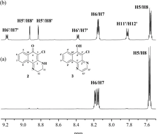
The 1H NMR data for dihydrobenzoquinoxaline 2
(Figures S5-S7 in the SI section) are in agreement with those previously reported26 and confirm that the compound
exists as the keto-amine tautomer, both in CDCl3 and in
CD3OD. The 13C NMR spectrum of 2 (CDCl3, Figure S8
in the SI section) exhibits all the expected signals, those corresponding to C4 (C=O) and C1 (C=N), at d 176.3 and 152.2, respectively. The ATR-FTIR (attenuated total reflectance Fourier transform infrared spectroscopy) spectrum of 2 (Figure S4 in the SI section)indicates similar
structure in the solid state. It shows the amine νN-H band,
at 3324 cm-1, and the quinoxaline ν
CO low intensity band,
at 1604 cm-1, at lower frequency than previously reported
(1635 cm1-)26 probably due to intermolecular hydrogen
bonding. The presence of EtOAc (νCO = 1737 cm-1) was
confirmed by elemental analysis.
The 1H NMR spectrum of benzoquinoxaline 3 in CDCl 3
(Figures S10 and S11 in the SI section) shows hydrogens
H11 and H12 as a multiplet which is actually a partially superimposed pair of doublets at d 7.82; H6/H7 appear at d 8.37 and 9.18 as multiplets, and H5/H8 appear at d
8.83 and 8.93 as doublets. A peak around d 6.7, attributed to the OH hydrogen, is absent in the spectra in CD3OD and
DMSO-d6 (Figure S12 and S13 in the SI section). The 13C
NMR (APT) spectrum of 3 in DMSO-d6 (Figure S14 in the SI section) shows all the expected peaks and no evidence for the presence of the keto-amine tautomer in solution. The OH bonded to C4 appears at d 151.0, typically a phenol carbon chemical shift. The sp2 C1 and C2 carbons bonded
to the quinoxaline nitrogens appear at d 140.0 and 137.0, respectively.
The ATR-FTIR spectrum of microcrystaline 3 is shown
in Figure S9 in the SI section. The spectrum exhibits a broad band centered around 3200 cm-1, attributed to ν
O-H,
but the νC=N band(s) could not be unambigously located in
the spectrum, suggesting the presence of intermolecular hydrogen bonds. In order to investigate the solid state structure of 3, an XRD study was carried out and the results
are described below.
Molecular and crystal structure of 3
Crystals suitable for an XRD study were obtained by slow evaporation of an ethanol solution of 3. The
X-ray structure analysis of 3 revealed the enol-imine
tautomer, shown in Figure 3a. Selected bond lengths and angles are also given in Figure 3a. As suggested by the IR spectrum, this tautomer is stabilized by hydrogen bonds [O(1)-H(1)...O(1w) and O(2w)-H(1w2)…N(1)] (Figure 3b). To our knowledge, this is the first example of a structurally characterized molecule containing the benzo[f]quinoxalin-6-ol nucleus.
The compound crystallizes in the P21/n space group
with three independent water molecules in the asymmetric
Scheme 1. Reactants and conditions: (i) K2CO3, MeCN, reflux, 5 h, (ii) CF3COOH, CH2Cl2, 24 h, room temperature, (iii) sat. Na2CO3(aq)/EtOAc extraction,
unit. It exhibits a planar structure with all ring atoms sp2 hybridized and bond distances and angles typical of
conjugated π-systems (see Tables S1 and S2 in the SI section).21,22 The benzoquinoxalines are packed in an
ABAB pattern formed by π-π interactions and hydrogen bonds along the b-axis (Figure 3b). The interactions between the water molecules result in double chains of packed molecules. These chains are interconnected by benzoquinoxaline molecules along the 2(1) screw axis. The observed distances between the parallel π-stacked benzoquinoxaline rings are shorter than 3.4 Å.54 Other
interactions between benzoquinoxalines and water molecules {[Q…O(1w)…O(2w)½+x,½–y,½+z...O(3w)x,y,1+z…Qx,y,1+z]
Figure S15a and [(Q...O(2w)...O(1w)-½+x,½–y,-½+z...Q-½+x,½–y,-½+z)]
Figure S15b and Table S3 in the SI section} result in the packing along the c-axis and [101] direction, respectively, and stabilize the 3D crystalline arrangement.
Mechanistic aspects of the dehydrogenation of 2
Pure dihydrobenzoquinoxaline 2 is air-stable in CH2Cl2
solution in the absence of acid. Nevertheless, it undergoes slow dehydrogenation when chromatographed on a silica gel column with the same solvent system used for its purification (see Experimental section and Figure 4).
Kallmayer and Seyfang43 reported in the eighties that
related dihydrobenzo[f]quinoxalin-6(2H)-ones (with
R = CH3, OCH3, CN and NHAc in place of Cl) also undergo
dehydrogenation to the respective benzoquinoxalin-6-ol compounds analogous to 3. When promoted by base, the
products were obtained in high yields. The acid promoted
reaction (aqueous HCl/EtOH solution), however, gave only trace amounts of the benzoquinoxalin-6-ol derivatives besides the respective hydrolysis products, 2-hydroxy-3-R-1,4-naftoquinones. The mechanism for the acid promoted dehydrogenation was suggested to involve, as the first step, reversible protonation of the carbonyl group, followed by proton migration and, presumably, H2 elimination (see
Figure S16 in the SI section).43
Herein, we suggest that the dark purple product formed under the conditions used for the N-Boc deprotection of
1 is the protonated form of the dihydrobenzoquinoxaline 2 proposed by Kallmayer and Seyfang 43 (see Scheme 2).
In other words, nucleophilic attack of the deprotected amine at the carbonyl carbon atom to yield 2 is a facile
process. Moreover, purple protonated 2 is stable in
CH2Cl2/CF3COOH. This would account for the fact
that when this purple species is treated with base, the dihydrobenzoquinoxaline 2 is formed in good yields (see
Scheme 1), but under acid conditions, in the presence of water, the reaction proceeds to give the dehydrogenated product 3.
Relative energies of a series of dihydrobenzoquinoxalines and benzoquinoxalines tautomers related to 2 and 3
Benzophenazines (e.g., Figure 1a) and dihydrobenzo-quinoxalines can exist in either the keto-amine form, such as 2 (see Figure 1b), or in the enol-imine form
exhibited by 3. In view of our continuing interest in the
tautomerism of naphthoquinone derived compounds,38,55,56
we decided to undertake a detailed investigation of the
relative energies of the tautomers of 2 and tautomers of 3 and of several related compounds. In addition, it was
also investigated possible correlations, e.g., with the nature of the substituent at position 3 and the extension of aromaticity. Solvation effects using the polarizable continuum solvation approach were included to investigate the effect of non-polar (CHCl3), polar protic (CH3OH) and
polar aprotic (DMSO) solvents.
From the two alternative conformations for the hydroxyl hydrogen of the enol-imine tautomer of compounds 2 and 3 and of related compounds containing H or CH3 in place of
the Cl atom at position 3, the most stable is that in which the hydroxyl hydrogen atom is directed towards the substituent.
This conformation is the one adopted in the solid state structure of 3 (see Figure 3a and Table S4 in the SI section).
Therefore, all computational results reported below are for this conformation. Calculated bond lengths and angles for
3 are in good agreement with the experimental (XRD) data
(see Tables S1 and S2 in the SI section).
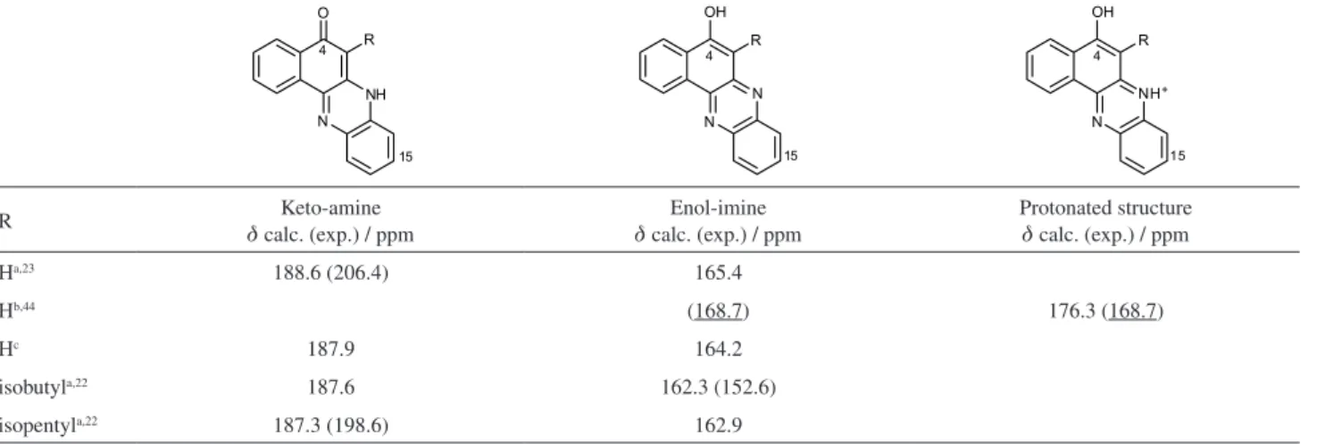
The relative energies calculated at the B3LYP/ 6-311++G(d,p) level for the tautomers of the dihydrobenzo-quinoxalines, benzoquinoxalines and benzophenazines are given in Table 2. All values reported were obtained by subtracting the energy of the keto-amine form from that of the enol-imine one, i.e., the more negative the value, the more stable the enol-imine tautomer and vice-versa.
Figure 4. (a) 1H NMR (500 MHz) spectrum of 2 in CDCl
3 (obtained after neutralization of the crude solid with saturated Na2CO3(aq), extraction with
EtOAc and purification by flash chromatography-hexane/EtOAc, 10/1 to 2/1, v/v); (b) 1H NMR (500 MHz) spectrum of 2 in CDCl
3 after flash chromatography
showing formation of 3.
For the dihydrobenzoquinoxaline series (Table 2), the keto-amine tautomer is by far the most stable form, independent of the substituent and solvent. These results are in agreement with the experimental data, both in the solid state and in solution (see Experimental and reference 26).
For the benzoquinoxaline series, the relative energy of the enol-imine tautomer is lower than that of the keto-amine tautomer. For the tautomers of the structurally related benzophenazines, which contain an additional benzene fused to the pyrazine ring, the gas phase relative energies also indicate the enol-imine tautomer as the most stable one, although less so than in the benzoquinoxaline series (Table 2). The different substituents (R = H, Cl or CH3) have
only marginal effect on the relative energies. However, in all cases, the Cl and CH3 substituents decrease the relative
stabilities of the enol-imine forms as compared to R = H. Solvation has a strong effect on the relative stabilities, with the more polar DMSO and CH3OH solvents having a
higher effect than the less polar CHCl3. The three solvents
preferentially stabilize the keto-amine tautomers, mainly because these tautomers have higher dipole moments than the enol-imine tautomers (see Table S5 in the SI section, e.g., |µ| (D) = 5.45 × 1.61 (2) and 5.95 × 1.19 (3), respectively).
The preferential stabilization of the keto-amine form by the solvent is enough to reverse the stability order in the case of the benzophenazines. For the latter compounds, the keto-amine form, which is the least stable in the gas phase, becomes the most stable in all solvents. The relative effect of the solvents follows the corresponding dielectric constant order (Table 2), with CH3Cl < CH3OH ~ DMSO.
In summary, in the gas-phase, dihydrobenzoquinoxalines are most stable as the keto-amine tautomer, while for the benzoquinoxalines and benzophenazines the enol-imine
forms are the most stable. The three solvents studied reverse the stability order for the benzophenazines, making them more stable in the keto-amine form.
These results agree with the 1H and 13C NMR spectra
of 3 obtained in the three (deuterated) solvents (see
Figures S10-S14 in the SI section) and the 1H NMR
spectrum of the CH3 substituted derivative reported in
CDCl3.43 They indicate, therefore, that the relative stabilities
of the two tautomers are dominated by the extent of aromaticity in the benzoquinoxaline series. However, this aspect seems to be less important for the benzophenazines, making the solvent more relevant in this case.
The 13C NMR spectrum of the unsubstituted
benzophenazine (R = H) in DMSO-d6 (d 206.4 ppm)
has been interpreted in terms of the keto-amine tautomer in agreement with the relative energies reported above (Table 2).23 In CDCl
3/CF3COOD (d 168.7 ppm), however,
the enol-imine tautomer was proposed as the most stable, differently from the prediction based on the relative energies.44 In CHCl
3 the energy difference between
the two tautomers for this derivative is very low, only 0.37 kcal mol-1 in favor of the keto-amine tautomer,
certainly below the accuracy of the theoretical method employed. To see whether this could be accounted for by the entropic and thermal contributions to the equilibrium, it was also calculated the relative Gibbs free energies of the two tautomers at 298 K (Table S6 in the SI section). The Gibbs free energy difference between the two tautomers is reduced to 0.22 kcal mol-1, still favoring the
keto-amine tautomer, thus suggesting that both tautomers may be present under equilibrium conditions.
To look for the origin of the difference between the computed relative energies and the main tautomer found
Table 2. B3LYP/6-311++G(d,p) relative energies (kcal mol-1) calculated for the two tautomers of dihydrobenzoquinoxalines, benzoquinoxalines and
benzophenazines. The energy values were calculated as Eenol-imine – Eketo-amine forms
Solvent (ε)a R =
H Cl (2) CH3 H Cl (3) CH3 H Cl CH3
Gas phase 11.70 12.13 12.53 −9.78 −8.15 −8.56 −2.69 −1.61 −1.06
CHCl3 (4.7) 13.50 14.35 14.21 −5.20 −3.66 −4.48 0.37 1.56 1.21
CH3OH (36.2) 14.31 15.25 14.92 −3.46 −1.96 −2.92 1.52 2.71 2.21
DMSO (46.8) 14.36 15.31 14.96 −3.35 −1.86 −2.83 1.59 2.78 2.27
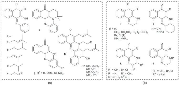
in solution for this benzophenazine, we simulated the 13C
NMR spectra of the two tautomers of this compound (Tables S7-S11 in the SI section). For the dihydrobenzoquinoxaline
2, which exists mainly as the keto-amine tautomer (d 176.3 ppm), and the benzoquinoxaline 3, which exists mainly
as the enol-imine tautomer (d 151.0 ppm), the C4 carbon chemical shifts (in CDCl3) are good probes to distinguish
between the two tautomers in solution.
The calculated C4 carbon chemical shift of the
benzophenazine keto-amine tautomer (d 188.6 ppm)
agrees (to within 8.6%) with the experimental value in DMSO-d6 (Table 3). However, the C4 carbon chemical shift measured in CDCl3/CF3COOD (d 168.7 ppm) has a much
lower value, indicating the prevalence of the enol-imine tautomer in this solvent. Indeed, the computed value for the C4 carbon chemical shift of this tautomer agrees with the experimental data to within 2.7%. Nevertheless, the experimental spectrum in CDCl3 was obtained in the
presence of CF3COOD,44 which could protonate either the
keto-amine or the enol-imine tautomer, leading in each case to the same structure with an OH bond, typical of the enol-imine tautomer. Calculation of the C4-carbon chemical shift for the protonated structure (optimized from the protonation of both tautomers, see data in Table 3) confirms that in CDCl3/CF3COOD, the experimental value
is close to that found for the enol-imine tautomer. Therefore, in this solvent system, the experimental data accounts for either the protonated benzophenazine or the enol-imine tautomer.
The reported 13C NMR data for benzophenazine
derivatives substituted at position 15 (see Figure 1a, g,
R5 = OMe, Cl and NO
2) in DMSO-d6 (d 206.4 for all
compounds)23 also agree with the proposed keto-amine
tautomer.
Likewise, the same keto-amine structure has been proposed in the literature for the 3-isopentyl- and 3-isobutyl-substituted benzophenazines22 (see Table 3)
in spite of the very different C4-carbon chemical shifts observed in the spectra of the two compounds in DMSO-d6 (R = isopentyl, d 198.6; R = isobutyl, d 152.6 ppm). As discussed above, chemical shifts in the 160-170 ppm range are typical for the enol-imine tautomer. It is suggested, therefore, that the observed chemical shift of d 152 ppm for the isobutyl substituted derivative indicates prevalence of the enol-imine tautomer for this derivative (see Tables S12-S15). This unexpected result might arise from intermolecular association which has been reported to lead to protomeric equilibrium shift in similar compounds.57,58
Studies of concentration effects on C4-carbon chemical shifts of this class of compounds would shed some light on the problem.
Conclusions
In the present study, we report on the tautomeric equilibrium in benzo[f]quinoxalin-6(4H)-ones derived from the 1,4-naphthoquinone nucleus. XRD analysis and DFT calculations confirm the enol-imine tautomer as the most stable form of these benzoquinoxalines. The higher stability of the enol-imine tautomer for these derivatives has been attributed to enhanced aromaticity in the enol-imine tautomeric form. For the enol-imine tautomer of dihydrobenzoquinoxalines, aromatization of the additional ring does not occur, which makes the keto-amine tautomer much more stable. This fact was also confirmed by means of the C4-carbon atom 13C chemical shift. The chemical
shift of this carbon was employed as a probe to conclude that benzo[a]phenazin-5(7H)-one exists mainly in the
Table 3. 13C NMR experimental and calculated C4 chemical shifts (d in ppm) for selected benzophenazines (see Tables S8-S16 in the SI section)
R Keto-amine
d calc. (exp.) / ppm
Enol-imine
d calc. (exp.) / ppm
Protonated structure
d calc. (exp.) / ppm
Ha,23 188.6 (206.4) 165.4
Hb,44 (168.7) 176.3 (168.7)
Hc 187.9 164.2
isobutyla,22 187.6 162.3 (152.6)
isopentyla,22 187.3 (198.6) 162.9
aIn DMSO (d
keto-amine form in DMSO, whereas in CHCl3/CF3COOH,
it exists mainly as the enol-imine form, although the B3LYP/6-311++G(d,p) calculations with inclusion of the solvent effects give a slight (0.22 kcal mol-1) preference for
the keto-amine tautomer.
Supplementary Information
Supplementary information associated with this paper contains IR and NMR spectra (1H and 13C), X-ray
structural parameters of 3 and the results of the theoretical
calculations. These data are available free of charge at http://jbcs.sbq.org.br as PDF file. Crystallographic data for the structural analysis of compound 3 have been deposited
with the Cambridge Crystallographic Data Center, CCDC No. 908496. Copies of this information may be obtained free of charge from CCDC, 12 Union Road, Cambridge, CB2 1EZ, UK.
Acknowledgments
The authors thank the Brazilian agencies Conselho Nacional de Desenvolvimento Científico e Tecnológico (CNPq), Coordenação de Aperfeiçoamento de Pessoal de Nível Superior (CAPES) and Fundação de Amparo à Pesquisa do Estado do Rio de Janeiro (FAPERJ) for financial support. FAPERJ-PRONEX (E-26/171.512.2010) is acknowledged. M. D. V. and J. W. M. C. are recipients of CNPq research fellowships. The authors thank LabCri at Universidade Federal de Minas Gerais, Brazil, for XRD data collection.
References
1. Welsch, M. E.; Snyder, S. A.; Stockwell, B. R.; Curr. Opin. Chem. Biol. 2010, 14, 347. 2.
2. Romeiro, N. C; Aguirre, G.; Hernández, P.; González, M.; Cerecetto, H.; Aldana, I.; Pérez-Silanes, S.; Monge, A.; Barreiro, E. J.; Lima, L. M.; Bioorg. Med. Chem. 2009, 17, 641. 3. Neves-Pinto, C.; Malta, V. R.; Pinto, M. C. F. R.; Santos, R. H.; De Castro, S. L.; Pinto, A. V.; J. Med. Chem. 2002, 45, 2112. 4. de Andrade-Neto, V.; Goulart, M.; da Silva Filho, J.; da Silva, M.;
Pinto, M.; Pinto, A.; Zalis, M.; Carvalho, L.; Krettli, A.; Bioorg. Med. Chem. Lett. 2004, 14, 1145.
5. Guillon, J.; Mouray, E.; Moreau, S.; Mullié, C.; Forfar, I.; Desplat, V.; Belisle-Fabre, S.; Pinaud, N.; Ravanello, F.; Le-Naour, A.; Léger, J.-M.; Gosmann, G.; Jarry, C.; Déléris, G.; Sonnet, P.; Grellier, P.; Eur. J. Med. Chem. 2011, 46, 2310.
6. Kumara, K. S.; Rambabua, D.; Sandra, S.; Kapavarapu, R.; Krishna, G. R.; Rao, M. V. B; Chatti, K.; Reddy, C. M.; Misra, P.; Pal, M.; Bioorg. Med. Chem. 2012, 20, 1711.
7. Carneiro, P. F.; Pinto, M. C. F. R.; Coelho, T. S.; Cavalcanti, B. C.; Pessoa, C.; de Simone, C. A.; Nunes, I. K. C.; de Oliveira, N. M.; de Almeida, R. G.; Pinto, A. V.; de Moura, K. C. G.; da Silva, P. A.; da Silva Júnior, E. N.; Eur. J. Med. Chem. 2011, 46, 4521.
8. Ghadage, R. V.; Shirote, P. J.; J. Chem. Pharm. Res. 2011, 3, 260.
9. Hazeldine, S.; Polin, L.; Kushner, J.; Paluch, J.; White, K.; Edelstein, M.; Palomino, E.; Corbett, T.; Horwitz, P.; J. Med. Chem. 2001, 44, 1758.
10. Wu, P.; Su, Y.;Guan, X.; Liu, X.; Zhang, J.; Dong, X.; Huang, W.; Hu, Y.; PloS One 2012, 7, e43171.
11. Chena, Q.; Bryanta,V. C; Lopez, H.; Kelly, D. L; Luo, X.; Natarajan, A.; Bioorg. Med. Chem. Lett. 2011, 21, 1929. 12. Ries, U. J.; Priekpe, H. W.; Hauel, N. H.; Handschuh, S.;
Mihm, G.; Stassen, J. M.; Wienen, W.; Nar, H.; Bioorg. Med. Chem. Lett. 2003, 13, 2297.
13. Kleim, J. P.; Bender, R.; Billhardt, U. M.; Meichsner, C.; Riess, G.; Rösner, M.; Winkler, I.; Paessens, A.; Antimicrob. Agents Chemother. 1993, 37, 1659.
14. Son, H.; Han, W.; Wee, K.; Yoo, D.; Lee, J.; Kwon, S.; Ko, J.; Kang, S.; Org. Lett. 2008, 10, 5401.
15. Mancilha, F.; Neto, B.; Lopes, A.; Moreira Jr, P.; Quina, F.; Gonçalves, R.; Dupont, J.; Eur. J. Org. Chem. 2006, 21,4924. 16. Kudo, K; Momotake, A.; Kanna, Y.; Nishimura, Y.; Arai, T.;
Chem. Commun. 2011, 47, 3867.
17. Ozdemir, S.; Sendur, M.; Oktem, G.; Dogan, O.; Toppare, L.;
J. Mater. Chem. 2012, 22, 4687.
18. Beaudoin, D. S.; Obare, S. O.; Tetrahedron Lett. 2008, 49, 6054.
19. Zapata, F.; Caballero, A.; Molina, P.; Tarraga A.; Sensors 2010,
10, 11311.
20. Wu, C.-D.; Lu, C.-Z.; Zhuang, H.-H.; Huang J.-S.; Inorg. Chem.
2002, 41, 5636.
21. da Silva, M.; Pinto, M.; de Simone, C.; Soares, J.; Reys, J.; de Souza Filho, J.; Harrison, W.; Carvalho, C.; Goulart, M.; da Silva Júnior, E.; Pinto, A.; Tetrahedron Lett. 2011, 52, 2415. 22. Carneiro, P.; Pinto, M.; Coelho, T.; Cavalcanti, B.; Pessoa, C.;
de Simone, C.; Nunes, I.; Oliveira, M.; Almeida, R.; Pinto, A.; de Moura, K.; da Silva, P.; da Silva Júnior, E.; Eur. J. Med. Chem. 2011, 46, 4521.
23. Rehberg, G. M.; Rutherford, J. L.; J. Heterocycl. Chem. 1995, 32, 1643.
24. Santos, M. D. F.; Litivack-Junior, J. T.; Antunes, R. V.; Silva, T. M. S.; Camara, C. A.; J. Braz. Chem. Soc. 2011, 22, 796. 25. Kallmayer, H.-J.; Arch. Pharm. 1974, 307, 806.
26. Kallmayer, H.-J.; Seyfang, K.; Arch. Pharm. 1980, 313, 603. 27. Kallmayer, H.-J.; Seyfang, K.; Arch. Pharm. 1984, 317, 743.
28. Kallmayer, H.-J.; Seyfang, K.; Arch. Pharm. 1984, 317, 855. 29. Kallmayer, H.-J.; Seyfang, K.; Arch. Pharm. 1984, 317, 329.
30. Kallmayer, H.-J.; Seyfang, K.; Arch. Pharm. 1985, 318, 360. 31. Kallmayer, H.-J.; Seyfang, K.; Arch. Pharm. 1985, 318, 607.
33. Kallmayer, H.-J.; Seyfang, K.; Dtsch. Apoth. Ztg. 1983, 123, 2147.
34. Kallmayer, H.-J.; Seyfang, K.; Arch. Pharm. 1986, 319, 52. 35. Cunha, A. S.; Lima, E. L. S.; Pinto, A. C.; Esteves-Souza, A.;
Echevarria, A.; Camara, C. A.; Vargas, M. D.; Torres, J. C.;
J. Braz. Chem. Soc. 2006, 17, 439.
36. Esteves-Souza, A.; Figueiredo, D. V.; Esteves, A.; Câmara, C. A.; Vargas, M. D.; Pinto, A. C.; Echevarria, A.; Braz. J. Med. Biol. Res. 2007, 40, 1399.
37. Cunha, A. S.; Vargas, M. D.; Gattass, C. R.; Pinto, A. C.; Camara, C. A.; Esteves, A. S.; Lima, E. L. S.; Oncol. Rep. 2008,
20, 225.
38. Francisco, A. I.; Casellato, A.; Neves, A. P.; Carneiro, J. W. D.; Vargas, M. D.; Visentin, L. C.; Magalhães, A.; Câmara, C. A.; Pessoa, C.; Costa-Lotufo, L. V.; Marinho Filho, J. D. B.; de Moraes, M. O.; J. Braz. Chem. Soc. 2010, 21, 169.
39. Neves, A. P.; Maia, K. C. B.; Vargas, M. D.; Visentin, L. C.; Casellato, A.; Novak, M. A.; Mangrich, A. S.; Polyhedron 2010, 29, 2884.
40. Neves, A. P.; Barbosa, C. C.; Greco, S. J.; Vargas, M. D.; Visentin, L. C.; Pinheiro, C. B.; Mangrich, A. S.; Barbosa, J. P.; da Costa, G. L.; J. Braz. Chem. Soc. 2009, 20, 712.
41. Neves, A. P.; Silva, G. B., Vargas, M. D.; Pinheiro, C. B.; Visentin, L. C.; Marinho Filho, J. D. B.; Araújo, A. J.; Costa-Lotufo, L. V.; Pessoa, C.; Moraes, M. O.; Dalton Trans. 2010,
39, 10203.
42. Resende, J. A. L. C.; Gomez, J. A.; Acta. Cryst. 2012, E68, o2361.
43. Kallmayer, H.-J.; Seyfang, K.; Arch. Pharm. 1985, 318, 865 44. Kaupp, G.; Naimi-Jamal, M. R.; Eur. J. Org. Chem. 2002, 1368.
45. Duclos, S.; Stoeckli-Evans, H.; Ward, T.; Helv. Chim. Acta, 2001, 28, 3148.
46. Tandon, V. K.; Maurya, H. K.; Mishra, N. N.; Shukla, P. K.;
Eur. J. Med. Chem. 2009, 44, 3130.
47. Agilent Technologies; CrysAlisPro SoftwareSystem, version 1.171.35.21, Xcalibur CCD System; Agilent Technologies UK
Ltd.: Oxford, UK, 2011,.
48. Sheldrick, G. M.; Acta Cryst. 2008, A64, 112.
49. Becke, A. D.; J. Chem. Phys. 1996, 104, 1040.
50. Lee, C.; Yang, W.; Parr, R. G.; Phys. Rev. B 1988, 37, 785. 51. Barone. V.; Cossi, M.; J. Phys. Chem. A 1998, 102, 1995.
52. Cossi, M.; Rega, N.; Scalmani, G.; Barone, V.; J. Comput. Chem.
2003, 24, 669
53. Frisch, M. J.; Trucks, G. W.; Schlegel, H. B.; Scuseria, G. E.; Robb, M. A.; Cheeseman, J. R.; Montgomery, J. A.; Vreen Jr., T.; Kudin, K. N.; Burat, J. C.; Millam, J. M.; Iyengar, S. S.; Tomsi, J.; Barone, V.; Mennucci, B.; Cossi, M.; Scalmani, G.; Rega, N.; Petersson, G. A.; Nakatsuji, H.; Hada, M.; Ehara, M.; Toyota, K.; Fukua, R.; Hasegawa, J.; Ishida, M.; Nakajim, T.; Honda, Y.; Kitao, O.; Nakai, H.; Klene, M.; Li, X.; Knox, J. E.; Hratchian, H. P.; Cross, J. B.; Adamo, C.; Jaramillo, J.; Gomperts, R.; Stratmann, R. E.; Yazyev, O.; Austin, A. J.; Cammi, R.; Pomelli, C.; Ochteski, J. W.; Ayala, P. Y.; Morokuma, K.; Voth, G. A.; Salvador, P.; Dannenberg, J. J.; Zakrzewski, V. G.; Dapprich, S.; Daniels, A. D.; Strain, M. C.; Faras, O.; Malick, D. K.; Rabu, A. D.; Raghavachari, K.; Foresman, J. B.; Ortiz, J. V.; Cui, Q.; Baboul, A. G.; Clifford, S.; Cioslowski, J.; Stefanov, B. B.; Liu, G.; Liashenko, A.; Piskorz, P.; Komaromi, I.; Martin, R. L.; Fox, D. J.; Keith, T.; Al-Laham, M. A.; Peng, C.; Nanayakkra, A.; Challacombe, M.; Gill, P. M. W.; Johnson, B.; Chen, W.; Wong, M. W.; Gonzalez, C.; Pople, J. A.; Gaussian 03, Revision B.02, Gaussian, Inc.: Pittsburg, P.A., USA, 2003.
54. Hunter, C. A.; Sanders, J. K. M.; J. Am. Chem. Soc. 1990, 112, 5525.
55. Francisco, A. I.; Fragoso, T. P.; Vargas, M. D.; Carneiro, J. W. D.; Casellato, A.; Silva, F. C.; Ferreira, V. F.; Barbosa, J. P.; Pessoa, Costa-Lotufo, C. L. V.; Marinho Filho, J. D. B.; Moraes, M. O.; Mangrich, A. S.; J. Braz. Chem. Soc. 2010, 21, 1293
56. Fragoso, T. P.; Carneiro, J. W. D.; Vargas, M. D.; J. Mol. Model.
2010, 16, 825.
57. Beak, P.; Covington, J. B.; Smith, S. G.; White, J. M.; Zeigler, J. M.; J. Org. Chem. 1980, 45, 1354.
58. Adamo, C.; Barone, V.; Loison, S.; Minichino, C.; J. Chem. Soc., Perkin Trans. 1993, 697.
Submitted: November 14, 2012
![Figure S15b and Table S3 in the SI section} result in the packing along the c-axis and [101] direction, respectively, and stabilize the 3D crystalline arrangement](https://thumb-eu.123doks.com/thumbv2/123dok_br/18997654.462706/6.892.100.764.113.366/figure-section-packing-direction-respectively-stabilize-crystalline-arrangement.webp)