O
RIGINALA
RTICLE Revista Brasileira de FisioterapiaStatic balance assessment among children and
adolescents with Down syndrome
Avaliação do equilíbrio estático de crianças e adolescentes com síndrome de Down
Meneghetti CHZ1, Blascovi-Assis SM2, Deloroso FT1, Rodrigues GM3
Abstract
Objectives: To evaluate static balance and the influence of visual information among children and adolescents with Down Syndrome (DS) by means of computerized biophotogrammetry. Methods: Eleven children and adolescents with DS took part in the study and 14 neurologically normal children and adolescents comprised the control group (both genders). During filming, the subjects remained in the orthostatic position with arms to the side of the body and feet parallel on a flat surface. Both groups were filmed in anterior view (frontal plane) and right lateral view (sagittal plane) with and without the eyes covered. While being filmed with eyes covered, the subjects wore fully blacked-out swimming goggles to eliminate all visual information. The instrument used was computerized biophotogrammetry, which served as an angular reference for verifying body sway in static stance. Results: The subjects with DS swayed more (p<0.05) than the control group. When the visual information was eliminated, the anterior-posterior and lateral sway showed significant differences in the balance of the subjects with DS, compared with the subjects of the control group (p<0.01).
Conclusion:The present study showed that children and adolescents with DS swayed more than the children in the control group with and without visual information and in both the anterior-posterior and lateral planes.
Key words: Down Syndrome; assessment; balance; photogrammetry.
Resumo
Objetivos: Avaliar o equilíbrio estático de crianças e adolescentes com Síndrome de Down (SD) pela Biofotogrametria Computadorizada e verificar a influência da visão nesta situação. Métodos: Participaram 11 crianças e adolescentes com SD e 14 crianças e adolescentes de ambos os gêneros, neurologicamente normais que compuseram o grupo controle. Durante as filmagens, os participantes se mantiveram na posição ortostática com os braços posicionados ao lado do corpo e com os pés paralelos sobre uma superfície plana. As crianças de ambos os grupos foram filmadas na vista anterior (plano frontal) e na vista de perfil direito (plano sagital) nas condições com visão e sem visão. Nas filmagens na condição de olhos fechados, foram utilizados óculos de natação totalmente vedados, com a finalidade do participante não ter nenhuma informação visual. O instrumento utilizado foi a Biofotogrametria Computadorizada, que serviu como referência angular para verificar as oscilações do corpo em equilíbrio estático. Resultados: As crianças e adolescentes com SD oscilaram mais (p<0,05) que as do grupo controle e, quando a informação visual foi manipulada, as oscilações ântero-posterior e latero-lateral mostraram a existência de diferenças significativas no equilíbrio nas crianças e adolescentes com SD quando comparadas com as crianças do grupo controle (p<0,01). Conclusão:O presente estudo mostrou que as crianças e adolescentes com SD oscilaram mais que as crianças do grupo controle com e sem a informação visual nos planos ântero-posterior e latero-lateral.
Palavras-chave: Síndrome de Down; avaliação; equilíbrio; fotogrametria.
Received: 21/07/2008 – Revised: 19/11/2008 – Accepted: 12/12/2008
1 Department of Physical Therapy, Centro Universitário Hermínio Ometto (Uniararas), Araras (SP), Brazil
2 Graduate Program in Developmental Disorders, Universidade Presbiteriana Mackenzie (UPM), São Paulo (SP), Brazil 3 Undergraduate Program in Physical Education, UPM
Correspondence to: Cristiane Helita Zorél Meneghetti, Rua das Nogueiras, 95, Jardim Nova, CEP 13601-291, Araras (SP), Brazil, e-mail: [email protected]
Introduction
Down Syndrome (DS) was clinically described for the irst time by English physician John Langdon Down in 1866, but it was not until 1959 that French geneticist Jerome Lejeune identi-ied its causes. DS is the most common of all genetic syndromes1
and it is caused by chromosomal changes, in this case the tri-somy of the 21st chromosome pair, resulting in physical and mental changes1,2. his syndrome has been studied by several
researchers and, with regard to aspects of child development, they have found that children with DS have a delay in motor skill development, indicating that these skills emerge at a diferent time compared to children with normal development3-6.
Some aspects have been suggested as causes for the delay in the acquisition of motor skills in DS children. he main causes of these diferences include an exacerbated weakness in the joints, muscle weakness, sensory-motor abilities, cerebel-lar hypoplasia, and hypotonia6-10. Dysfunctions in the postural
control are often described in DS children and associated with motor coordination diiculties, problems with sensory-motor integration or simply with awkward movements. Movements are considered awkward when the individuals are slow to adapt to a task and to changing conditions in the environment or are less capable of making anticipatory postural adjustments6,11.
To maintain balance in any posture, the human body must receive information about its position in space and about the en-vironment. he body receives this information through the neu-ral system, which integrates the sensory information to access the position and the movement of the body in space, and the musculoskeletal system, which generates forces to control the position of the body, known as postural control system12,13.
Pos-tural control has two behavioral aims: orientation and posPos-tural balance. Postural orientation is the positioning and alignment of body segments in relation to one another and in relation to the environment. Postural balance is the state of equilibrium be-tween all the forces that act on the body to maintain the desired position and orientation14. To ensure that the postural control
system achieves both behavioral objectives, namely orientation and postural balance, two elements are necessary: perception (the integration of the sensory information to analyze the posi-tion and the movement of the body in space) and acposi-tion (the capacity to produce forces to control the body’s positioning sys-tems). hus, postural control requires a continuous interaction between the musculoskeletal and the neural systems13,15.
Among the instruments used to assess balance is computer-ized biophotogrammetry, which applies the photogrammetric principle to photographic images obtained from body move-ments. Photointerpretation principles are then applied to these images, generating a new tool for the study of kinematics16-18.
hus, computerized photogrammetry is a resource that can be
used in assessments for functional physical diagnosis in several areas, having been used in many studies which demonstrated its validity16,19-21. herefore, the objectives of this study were to
evaluate the static balance of children and adolescents with Down Syndrome by means of computerized photogrammetry and to determine the inluence of sight on static balance.
Methods
his was a case-control study approved by the Research Ethics Committee of Centro Universitário Hermínio Ometto (UNIARARAS), under the protocol number 236/2007. Twenty-ive children and adolescents aged 7 to 14 years took part in the study. Eleven had DS and constituted the studied group (SG), and the remaining 14 were neurologically normal and com-posed the control group (CG). Both groups were homogeneous in gender, weight, height, and age. he subjects were recruited from a special education institution and from regular schools. A parent or guardian signed the consent form.
Inclusion criteria were DS children and adolescents aged 7 to 14 years and diagnosed by a karyotype test. he criteria for exclusion were DS children and adolescents with a diagnosis of autism or other diagnosed neurological dysfunctions, children and adolescents without the syndrome who had been diag-nosed with neurological dysfunctions, and those who could not remain in the orthostatic position during ilming.
he data collection for the assessment of the static balance occurred on the premises of the child’s educational institution as the methodology allowed the researchers to set up the equip-ment in diferent locations. Each subject had their body mass and height measured on a digital scale (Welmy digital) duly inspected by INMETRO(National Institute of Metrology, Normalization, and Industrial Quality). An adhesive marker measuring 19mm in diameter was then placed on the glabella for anterior view evalu-ation and on the euryonfor the sagittal view evaluation.
During filming, each subject was advised to assume a relaxed posture, with arms as stable as possible to the side of the body and feet parallel on a flat surface, previously marked for the plantar support. A plumb line was placed in the background to serve as a reference for the angle analy-sis. The children from both groups (SG and CG) were filmed in the anterior view ( frontal plane) and the right lateral view (sagittal plane). During filming with eyes open, the subject was asked to fix their gaze at a target on the front wall, at eye level. The target was a round piece of yellow paper. During filming with eyes covered, the subjects wore totally blacked-out goggles to eliminate all visual informa-tion. Three subjects were excluded from the studied group (SG) because they did not remain in the orthostatic position
and thus could not be filmed. The children were positioned so that the previously marked anthropometric points were aligned with the plumb line both in the anterior-posterior view (glabellar area) and in the right lateral view (euryon) and, to form the angle, a straight line was drawn to the ver-tex, which was perpendicular to the plumb line to determine the point of intersection.
he assessment rooms where the footage was taken had artiicial lighting and a working area of approximately 18m². Isolated rooms were chosen to minimize sound interference during data collection. A digital video recorder (Sony DSC-H2 digital 6.0 mega pixels) was placed on a leveled tripod itted with plumb-bob, at a distance of 2.70m and 1.00m above the ground. he camera remained in this position throughout ilm-ing. he time of exposure to the camera was 30 seconds for each posture: anterior view and right lateral view, with and without the eyes covered. Computerized biophotogrammetry was used to quantify angles and verify body sway in static balance, fol-lowed by the application of Romberg’s test. In order to obtain the moment of greatest anterior-posterior (sagittal) sway with and without the eyes covered, the images were analyzed frame by frame with the aid of the software Windows Movie Maker. At the moment of greatest sway in each plane, the image was selected and analyzed by computerized biophotogrammetry using the software Corel Draw and then calculated in degrees, as shown in Figure 1.
Data analysis
To analyze the efect of sight, we proposed the calculation of the diference between each child’s number of sways with and without eyes covered.
Diference = Without eyes covered - With eyes covered
With this kind of subtraction, negative values indicated a greater sway in the “eyes covered” condition, and positive val-ues indicated greater sway “without eyes covered”. To quantify the degrees of anterior-posterior sway, we calculated the sum of the anterior and posterior sway deviations, and to quantify the degrees of lateral sway we calculated the sum of the sways to the left and to the right.
Statistical analysis
he paired t test was used to verify the efect of sight on sway in the frontal and sagittal planes in the SG and CG. To evaluate the normality of the sample, the Shapiro-Wilk test was applied. To compare the means of the groups, we used the analysis of variance (ANOVA) for each of the deined experimental condi-tions: frontal sway without eyes covered, frontal sway with eyes covered, sagittal sway without eyes covered, and sagittal sway with eyes covered. Turkey’s test was then applied to compare the means. he level of signiicance adopted for all the statisti-cal analyses was p<0.05.
Results
The paired t test was used to verify the effect of sight on sway in the frontal and sagittal planes. With regard to frontal sway, the SG had greater sway with eyes covered (p<0.05). Nevertheless, the CG had no significant difference, as shown in Table 1. In the sagittal plane, there was no sig-nificant difference as the p value was greater than 0.05, as shown in Table 2.
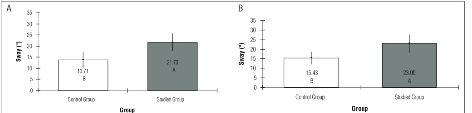
he results of the ANOVA test revealed that the SG children had a greater sway in the frontal plane without eyes covered (p<0.05) compared to those of the CG. Similarly, in the frontal sway with eyes covered, the SG had a signiicance level of p<0.01 compared to the CG, as illustrated in Figure 2. In the sagittal plane with and without eyes covered, the SG had greater mean sway (p<0.01) compared to the CG. he means can be observed in Figure 3.
Discussion
he understanding and quantiication of human body move-ments have attracted great interest in various ields of knowl-edge. he search for eicient and precise evaluation methods has been a constant concern when planning and programming efective interventions. Computerized biophotogrammetry is Figure 1. Sway angles (°) in the frontal (right-left) plane measured
with Corel Draw.
A B
A. Sway angles (°) in the frontal (right-left) plane measured with Corel Draw; B. Sway angles (°) in the sagittal (anterior-posterior) plane measured with Corel Draw.
not only a reliable method but also highly precise, and it also allows image storage in a ile for comparisons and measure-ments whenever necessary16-18. For static balance evaluation,
the instrument was easily applied16,19-21.
Overall, some changes were observed in the sway of chil-dren with DS in the anterior-posterior and lateral directions, both with and without eyes covered. For some authors22-24, the
fact that children with DS sway more can be explained by the diiculty in capturing the sensory information which deter-mines the position of the body in space and the speed at which the body is moving. his seems to happen particularly when the information from one of the sensory systems is removed or manipulated, further increasing the body sway in individuals with DS11,24.
The dynamic postural control system attributes a weight or value of importance to each type of sensory information. This sensory information basically relies on the context of the postural task to generate more precise information con-cerning the position of the body segments and the body’s center of mass in space25.Thus, depending on the task, a
particular type of sensory information may prevail over the others; however, in a different context, this preponderance may be altered or even inverted25. Based on this
perspec-tive, children with DS are less efficient in selecting and using sensory information according to the context in which the task is being executed.
he results of the present study show that, during the tests in which no visual information was available, the evaluated groups had a greater sway than when the visual information was preserved. In fact, both anterior-posterior and lateral sway was more signiicant with eyes covered than without eyes cov-ered. Studies have detected an increase in body sway when the sensory information is removed or manipulated26,27. his
difer-ence may be due to the context in which the task is executed. Maintaining an erect posture in a context where there is no interference with sensory information is apparently simpler and requires less adaptation from the postural control system. However, when visual information is removed or manipulated, the context becomes more complex and requires a more active participation from this system28,29.
Group Difference
mean
Standard
deviation P value
CG -2.5714 4.8787 0.070257 ns
SG -13.5455 14.2784 0.010397 *
Table 1. Sway values in the frontal plane.
* Significance level (p<0.05); ns (non-significant).
Group Difference
mean
Standard
deviation P value
CG -1.71429 7.83904 0.42796 ns
SG -1.27273 7.77291 0.59898 ns
Table 2. Sway in the sagittal plane.
ns (non-significant).
Figure 2. Frontal sway values for SG and CG without eyes covered (A) and for SG and CG with eyes covered (B).
10.14 B 17.00 A 0 5 10 15 20 25 30 35
Control Group Studied Group
Group Sway (º ) 12.71 B 30.55 A 0 5 10 15 20 25 30 35
Control Group Studied Group
Group Sway (º ) A B 15.43 B 23.00 A 0 5 10 15 20 25 30 35
Control Group Studied Group
Group
Sway (º)
Figure 3. Sagittal sway values for SG and CG without eyes covered (A) and for SG and CG with eyes covered (B). 13.71 B 21.73 A 0 5 10 15 20 25 30 35
Control Group Studied Group
Group
Sway (º)
A B
Another result concerned the sagittal plane, in which there was no signiicant diference in sway between the groups. Oliveira and Barreto30 evaluated individuals with acquired
vi-sual impairment and individuals with normal sight on a force platform. he authors observed that the visually-impaired in-dividuals had signiicantly greater lateral sway; nevertheless, in the anterior-posterior direction, they found no signiicant diference between the groups30.
A second point to be discussed regarding the diferences in the static balance of DS children and normal children is the pos-sible delay in motor development in children with DS. Studies have suggested developmental changes such as sensory-motor abilities, muscle fatigue, exacerbated joint fatigue, hypotonia and cerebellar hypoplasia6-10.
Kokubun et al.23 compared balance with unilateral
sup-port in DS children to that of children with other kinds of mental impairment. he authors observed that the frequen-cies of sway waves were higher in children with DS, suggest-ing that higher frequencies of sway wave may be related to muscle hypotonia.
Conclusion
Computerized biophotogrammetry was eicient in the assessment of balance in DS individuals, establishing itself as an important tool for the evaluation procedures in physi-cal therapy practice. he group composed of DS children and adolescents (SG) and assessed by means of this instrument had a greater sway in static balance when compared to the CG. Likewise, when visual information was removed, the SG had greater anterior-posterior and lateral sway compared to the CG. However, other studies on balance in this population are needed to carry on this investigation given the limited number of subjects who took part in this study.
Acknowledgements
Coordenação de Aperfeiçoamento de Pessoal de Nível Su-perior (CAPES) for the scholarship, and to Mackpesquisa, for the inancial support.
1. Nussbaum RL, Mcinnes RR, Willard HF. Thompson e Thompson Genética Médica. 6ª ed. Rio de Janeiro: Guanabara Koogan; 2002.
2. Moreira LMA, El-Hani CN, Gusmão FAF. A síndrome de Down e sua patogênese: considerações sobre o determinismo genético. Rev Bras Psiquiatr. 2000;22(2):96-9.
3. Wang WY, Ju YH. Promoting balance and jumping skills in children with Down syndrome. Percept Mot Skills. 2002;94(2):443-8.
4. Mancini MC, Silva PC, Gonçalves SC, Martins SM. Comparação do desempenho funcional de crianças portadoras de síndrome de Down e crianças com desenvolvimento normal aos 2 e 5 anos de idade. Arq Neuropsiquiatr. 2003;61(2-B):409-15.
5. Pueschel S. Síndrome de Down: guia prático para pais e educadores. 9ª ed. Campinas: Papirus; 2005.
6. Polastri PF, Barela JA. Perception-action coupling in infants with Down syndrome: effects of experience and pratice. Adapt Phys Activ Q. 2005;22(1):39-58.
7. Soares MPS, Lemos SS, Barros JF. Detecção de características específicas na articulação do joelho e do quadril que dificultam a marcha em indivíduos portadores de síndrome de down. Revista Alvorada. 2003;1(2):41-64.
8. Vieregge P, Schulze-Rava H, Wessel K. Quantification of postural sway in adult Down’s syndrome. Dev Brain Dysfunct. 1996;9:211-4.
9. Davis WE, Kelso JA. Analysis of “invariant characteristics” the motor control of Down’s syndrome and normal subjects. J Mot Behav. 1982;14(3): 194-212.
10. Kanode JO, Payne VG. Effects of variable practice on retention and motor schema development in Down syndrome subjects. Percept Mot Skills. 1989;69(1):211-8.
11. Webber A, Virji-Babul N, Edwards R, Lesperance M. Stiffness and postural stability in adults with Down syndrome. Exp Brain Res. 2004;155(4):450-8.
12. Mochizuki L, Amadio AC. As funções do controle postural durante a postura ereta. Rev Fisioter Univ São Paulo. 2003;10(1):7-15.
13. Shumway-Cook A, Woollacott MH. Controle motor: teoria e aplicações práticas. 2ª ed. Barueri: Manole; 2003.
14. Horak FB, Macpherson JM. Postural Orientation and equilibrium. In: Rowell LB, Sherpherd JT, editores. Handbook of physiology: a critical, comprehensive presentation of physiological knowledge and concepts. New York: Oxford American Physiological Society; 1996. p. 255-92.
15. Barela JA. Estratégias de controle em movimentos complexos: ciclo percepção-ação no controle postural. Rev Paul Educ Fís. 2000;Supl3: S79-88.
16. Baraúna MA, Duarte F, Sanchez HM, Canto RST, Malusá S, Campelo-Silva CD, et al. Avaliação do equilíbrio estático em indivíduos amputados de membros inferiores através da biofotogrametria computadorizada. Rev Bras Fisioter. 2006;10(1):83-90.
17. Ricieri DV. Validação de um protocolo de fotogrametria computadorizada e quantificação angular do movimento toracoabdominal durante a ventilação tranqüila. [Dissertação]. Uberlândia: UNITRI – Centro Universitário do Triângulo; 2000.
234
18. Ricieri DV. Biofotogrametria: análise cinemática angular dos movimentos. 2ª ed. Curitiba: Revisada e Ampliada; 2005.
19. Baraúna MA, Canto RST, Oliveira AS, Soares AB, Silva CDC, Cardoso FAG. Avaliação do equilíbrio estático do portador de diabetes mellitus pela biofotogrametria. Diabetes Clínica. 2003;7(1):57-62.
20. Baraúna MA, Barbosa SRM, Canto RST, Silva RAV, Silva CDC, Baraúna KMP. Estudo do equilíbrio estático de idosos e sua correlação com quedas. Fisioter Bras. 2004;5(2):136-41.
21. Guimarães EA. Avaliação do equilíbrio estático de indivíduos normais através da biofotogrametria computadorizada e da oscilometria. [Dissertação]. Uberlândia: UNITRI – Centro Universitário do Triângulo; 2003.
22. Butterworth G, Cicchetti D. Visual calibration of posture in normal and motor retarded Down’s syndrome infants. Perception. 1978;7(5):513-25.
23. Kokubun M, Shinmyo T, Ogita M, Morita K, Furuta M, Haishi H, et al. Comparison of postural control of children with down syndrome and those with other forms of mental retardation. Percep Mot Skills. 1987;84(2): 499-504.
24. Vuillerme N, Marin L, Debu B. Assessment of static postural control in teenagers with Down syndrome. Adapt Phys Activ Q. 2001;18:417-33.
25. Jeka J, Oie KS, Kiemel T. Multisensory information for human postural control: Integrating touch and vision. Exp Brain Res. 2000;134(1):107-25.
26. Paulus W, Straube A, Krafczyk S, Brandt T. Differential effects of retinal target displacement, changing size, and disparity in control of anterior posterior and lateral body sway. Exp Brain Res. 1989;78(2):243-52.
27. Paulus WM, Straube A, Brandt T. Visual stabilization of posture: physiological stimulus characteristics and clinical aspects. Brain. 1984;107(Pt 4): 1143-63.
28. Shumway-Cook A, Woollacott MH. The growth of stability: postural control from a developmental perspective. J Mot Behav. 1985;17(2):131-47.
29. Riach CL, Starkes JL. Stability limits of quiet standing postural control in children and adults. Gait Posture. 1993;1(2):105-11.
30. Oliveira DN, Barreto RR. Avaliação do equilíbrio estático em deficientes visuais adquiridos. Revista Neurociências. 2005;13(3):122-7.