Article
*e-mail: [email protected]
Exchange Interactions in the Copper(II)-
N
-Benzoylglycine (Hippuric Acid) Complex
Ernanni D. Vieira,a Gianella Facchin,b Maria H. Torreb and Antonio J. Costa-Filho*,a
aDepartamento de Física e Informática, Instituto de Física de São Carlos, Universidade de São Paulo,
CP 369, 13560-970 São Carlos- SP, Brazil
bCátedra de Química Inorgânica, Universidad de la República, Montevideo, Uruguay
O complexo CuII-N-benzoilglicina provê um sistema conveniente para se estudar interações
de troca fracas entre elétrons desemparelhados que são transmitidas por caminhos químicos de
interesse biológico. A molécula existe como um dímero onde cada CuII está coordenado a cinco
ligantes, formando uma pirâmide de base quadrada distorcida. O único caminho químico entre
íons CuII magneticamente não-equivalentes envolve 17 átomos diamagnéticos e uma fraca ligação
de hidrogênio putativa. Esta interação foi estudada por Ressonância Paramagnética Eletrônica de amostras monocristalinas a 9,5 e 34,5 GHz, tendo sua magnitude sido calculada em 4(1) mK. Esse valor é discutido em termos de valores obtidos para caminhos similares em outros compostos-modelo e proteínas.
The complex CuII-N-benzoylglycine provides a convenient system to study weak exchange
interactions between unpaired spins transmitted through a biologically relevant long chemical bridge.
The molecule exists as a dimer with each CuII ion coordinated to five ligands, forming a distorted
square–based pyramid. The only chemical connection between magnetically non-equivalent copper ions is a very long path comprising 17 diamagnetic atoms and a weak putative hydrogen bond. This interaction was studied using Electron Paramagnetic Resonance in single-crystal samples at 9.5 and 34.5 GHz and its magnitude was calculated as 4(1) mK. This value is discussed in terms of values obtained for similar paths in other model compounds and in proteins.
Keywords: electron paramagnetic resonance, exchange interactions, model systems
Introduction
Toluene and other volatile organic compounds (VOCs)1
are widely used as organic solvents in pharmacological, plastic and rubber industries, as well as in various materials such as paints, adhesives and combustion materials.2 The
exposure to high concentration of toluene and other VOCs causes adverse health effects in nervous system,3 mucous
and dermal irritations,4 and chromosome aberrations.5,6
Toluene is metabolized in human liver microsomes7,8 or in
rat hepatic microsomes9,10 by the hepatic cytochrome
P450-mediated mixed function oxidase system to benzyl alcohol, to benzoic acid and finally to hippuric acid, by conjugation with glycine to yield hippuric acid or N-benzoylglycine, which is then excreted in urine.11
Copper is a bioessential element with relevant oxidation states.12,13 Due to the complexity of metalobiomolecules
and the associated problems, a very useful way of gaining information is examining the behavior of simpler model compounds. The study of ion complexes of copper-amino acids14-26 (or peptides)27-31 and copper-nucleoside (or related
bases)32,33 has been a topic of increasing interest in the last
few years, manly due to their relevance in the development of new reagents for biotechnology and medicine.34
N-benzoylglycine is structurally similar to a dipeptide except for the absence of the N-terminal group. Due to its high affinity for CuII, the dimeric complex
CuII-N-benzoylglycine could form in vivo, especially in
cases of increased concentrations of hyppuric acid, and could then be used as a means of controlling hyppuric acid concentration.
Complexes of N-benzoylglycine (hippuric acid) with transition metal ions have already been studied by different physical chemistry techniques.35-40 In many cases, the
exchange interaction between those ions. In this case, there is a greater possibility of formation of antiferromagnetically (AFM) coupled dimers, where the singlet (S = 0) state is the ground state and the triplet is thermally populated, than the ferromagnetic (FM) interaction at different site symmetries in bimetallic systems,41–46 as here reported.
Electron Paramagnetic Resonance (EPR) has been extensively used to study the electronic structure and the local properties of metal ions,47 radicals,48 and other
systems bearing an unpaired electron spin.49 EPR enables
the evaluation of exchange interactions between coupled metal ions or radicals. In this way, the EPR technique has been used to measure the magnitudes of those interactions, whose nature is related to the electronic structure of the chemical path connecting the copper ions. Their study provides information about pathways that is difficult to obtain when one is dealing with long covalent and noncovalent bonds, whose weak contributions are masked by stronger interactions. On the other hand, the magnitudes of the exchange interactions between unpaired spins are related to the matrix elements for electron transfer between redox centers in proteins,50 and thus measurements of exchange
couplings give information about electron transfer properties of the path that are difficult to obtain from other sources.51-53
In combination with crystal structure data, EPR studies of model compounds allow one to study the magneto-structural correlations needed to characterize properties of weak bonds. This information may be translated to similar chemical paths occurring in macromolecules.
To evaluate the exchange interaction parameters from EPR data, detailed single-crystal measurements are required. The magnitudes of the exchange couplings (J) are calculated from the positions and line widths of the spectra obtained from systems having two or more anisotropic metal ions per unit cell.20,21,54-56 Since exchange narrowing,
in general, is essentially a temperature-independent phenomenon,57-60 the great advantage of these experiments
is that exchange interactions having very small magnitudes can be measured at room temperature.
In the present paper we report studies on the magnetic properties of di-(µ-hippurato-O)-bis[diaqua(hippurato-O) copper(II)] studied using EPR spectroscopy in single crystal samples at two microwave frequencies, 9 and 34.5 GHz, and at room temperature. The synthesis of a copper complex with N-benzoylglycine (hippuric acid), the X-ray structure and the coordination properties of the complex were first studied by Brown at al.61 The EPR results are analyzed
using the theories of Anderson,57 and Kubo and Tomita59
for the exchange narrowing phenomenon. The magnitudes of the exchange interactions between neighbor copper ions are estimated and discussed in terms of bond properties.
Experimental
Synthesis
The complex di-(µ-hipurato-O)-bis[diaqua(hippurato-O) copper(II)],1 or [{Cu(hippurato)
2(H2O)2}2], was synthesized
from the mixture of aqueous solutions of CuCl2⋅2H2O (Merck, 43 mg in 5 mL of water) and sodium hippurate (Sigma, 100 mg in 30 mL of water). The final solution was slowly evaporated at room temperature to yield, after several days, light blue crystals. The composition of the complex was confirmed by elemental analysis, using a Carlo Erba 1108 Elemental Analyser. [{Cu(hippurato)2(H2O)2}2] (Found: C, 43.64; H, 5.2; N, 5.74. Calc. for Cu[(C6H5) CONHCH2COO]2.4H2O, C18H24CuN2O10: C, 43.95;
H, 4.93; N, 5.70%).
EPR measurements
Room temperature EPR spectra of single crystals were recorded at 9.5 (X-band) and 34.5 GHz (Q-band), using a Varian E-109 spectrometer. The angular variation was achieved either by rotating the sample at X-band or rotating the magnet at Q-band. At both frequencies we used 100 kHz magnetic field modulation and CrIIIMgO (g = 1.9797)as
a field marker. The single crystal sample with dimensions (2.5mm × 1.0mm × 1.0mm) was fixed with vacuum grease to the xz face of a cubic sample holder made of cleaved KCl crystal, which defined the xyz laboratory coordinate system. The single crystal was then oriented so that the abc crystal system was aligned with the xyz laboratory coordinate system. The sample holder was positioned on a pedestal in the center of the rectangular (X-band) or cylindrical (Q-band) cavities. The magnetic field B = Bh
was then applied in the ab, c*a and c*b crystal planes, where h = (sinθcosφ, sinθsinφ, cosθ) denotes the direction
of the external field in the abc* axes system. Single crystal spectra were obtained at five degree intervals in those three orthogonal planes. A single resonance line was observed for all orientations of the magnetic field B in the plane
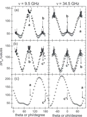
c*a, while two resonances were observed in the ab and c*b planes, except for directions close to the crystal axes, where they collapse to a single resonance (Figure 1).
Results and Discussion
Exchange pathways
The compound Cu[(C6H5)CONHCH2COO]2⋅4H2O reported by Brown at al.61 crystallizes in the monoclinic
cell, a = 7.253(1) Å, b = 40.169(3) Å, c = 7.466(1) Å, and β = 102.81(1) Å. The molecule exists as a dimer with each CuII ion coordinated to five ligands forming a distorted
[4 + 1] square–based pyramid. The coordination sphere of each CuII ion is constituted by an oxygen atom from a
hippurate ion (O(14)), two water molecules (W1 and W2), and two other hippurate ions, whose oxygen atoms (O(1) and O(1*)) act as µ-oxi bridges in the dimer (Figure 2).
The P21/c symmetry operations relate the four CuII ions
as follows: from Cu(A) = (x;y;z), the other three copper sites are Cu(B) = (–x;1/2+y;1/2–z), Cu(C) = (–x;–y;–z), and Cu(D) = (x,1/2–y;1/2+z). The crystal packing shows two molecular chains: chain 1 is formed by molecules containing copper ions Cu(A) and Cu(C) and chain 2 is composed by molecules with Cu(B) and Cu(D), each forming a helix-like structure along the crystalline axes a and c,respectively.
Due to the chain structure observed for the CuII
-N-benzoylglycine, the chemical connections between CuII ions are better discussed when presented as intra
and inter-chain connections. Many chemical paths can be identified as intra-chain connections and all involve magnetically equivalent CuII sites. The contacts between
Cu(A) and Cu(C) (or Cu(B)and Cu(D)) are supported by two µ-oxi bridges (Figure 2). Other possible paths that connect the same type of ion in different molecules (for example, Cu(A) in one molecule and Cu(A) in a symmetry-related molecule) are mediated by several weak hydrogen bonds. These contacts along with the strong µ-oxi bridges couple magnetically equivalent copper ions, which makes the determination of the associated coupling magnitudes not feasible from EPR experiments.
As for inter-chain contacts, the only significant connection found in the structure comprises a very unusual path involving hydrogen H(2) of the aromatic ring of one molecule of the hippuric acid in chain 1 and the carbon C(3) of the aromatic ring of the hippuric acid ofchain 2 (Figure 3). The distance d[C(11)-H(2)] is 3.550 Å and the angle between atoms C(3)::::H(2)−C(11) is 103.70o. This connection
is long, involving 17 diamagnetic atoms with total bond distances d[Cu(A)---Cu(B)] = 28.332 Å and d[Cu(A)---Cu(D)] = 27.890 Å. The spatial separation between ions is d[Cu(A)---Cu(B)] = 20.173 Å and d[Cu(B)---Cu(D)] = 19.671 Å. This chemical Figure 1. EPR spectra at Q-band (34.5 GHz) of the compound
CuII-N-benzoylglycine, showing the phenomenon of collapse and line
narrowing by exchange in the ab (b) and c*b (c) crystal planes. The c*a (a) plane presents only one line as a consequence of the symmetry of the spatial group P21/c.
path has extremely weak character, since it encompasses long contacts through weakly electronegative atoms. One possible explanation for the establishment of such path would be the existence of a water molecule in the crystal structure which has not been detected when the structure was first determined.
EPR results
A single resonance line was observed for all orientations of the magnetic field B in the plane c*a, while two resonances were observed in the ab and c*b planes, except for directions close to the crystal axes, where they collapse to a single resonance (Figure 1). The positions and peak-to-peak line widths (∆Bpp) of the single resonance for the c*a plane, or the two resonances in some orientations of the ab and c*b planes, were obtained from least-squares fits of the field derivative of one or two Lorentzian lines to the observed signal (Figure 1). The angular variation of the line positions and line widths calculated from the EPR spectra are shown in Figures 4 and 5.
The data in Figures 4 and 5 can be analyzed in terms of a spin Hamiltonian written as 21,23,28
(1),
where HZ and Hex are the contributions appearing from the Zeeman and exchange interactions, Siα is the spin operator corresponding to a copper ion in the (α = A, B, C, or D) position of the ith unit cell (S = 1/2), and µB is the Bohr magneton. As described in the previous section, the compound CuII-N-Benzoylglycine belongs to space group
P21/c with two dimer units per unit cell. The distances between copper ions in the crystallographic structures are greater than 7.5 Å, and thus dipole-dipole interactions were disregarded in equation 131. The g
α are the symmetry-related
molecular g-tensors associated to each copper ion in the
sites defined before. Thus, gA≡gB and gC≡gD, with gA, gC and gB and gD related by a C2 rotation around the b axis.
Considering only exchange interactions between nearest neighbor copper ions and the symmetry conditions relating the exchange interaction parameters Jiα,iβ in equation 1, only few different parameters contribute, which are discussed below in terms of the crystal structure of the compound.
Figure 3. Two neighbor copper ions separated by path containing the hydrogen bond discussed in the text are displayed. The distance and the angle of the putative hydrogen bond between molecules are given in Angstroms (Å) and degrees (o), respectively.
Figure 4. Angular variation of the molecular g2
α(θ,ϕ) tensors of each copper site (solid line) in three crystal planes of single crystals of CuII-N-benzoylglycine. Open triangles give the experimental data at 9.5
and 34.5 GHz. The solid lines were obtained with the components of g2
Based on the previous discussion, the Zeeman and exchange interactions can be written in this case as55
(2)
(3)
where and , and the exchange paths associated with the exchange parameters JAA, JAB, and JAD were described above. The sum over m in the first term is over the nearest neighbor copper ions in cell i. Since gA = gB and gC = gD in equation 2, the contributions to Hex proportional to JAA and JAB in equation 3 do not produce any effect on the spectra.
The angular variation of the g2-tensor (Figure 4) shows
that, for some orientations of the magnetic field, two resonance lines are resolved in the EPR spectra, which in turn collapse for directions around the crystal axes. This scheme can be rationalized in terms of a competition
between the residual Zeeman interaction, the difference in Zeeman energy of two magnetically non-equivalent CuII sites, and the respective exchange interaction. At
some orientations, Jex is greater than the residual Zeeman interaction and then a single line is observed. This situation allows us to use a perturbation approach and divide the contributions to the Spin Hamiltonian in equation 1 in H0 and H1, where H1 represents the weaker interactions perturbing the term H0. Two resonance regimes arise (split and collapsed regimes), whose differences rely on the interplay between exchange and residual Zeeman contributions to H1.
Split resonance regime
In the split resonances regime, where two resonance lines are observed (planes ab and c*b; see Figures 4 and 5), the Hamiltonians H0 and H1may be written as
H0 = Hz (4)
H1 = Hex (5)
Each one of the lines observed in the planes ab and c*b corresponds to a magnetically non-equivalent CuII site
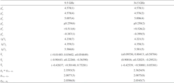
(Cu(A) or Cu(B)). The molecular gα-tensor is obtained directly from the data in Figure 5 in the regions where the lines are well-resolved, without the necessity of the use of decomposition methods. A least-squares fitting of the experimental data in those regions, for all orientations in the c*a plane against the function g2 =⋅g⋅g⋅, leads to
the components of the gα tensors given in Table 1. These results are used to draw the solid lines in Figure 4.
Collapsed resonances regime
In the collapsed resonances regime (ab and c*b planes near the crystal axes), the unperturbed Hamiltonian H0 should be written as
(6)
where S =
S
Siα is the total spin of the system and g = (gA + gB + gC + gD)/4 is the average g tensor. The exchange interaction Hex commutes with the total spin S, and thus H0 gives rise to a single EPR line at the average g factor. The perturbation can be written in this case as55(7)
and the line broadening is62
Figure 5. Angular variation of the peak to peak EPR line width observed at 9.5 and 34.5 GHz for a magnetic field applied in the three crystalline planes ab (a), c*b (b), and c*a (c) of the single crystal sample CuII-N-benzoylglycine. The solid lines are obtained with equation (9) and
(8)
where Gu accounts for the differences among the four molecular g factors. ∆Bpp(0) is a residual line width that considers the small line width observed for the magnetic field along the crystal axes (Figure 5). Due to the space symmetry of the compound, and neglecting the interaction between A and C type copper electron spins, the sum over u contains a single term28,67
(9)
A least-squares fitting of the line width data against the function defined in equation 9 for the regions around the crystal axes in the ab and c*b planes of the compound was performed. The values of the off-diagonal components of the molecular g tensors gA and gD presented in Table 1 were used in this calculation. The results of these fittings are shown as solid lines in Figure 5. These values and equation 9 were used to obtain the exchange frequencies and the values of the exchange coupling parameters
| JAD/k | = 4(1)mK (10)
The magnitude of JAD (4 mK, ca. 0.0028 cm-1) is very
weak, as expected from the exchange pathway analysis
described above. Similar magnitudes of exchange coupling between copper ions have been found in several complexes. In dichlorobis(1-phenyl-3,5-dimethylpyrazole)copper(II), an exchange coupling of 0.00386 cm-1 (ca. 5.5 mK) was
found, mediated by a long path (bond distance 13.60 Å) constituted by the pyrazole ring and a hydrogen-halogen bond C−H···Cl64. In the complex Cu(Gly)
2⋅H2O, Hoffman et al.54
determined an exchange interaction of 0.004 cm-1
(ca. 5.7 mK) between magnetically non-equivalent copper ions connected through a hydrogen bond between a coordinated water molecule and a glycine oxygen atom. More recently, Santana et al.65 calculated the coupling
magnitude between copper ions in the compound CuII(L-arginine)
2(SO4).(H2O)6, finding a weak coupling of
9 mK assigned to a long path with 12 atoms (total bond distance 19.789 Å) and two hydrogen bonds. Furthermore, in the complex aqua(glycyl-L-tryptophanate)-copper(II) dehydrate, an exchange coupling of 3 mK was found and also assigned to a long path with 10 diamagnetic atoms, including a moderate hydrogen bond (total bond distance 16.69 Å). All these model systems are very nicely related to the biological electron transfer system in the bacterial photosynthetic reaction center protein of Rhodobacter sphaeroides, where the coupling between the two quinone acceptors at 17.3 Å is 3.8 mK.66 Following the classification
proposed by Vieira et al.,31 the exchange coupling
determined in the present work would be in their group III, Table 1. Values of the components of the crystal and molecular g2 tensor obtained by least-squares analyses of the data at each microwave frequency. (g2)
1, (g2)2, and (g2)3 and n
1, n
2 and n
3 are the eigenvalues and eigenvectors of the g2 tensor in abc* coordinate system(a)
9.5 GHz 34.5 GHz
g2
xx 4.578(1) 4.578(1)
g2
yy 4.578(4) 4.576(2)
g2
zz 5.007(4) 5.006(4)
g2
xy +0.259(6) +0.250(2)
g2
zx 0.511(6) 0.526(2)
g2
zy –0.387(1) –0.399(5)
(g2)
1 4.238(7) 4.221(3)
(g2)
2 4.359(3) 4.358(3)
(g2)
3 5.566(6) 5.581(5)
n
1 (0.01485; 0.83602; ±0.054849) (±0.09356; 0.80413; ±0.58704)
n
2 (–0.90443; ±0.22268; –0.36390) (0.90016; ±0.32025; –0.29521)
n
3 (–0.42637; 0.50148; 0.75281) (–0.42539; 0.50081; 0.05381)
g|| = gCu – O1 2.3593(5) 2.3624(9)
gO14 – O1* 2.0877(3) 2.0875(8)
gW1 – W2 2.0586(8) 2.0545(7)
(a) The values of g
O14 – O1*, gW1 – W2 and g|| = gCu – O1 represent the molecular g value for each compound in oxygen-oxygen, water-water, and parallel directions,
which includes long paths with more than 10 diamagnetic atoms and magnitudes of the order of few milliKelvin.
Conclusions
In this article we studied the magnetic properties of the CuII-N-benzoylglycine compound, whose interest is
associated with its toxicology or ecotoxicology. The EPR technique, along with X-ray diffraction data, supplies information on the possible chemical path and the magnetic interactions established in model metal-ligand complexes. From the analysis of the experimental data, it is possible to calculate the coupling constants between the paramagnetic sites. These couplings usually present very weak magnitudes and are thought to model biologically-relevant interactions. One can readily see that room-temperature EPR measurements can thus offer unique possibilities of evaluating very small magnitude interactions, which would be directly detected by other experimental techniques only by means of very low temperature measurements.
In the CuII-N-benzoylglycine complex, the copper
coordination sphere in each site is a slightly distorted square-based pyramid with values of g|| > g⊥, which indicates that the ground-state orbital of the copper ions is essentially a dx2–y2 state. The values obtained from the data at 34.5 GHz in
the Table 1, g|| = 2.3624(9), gO(14)–O(1) = 2.0875(8), gW(1)–W(2) = 2.0545(7), indicate that the electron ground-state orbital is in the plane containing the copper ion and the hippurato oxygen and water ligands. The only interactions that we can quantify using the EPR technique are the inter-chain contacts that relate copper ions in the two different chains observed in the crystal structure. The evaluated magnitude |J/k| is 4(1) mK. This low value of |J/k| agrees well with the described chemical path discussed in the text.
Acknowledgments
The authors thank the Brazilian Agencies CNPq, FAPESP, CAPES and FINEP for financially supporting this work. AJCF thanks CNPq for a Research Fellowship (307102/2006-8). This work is part of a joint program PRONEX/FAPESP/CNPq (Grant number 03/09859-2).
References
1. Ohashi, Y.; Mamiya, T.; Mitani, K.; Wang, B. L.; Takigawa, T.; Kira, S.; Kataoka, H.; Anal. Chim. Acta2006, 566, 167. 2. Saijo, Y.; Kishi, R.; Sata, F.; Katakura, Y.; Urashima, Y.;
Hatakeyama, A.; Kobayashi, S.; Jin, K.; Kurahashi, N.; Kondo, T.; Gong, Y. Y.; Umemura, T.; Int. Arch. Occup. Environ. Health
2004, 77, 461.
3. Harkonen, H.; Lindstrom, K.; Seppalaimen, A. M.; Asp, S.; Hernberg, S.; Scand. Work Environ. Health1978, 4, 53. 4. Uchida, Y.; Nakatsuka, H.; Ukai, H.; Watanabe, T.; Liu, Y.T.;
Huang, M. Y.; Wang, Y. L.; Zhu, F. Z.; Yin, H.; Ikeda, M.; Int. Arch. Occup. Environ. Health1993, 64, 597.
5. Bilban, M.; Am. J. Ind. Med. 2004, 45, 468.
6. Celik, A.; Akbas, E.; Ecotox. Environ. Safe. 2005, 60, 106. 7. Kim, H.; Wang, R. S.; Elovaara, E.; Raunio, H.; Pelkonen, O.;
Aoyama, T.; Vainio, H.; Nakajima, T.; Xenobiotica1997, 27, 657.
8. Tassaneeyakul, W.; Birkett, D. J.; Edwards, J. W.; Veronese, M. E.; Tassaneeyakul, W.; Tukey, R. H.; Miners, J. O.;
J. Pharmacol. Exp. Ther.1996, 276, 101.
9. Foureman, G. L.; Harris, C.; Guengerich, F. P.; Bend, J. R.;
J. Pharmacol. Exp. Ther. 1989, 248, 492.
10. Waxman, D. J.; Walsh, C.; Biochemistry1983, 22, 4846. 11. Siqueira, M. E. P. B.; Paiva, M. J. N.; Rev. Saúde Publ.2002,
36, 723.
12. Holm, R. H.; Kennepohl, P.; Solomon, E. I.; Chem. Rev. 1996,
96, 2239.
13. Lippard, J. S.; Berg, J. M.; Principles of Bioinorganic Chemistry, University Science Books: Mill Valley, CA, 1994, ch 2. 14. Fujimoto, M.; Janecka, J.; J. Chem. Phys.1971, 55, 1152. 15. Steren, C. A.; Gennaro, A. M.; Levistein, P. R.; Calvo, R.;
J. Phys. Condens. Matter1985, 1, 637.
16. Gennaro, A. M.; Calvo, R.; J. Phys. Condens. Matter1989, 1, 7061.
17. Calvo, R.; Passeggi, M. C. G.; J. Phys. Condens. Matter1990,
2, 9113.
18. Calvo, R.; Passeggi, M. C. G.; Novak, M. A.; Symko, O. G.; Oseroff, S. B.; Nascimento, O. R.; Terrile, M. C.; Phys. Rev. B
1991, 43, 1074.
19. Calvo, R.; Passeggi, M. C. G.; Phys. Rev. B1991, 44, 5111. 20. Brondino, C. D.; Cassado, M. M. C.; Passeggi, M. C. G.; Calvo,
R.; Inorg. Chem.1993, 32, 2078.
21. Martinho, D. M. ; Passeggi, M. C. G.; Calvo, R.; Phys. Rev. B
1995, 52, 9466.
22. Rapp, R. E.; Souza, E. P.; Godfrin, H.; Calvo, R.; J. Phys. Condens. Matter1995, 7, 9595.
23. Martinho, D. M.; Passeggi, M. C. G.; Calvo, R.; Nascimento, O. R.; Physica B1996, 225, 63.
24. Massa, M. B.; Dolasto, S. D.; Ferreyra, M. G.; Labadil, G.; Calvo, R.; J. Phys. Chem. A1999, 103, 2606.
25. Dolasto, S. D.; Ferreyra, M. G.; Calvo, R.; Piro, O. E.; Castellano, E. E.; J. Inorg. Biochem.1999, 73, 151.
26. Hoffmann, S. K.; Hilczer, W.; Goslar, J.; Massa, M. M.; Calvo, R.; J. Magn. Reson.2001, 153, 92.
27. Cassado, M. M. C.; Isaacson, R. A.; Calvo, R.; J. Inorg. Biochem.2001, 84, 201.
28. Costa-Filho, A. J.; Nascimento, O. R.; Chivelder, L.; Calvo, R.;
29. Facchin, G.; Torre, M. H.; Kremer, E.; Baran, E. J.; Mombrú, A.; Pardo, H.; Araujo, M. P.; Batista, A. A.; Costa-Filho, A. J.;
Inorg. Chim. Acta2003, 355, 408.
30. Costa-Filho, A. J.; Nascimento, O. R.; Calvo, R.; J. Phys. Chem. B2004, 108, 9549.
31. Vieira E. D.; Casado N. M. C.; Facchin, G.; Torre, M. H.; Costa-Filho, A. J.; Calvo, R.; Inorg. Chem. 2006, 45, 2942. 32. Nagane, R.; Koshigoe, T.; Chikira, M.; J. Inorg. Biochem.2003,
93, 204.
33. Brotschi, C., Leumann, C. J.; Nucleosides Nucleotides Nucleic Acids2003, 22, 1195.
34. Vaidyanathan, V. G.; Nair, B. U.; J. Inorg. Biochem.2003, 93, 271.
35. Marcotrigiano, G.; Pellacani, G. C.; Z. Anorg. Allg. Chem.1975,
415, 268.
36. Marcotrigiano, G.; Pellacani, G. C; Inorg. Nucl. Chem. Lett.
1975, 10, 643.
37. Marcotrigiano, G.; Menabue, L.; Pellacani, G. C; J. Inorg. Nucl. Chem. 1977, 39, 1897.
38. Andreoli, R.; Gviolli, B. G.; Benedetti, L.; Grandi, G.; Marcotrigiano, G.; Menabue, L.; Pellacani, G. C.; Inorg. Chim. Acta1980, 46, 215.
39. Sgarabotto, P.; Bisceglie, F.; Pelosi, G.; Abdel-Rahman, L.;
Polyhedron 1999, 18, 2505.
40. Capllonch, M. C.; Garcia-Raso, A.; Terron, A.; Apella, M. C.; Espinosa, E.; Molin, E.; J. Inorg. Biochem.2001, 85, 173. 41. Lippard, J. S.; Berg, J. M.; Principles of Bioinorganic Chemistry,
University Science Books: Mill Valley, CA, 1994, ch.4. 42. Bencini, A.; Gatteschi, D.; Electron Paramagnetic Resonance
of Exchange Coupled Systems, Springer-Verlag: Berlin Heidelberg, 1990, ch. 1.
43. Kahn, O.; Molecular Magnetism, VCH Publications: New York, 1993, ch. 6.
44. Sreehari, N.; Varghese, B.; Manoharan, P. T.; Inorg. Chem.1990,
29, 4011.
45. Mohanta, S.; Nanda, K. K.; Thompson, L. K.; Florke, U.; Nag, K.; Inorg. Chem.1998, 37, 1465.
46. Pardo, E.; Bernot, K.; Julve, M.; Lioret, F.; Cano, J.; Ruiz-Garcia, R.; Delgado, F. S.; Ruiz-Perez, C.; Ottenwaelder, X.; Journaux, Y.; Inorg. Chem. 2004, 43, 2768.
47. Wertz, J. E.; Bolton, J. R.; Electron Spin Resonance – Elementary Theory and Practical Applications, McGraw-Hill: New York, 1972, ch. 11 and 12.
48. Atherton, N. M.; Principles of Electron Spin Resonance, 2nd
ed., Ellis Horwood: New York, 1993, ch. 3.
49. Campbell, I. D.; Dwek, R. A.; Biological Spectroscopy, Benjamin Cummings Company: California, 1984, ch. 6. 50. Calvo, R.; Abresch, E. C.; Bittl, W.; Feher, G.; Hofbauer, W.;
Isaacson, R. A.; Lubitz, W.; Okamura, M. Y.; Paddock, M. L.;
J. Am. Chem. Soc.2000, 122, 7327.
51. Moser, C. C.; Keske, J. M.; Warncke, K. R.; Farid, S.; Dutton, P. L.; Nature1992, 355, 796.
52. Dutton, P. L.; Moser C. C.; Proc. Natl. Acad. Sci. U.S.A.1994,
91, 10247.
53. Moser, C. C.; Page, C. C.; Farid, R.; Dutton, L.; J. Bioener. Biomem.1995, 27, 263.
54. Hoffmann, S. K.; Groslar, J.; Szezepaniak, L. S.; Phys. Rev. B
1988, 37, 7331.
55. Passeggi, M. C. G.; Calvo, R.; J. Magn. Reson. A1995, 114, 1. 56. Brondino, C. D.; Calvo, R.; Atria, A. M.; Spodine, E.; Peña,
O.; Inorg. Chim. Acta1995, 228, 261.
57. Anderson, P.W.; J. Phys. Soc. Jpn. 1954, 9, 316.
58. Abragam, A.; The Principles of Nuclear Magnetism, Claredon Press: Oxford, U.K., 1961, ch. 4 and 10.
59. Kubo, R.; Tomita, K. J. Phys. Soc. Jpn. 1954, 9, 888. 60. Abragam, A.; Bleaney, B.; Electron Paramagnetic Resonance of
Transition Ions, Clarendon Press: Oxford, U.K., 1970, ch. 9. 61. Brown, J. N.; Trefonas, L. M.; Inorg. Chem.1973, 12, 1730. 62. Costa-Filho, A. J.; Munte C. E.; Barberato, C.; Castellano, E. E.;
Mattioli, M. P. D.; Calvo, R; Nascimento, O. R.; Inorg. Chem.
1999, 38, 4413.
63. Yokota, M.; Koide, S.; J. Phys. Soc. Jpn. 1954, 9, 953. 64. Goslar, J.; Hilkzer, W.; Hoffmann, S. K.; Phys. Stat. Sol.B
1993, 175, 465.
65. Santana, R. C.; Cunha, R. O.; Carvalho, J. F.; Vencato, I.; Calvo, R.; J. Inorg. Biochem. 2005, 99, 415.
66. Calvo, R.; Isaacson, R. A.; Paddock, M. L.; Abresch, E. C.; Okamura, M. Y.; Maniero, A. L.; Brunel, L. C.; Feher, G.;
J. Phys. Chem. B. 2001, 105, 4053.
Received: February 2, 2008 Web Release Date: October 10, 2008