Licenciado sob uma Licença Creative Commons DO): http://dx.doi.org. . / - . . .AO
[T]
Nocturnal oxyhemoglobin desaturation during
sleep in congestive heart failure patients
[)]
Dessaturação da oxihemoglobina durante o sono em
pacientes com insui ciência cardíaca congestiva
[A]
Jéssica Julioti Urbano[a], Lilian Nanami Uchiyama[b], Anderson Soares Silva[a],
Roger André Oliveira Peixoto[a], Sergio Roberto Nacif[a], Luis Vicente Franco Oliveira[a]*
[a] Universidade Nove de Julho UN)NOVE , São Paulo, SP, Brazil
[b] Universidade do Vale do Paraíba UN)VAP , São José dos Campos, SP, Brazil
[R]
Abstract
Introduction: Sleep breathing disorders occur in % of patients with heart failure, with %– %
manifes-ting Cheyne-Stokes respiration with central sleep apnea and % exhibimanifes-ting obstructive sleep apnea. Several studies have shown that sleep pathophysiology may negatively affect the cardiovascular system and that car-diac dysfunction alters sleep and respiration. Objective: The aim of this study was to examine
oxyhemoglo-bin desaturation during sleep in patients with congestive heart failure C(F using overnight pulse oximetry.
Methods: Overnight pulse oximetry was conducted in the patients’ homes with wrist pulse oximeters and
inger probes that were placed around the fore ingers of patients with C(F and ejection fractions less than %, who were classi ied as New York (eart Association functional classes )) and ))). Results: The patients
were divided into two groups. The irst group consisted of seven patients with oxyhemoglobin desaturation
*JJU: Master Student, e-mail: jjulioti@yahoo.com.br LNU: MS, e-mail: uchiyama.ln@gmail.com
ASS: BS, e-mail: andersonsamaniego@yahoo.com.br
RAOP: MS, e-mail: roger.peixoto@santacatarinaoxigenio.com.br SRN: PhD, e-mail: pro_ar@uol.com.br
indices of over events/h, and the second group contained eight patients with oxyhemoglobin desaturation indices of or less events/h. Student’s t-tests did not show any signi icant differences between the groups. The patients’ body mass indices correlated positively with the total desaturation episodes and desaturation time less than % and correlated negatively with the arterial oxygen saturation nadir. Conclusion: Pulse oximetry
monitoring during sleep can be used to detect sleep breathing disorders in stable patients with C(F.
Keywords: Sleep Disorders. Obstructive Sleep Apnea. Oximetry.
Resumo
Introdução: Os distúrbios respiratórios do sono ocorrem em 45% dos pacientes com insuϔiciência cardíaca, com 36%-50% manifestando respiração Cheyne-Stokes com apneia do sono central e 12% exibindo apneia obstrutiva do sono. Vários estudos têm demonstrado que a ϔisiopatologia do sono pode afetar negativamente o sistema cardiovas-cular e que a disfunção cardíaca altera o sono e a respiração. Objetivo: Examinar a dessaturação da oxihemoglobina durante o sono em pacientes com insuϔiciência cardíaca congestiva (ICC), utilizando a oximetria de pulso durante a noite. Métodos: A oximetria de pulso noturna foi realizada nas casas dos pacientes com oxímetros de pulso acoplados ao redor dos dedos indicadores de 15 pacientes com ICC e fração de ejeção menor que 50%, sendo classiϔicados pelo New York Heart Association como classes funcionais II e III. Resultados: Os pacientes foram divididos em dois grupos. O primeiro grupo era composto por sete pacientes com índices de dessaturação da oxihemoglobina (IDO) maior que 5 eventos/h e o segundo grupo continha oito pacientes com IDO igual ou menos que 5 eventos/h. Testes t de Student não apresentou diferenças signiϔicativas entre os grupos. Os índices de massa corporal dos pacientes foram positivamente correlacionados com o total de episódios de dessaturação e tempo de dessaturação inferior a 90% e negativamente com a saturação de oxigênio arterial. Conclusão: O monitoramento da oximetria de pulso durante o sono pode ser usado para detectar distúrbios respiratórios do sono em pacientes estáveis com ICC.
Palavras-chave: Transtornos do Sono. Apneia do Sono Tipo Obstrutiva. Oximetria.
Introduction
Over years ago, irregular breathing patterns
were observed in patients with congestive heart fail-ure C(F . These breathing pattern disorders have only been considered clinically signi icant since the s, when sleep-disordered breathing SDB was shown to be related to worsening heart function . )s observed in patients with more severe SDB a higher prevalence of left atrial enlargement LAE , suggest-ing that SDB may cause LAE. The SDB leads to noctur-nal hypoxemia, excitement reactions and consecutive repetitive bursts of sympathetic activity .
SDB occurs in approximately % of patients with heart failure (F , with % exhibiting Cheyne-Stokes respiration CSR , % demonstrating obstructive sleep apnea OSA , and the rest having a mixed form . CSR is more common in male patients with (F than in females patients with (F, and its pathophysi-ology is not yet understood.
OSA and CSR with central sleep apnea are the two main types of SDB in patients with C(F . OSA,
which is characterized by repetitive episodes of com-plete or partial closure of the upper airway during sleep, produces sleep fragmentation and oxygen de-saturation . )n contrast, central apnea is associated with no respiratory efforts for at least s .
Currently, (F is a major public health problem that has an increasing incidence and prevalence due to the increased average life spans and improved therapies of ischemic coronary artery disease and hypertension, which are the most common risk factors for (F. )t is estimated that . % - % of the population of the United States has some form of (F and that its prevalence in-creases to approximately % - % in individuals over
years of age. Of the patients with (F from ventricular systolic dysfunction, at least % have an apnea-hypop-nea index A() of or more events/h .
OSA has several pathophysiologic effects on the after-load, hypoxia, and activation of the sympathetic nervous system. These effects can be presumed to result from the cumulative in luence of hundreds of obstructive apneas that occur each night over a period of months to years and that contribute to the development and/or aggravation of left ventricular dysfunction in patients with (F .
Oxygen desaturation from SDB contributes to the worsening of C(F and is associated with poor
progno-ses . Nakano et al. examined oxyhemoglobin
desatu-ration by monitoring nocturnal pulse oximetry NPO during sleep in patients with suspected sleep apnea, and they then proposed that NPO can be used as a low-cost
screening test of SDB .
A study on the use of the WristOx™ wrist pulse
oximeter has shown that monitoring the NPO of patients with OSA is extremely important, especially in regions where polysomnography PSG is dif icult to access. )n patients with suspected sleep apnea/hypopnea syn-drome SA(S , a negative oximetry result is de ined as an adjusted O desaturation index which is the mean number of O desaturations of % or less/h of total re-cording time of . or less, with the exclusion of SA(S, which is de ined by an A() of or more, with a
sensi-tivity of %. A positive oximetry result is de ined as
an adjusted O desaturation index which is the mean number of O desaturations of % or less/h of total re-cording time of over SA(S is de ined by an A() of
or more , with a speci icity of %. The results of
that study suggested that the WristOx™ might be
a valuable tool for the diagnosis or exclusion of SA(S. (owever, additional studies are necessary to determine if the results found in their study are applicable to the
use of the WristOx™ at home .
Few studies have examined the use of NPO to screen for OSA in patients with C(F. Thus, the aim of this study was to examine oxyhemoglobin saturation with NPO in patients with C(F who were classi ied as functional classes )) or ))) according to the New York (eart Association during sleep. Our secondary objectives were to examine the relationships of the values obtained with the NPO with the anthropomet-rics data and Epworth Sleepiness Scale scores and to verify the possibility of using NPO as a screening test of the presence of a sleep breathing disorder SBD .
Methods
The present cross-sectional study was conducted at the Sleep Laboratory of Nove de Julho University. The protocol was approved by the Research Ethics Committee
of Nove de Julho University protocol number ,
and informed consent was obtained from each patient. Data collection started after the approval of the ethics
committee and was inalized in November/ . The
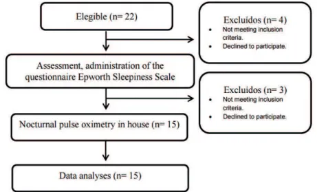
subjects consisted of patients with C(F eight men and seven women from the Cardiology Service from Sao Paulo. Figure shows the study design.
The inclusion criteria were BM) < kg/m , and the patients had been clinically stable for at least one month. Exclusion criteria were renal insuf iciency, unstable an-gina, myocardial infarction, cardiac surgery or acute heart failure decompensation within the previous months. Patients were instructed about the details of the study, including the bene its and risks. All patients signed and received a copy of the informed consent.
Clinical Evaluation
The patients with C(F provided information on their medical and surgical histories, including their concomitant medications, demographic data, anthro-pometric measures, and physical examinations. The measurements included body weight kg , height cm , body mass index BM) , heart and respiratory rates, and peripheral blood pressure.
Epworth Sleepiness Scale
The patients completed the Epworth Sleepiness Scale, which is a simple and self-administered ques-tionnaire that is used to assess recent daytime sleepi-ness with eight questions that refer to eight situations that are based on their usual way of life. They were asked to rate each situation on a scale of - , no chance of napping; , small chance of napping; , moderate chance of napping; and , strong chance of napping according to their felt or estimated degree of sleepiness. Total scores of or more were used to identify clinically relevant levels of sleep-related
daytime dysfunction .
Nocturnal pulse oximetry
NPO was monitored with a WristOx™ Nonin
Medical, )nc., Plymouth, MN, USA and a inger probe that was placed around the patient’s fore inger. The equipment was set at s/sample, which was the short-est measurement time interval. Each desaturation epi-sode was de ined as a decline of the baseline oxygen saturation SaO of % during a period of at least s. )n addition to the SaO data, the oximeter simul-taneously recorded heart rate (R . An (R variation episode was de ined as an (R alteration of at least beats/min during a period of s or more. For the data analysis, the patients were divided into two groups. Patients with an oxyhemoglobin desatura-tion index OD) of or more events/h, which is con-sidered abnormal, were assigned to Group , while Group consisted of patients with an OD) of less than events/h, which is considered normal. The readings will be performed manually by a specialized techni-cian. A report of the results will be prepared by a doctor specializing in sleep medicine at the Sleep Laboratory of Nove de Julho University.
)n general, abnormal OD) values have three lev-els that appear to mirror the definition of abnor-mal A() apnea/hypopnea events/sleep h values. The levels for abnormal OD) values are or more desaturation events/h, or more desaturation events/h, and or more desaturation events/h . For A(), an index of or more events/h has been used to define a significant number of SBD events in OSA in population studies of subjects who
do not have (F .
Statistical Analysis
The Shapiro-Wilk test was used to test the nor-mality of the data, which are described as mean ±
standard deviation. Student’s t-tests were used to
compare the means and identify signi icant differ-ences between the groups. Pearson correlation coef-icients were used to assess the relationships among the measures. P values less than . were considered statistically signi icant in all of the analyses.
Results
Table lists the anthropometric data of the pa-tients, their medications, and their ejection fractions that were veri ied with echocardiography.
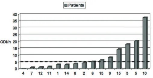
Of all of the patients, seven . % had OD)
val-ues of or more events/h, and these patients were
assigned to Group igure . Student’s t-tests did
not ind statistically signi icant differences between the groups, except for the left atria systolic diameter
LASD; Group : . ± . ; Group : . ± . ,
which was signi icantly decreased in Group . Some of the echocardiographic values were collected from the patient’s medical records with authorization of the responsible doctor because of the lack of echo-cardiography results. Thus, the LASD values were missing for two patients, and the left ventricular end-diastolic diameter LVEDD values were missing for two patients.
Table 1 - Anthropometric variables, type of neurological damage and classification of foot deformities in children with chronic non-progressive encephalopathy, Jequié, Bahia, Brazil, 2014
VARIABLES (n = 15) ODI > 5/h (n = 7) ODI ≤ 5/h (n = 8)
Age (years) 67.71 ± 11.44 58.88 ± 10.13
Weight (Kg) 79.29 ± 20.32 67.63 ± 12.33
Height (cm) 167.29 ± 6.24 164.13 ± 8.87
BMI (Kg/m²) 28.20 ± 6.54 24.90 ± 2.34
EF (%) 40.07 ± 9.40 38.75 ± 5.89
Functional class (NYHA)
II – n = 4 III – n = 7 II-III – n = 4
-
-Schemic myocardiopathy 7 -
-Dilated myocardiopathy 4 -
-Idyopathic myocardiopathy 3 -
-Schemic dilated myocardiopathy 1 -
-Medications in use -
-Digitalis (%) 66.7 -
-Diuretics (%) 66.7 -
-Anti-hypertensives (%) 40 -
-Vasodilators (%) 13.3 -
-ACE inhibitors (%) 6.7 -
-Angiotensin-1 receptor
antagonist (%) 13.3 -
-Beta blockers (%) 13.3 -
-Note: NYHA = New York Heart Association; Kg = kilogram; cm = centimeters; Kg/m² = kilograms per meters squared; EF = ejection frac-tion; ACE = Angiotensin conversor enzyme.
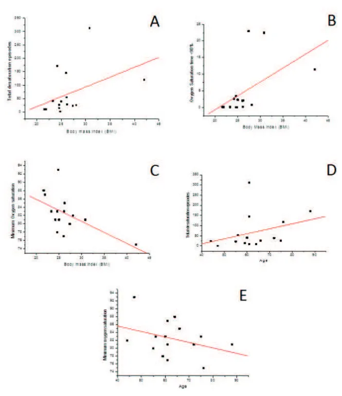
Figure 3 - A = Correlation coefficient between BMI and total desaturation episodes (r = 0.419);
B = Correlation coefficient between BMI and SaO2 time < 90% (r = 0.522); C = Correlation coefficient between BMI and
increases in the A() values and LA areas. Patients who were at a very high risk for a fatal outcome were
identi-ied by A() values of or more events/h and LAs of or more cm . Nevertheless, in patients with isolated indings of enlarged LAs without SBDs and vice versa, the risk was low for patients with A() values of or more events/h and small LAs.
)n the present study, the means ± standard devia-tions of the LASD and LVEDD were . ± . mm and . ± . mm respectively, which are both considered abnormal, and that of OD) was . ± . , which suggested the existence of a group of patients with a low to moderate risk of cardiac death, except for one patient who presented an OD) of events/h, an LASD of mm, and an LVEDD of mm . (owever, in order to con irm these results, more long-term studies on the prognostic value of SBD and cardiac dysfunction, sleep architecture, and arousals with PSG are needed.
The signi icantly greater LASD mean in Group can be explained by the observations that two pa-tients in Group one had a pacemaker had normal LASD values, even though they had more disturbed sleep, as shown by their exhibiting more than de-saturation episodes during sleep, and one patient in this group did not have a LASD value.
Javaheri et al. studied ambulatory patients with stable C(F who had ejection fractions of % of less. The patients underwent basic tests, pulmonary func-tion tests, blood gas analyses, PSG, and (olter heart monitoring. They found that % of the patients with stable C(F who were subjected to the optimized treat-ment conditions presented mean A()s of approximately
events/h, and that the prevalence of severe occult respiratory disorder was high in these patients with stable C(F. )n addition, they reported that the respira-tory disorder was associated with excessive awakenings
and severe arterial oxyhemoglobin desaturation .
)n a prospective study of stable male patients with (F due to systolic dysfunction and left ventricu-lar ejection fractions of % or less, Javaheri et al. found that % of the patients had moderate to se-vere respiratory disorder. )n addition, the patients with (F and sleep apnea had a high prevalence of atrial ibrillation, ventricular tachycardia, and low ejection fractions compared with patients with no
SBD . Javaheri observed that an interaction
be-tween SDB and left ventricular dysfunction can result in a vicious circle that increases the morbidity and
mortality of patients with (F .
Discussion
The incidence and prevalence of (F, which has become one of the main cardiovascular disorders, have been increasing, which has resulted in excessive morbidity and mortality. (F is therefore one of the major risk factors for SDB, and it adversely affects cardiovascular function and contributes to morbidity and mortality. The different prevalence rates of SBDs that have been reported in patients with systolic (F can be attributed to differences in the studies, the various thresholds used to de ine the disorders, and the several de initions of hypopnea .
)n our study, we observed that . % of the pa-tients with C(F had OD) values of or more events/h, which is considered abnormal. Thus, the OD) mir-rored the presence of SBD. Our results were in ac-cordance with the majority of studies that have been
performed on patients with C(F - .
The OD) values of the seven patients that exhib-ited a number of important desaturation episodes varied from . to . events/h, with a mean of . events/h. These results for a sample of patients with a severe number and degree of oxyhemoglobin de-saturation episodes were similar to the results of a number of previous studies, as described above.
These results do not dismiss the need for studies of SDB in patients with C(F because the repetitive oxyhemoglobin desaturations that accompany apnea episodes contribute to the progression of myocardial failure due to increased left ventricular afterload. The arousals and increased sympathetic nervous system activity, with the consequent increases in (R and blood pressure, contribute to a greater need for
car-diac O supply, which is not available .
Chung et al. examined OD) in surgical patients who were monitored with NPO and demonstrated strong correlations with PSG parameters. The OD) levels of over , over , and over were good predictors of A() values of over , over , and over , respec-tively, and OD) effectively identi ied surgical patients
with moderate and severe OSA .
Tkacova et al. showed that patients with C(F
that is associated with CSR have greater left ventricular volumes than patients with C(F without CSR, which is consistent with a higher illing pressure. )n addition,
Lanfranchi et al. found an association of the area
. % with OD) values of or more events/h in our study, ive had BM)s of or less. Nevertheless, it is necessary to emphasize that these patients had C(F, and this characteristic differs from the patients with
OSA who were examined in the previous study .
)n order to clarify if the patients with C(F having OD) values of or more events/h and BM)s of or more were desaturated more because of the higher sensitiv-ity of oximetry, PSG is required to con irm the A() data. The positive correlation between age and mini-mum SaO that was found in this study was not in accordance with the results of the study of Javaheri
et al. . (owever, Quan et al. have suggested
that age is a risk factor for OSA and CSR in patients
with (F, and Kenchaiah et al. have shown that
age and being male have consistently been identi ied as risk factors for (F. The increased incidence of (F in men is due in part to the greater prevalence and incidence of coronary heart disease in men.
Of the patients examined in this study, seven were women, and eight were men. Of the seven
pa-tients . % with OD) values of or more events/h,
ive were men. (owever, the anthropometrics values and ejection fractions did not differ signi icantly be-tween the groups. The TOD and the total episodes of (R variations did not correlate. Moreover, four patients had pacemakers for at least years. Thus, a lower average number of total episodes of (R varia-tion was found in these patients compared to those patients who did not have pacemakers. This did not
change the observation that the TOD average .
± . of the patients with pacemakers was
great-er than the TOD . ± . of the patients who
did not have pacemakers, which suggested that the oxyhemoglobin desaturation episodes did not depend on (R variations during sleep.
(R variability correlated positively and direct-ly proportionaldirect-ly with OD) and A() in a study by Tateishi, but they did not ind any correlations with either desaturation time or mean SaO , which indi-cated that (R variability can be regarded as a predic-tor of oxyhemoglobin desaturation but that it does
not re lect its degree .
The Epworth Sleepiness Scale scores did not cor-relate signi icantly with any of the analyzed param-eters. This might have been because the treating of a work group with desaturation degree minus severe and due to the small number of patients examined
compared with other studies , .Pulse
oxim-etry is conducted as a component of PSA for OSA )n contrast, in Group , all of the patients
pre-sented with abnormal LASD values, even though they did not have desaturation episodes and one patient in this group did not have a LASD value. These results suggested that cardiac remodeling might be unaf-fected by the number of oxyhemoglobin desaturation episodes during sleep and that it is therefore affected by other factors. )n addition, the degree of desatura-tion in the patients in Group could not have been severe enough to cause greater overload in the heart.
Being overweight and obese are well-established major risk factors for (F. The probable mechanisms by which obesity increases the risk of (F include the promotion of atherogenic risk traits, alterations in car-diac loading conditions, the potentiation of structural and functional changes, neurohormonal activation,
natriuretic handicaps, and predisposition to SBD .
We observed that BM) was positively correlated with the TOD and SaO time less than % and nega-tively correlated with the minimum SaO . Therefore, our study found that the greater the BM), the greater was the damage from the SaO during sleep.
The most important risk factors for OSA in pa-tients with (F are obesity and age over in women
. The degree of desaturation in an apnea event is correlated with the degree of obesity expressed by the BM). Nakano et al. have hypothesized that the diagnostic sensitivity of oximetry for OSA is lower in nonobese patients . Those authors classi ied patients with OSA so that the OSA was the dominant type and then divided them into three groups
accord-ing to BM): normal-weight BM) < , overweight
BM) < , and obese BM) . The A() values
did not differ among the groups, but the parameters related to SaO were worse in the overweight and obese groups, which suggested a high sensitivity of oximetry in the obese group.
This might have been related to the observation that the OD) and A() values were signi icantly greater in the overweight and obese groups. The higher sen-sitivity of the oximetry might be because the rate of oxygen desaturation in an apnea event is exagger-ated by a number of factors, such as low baseline oxygen saturation, low lung volume, and high oxygen expenditure, all of which are expected to be present in obese subjects.
Javaheri et al. observed a positive correlation be-tween BM) and obstructive A(), but not with central A(), and age did not correlate with any episodes of
Financial support
This work was partially supported by Coordenação de Aperfeiçoamento de Pessoal de Nível Superior / CAPES JJU , Fundação de Amparo à Pesquisa do Estado de São Paulo local acronym FAPESP and Conselho Nacional de Desenvolvimento Cienti ico e Tecnologico local acronym CNPq - Research Productivity modality – PQ)B, Luis Vicente Franco
de Oliveria; process number / - .
References
. Thalhofer S, Dorow P. Sleep-Breathing Disorders and (eart Failure. Sleep Breath. ; : - . . Mäuser W, Sandrock S, Demming T, Kotzott L,
Bonnemei-er (. Sleep disordBonnemei-ered breathing is an independent risk factor for left atrial enlargement in patients with con-gestive heart failure. )nt J Cardiol. ; : - . . Lipkin DP. Sleep-disordered breathing in chronic
stable heart failure. Lancet. ; : - . . Andreas S. Nocturnal insights in chronic heart failure.
Eur (eart J. ; : - .
. The Report of an American Academy of Sleep Medicine Task Force Sleep-Related breathing disorders in adults: Recom-mendations for syndrome de inition and measurement techniques in clinical research. Sleep. ; : - . . Javaheri S. (eart failure and sleep apnea: emphasis on prac-tical therapeutic options. Clin Chest Med. ; : - . . Naughton MT, Respiratory sleep disorders in pa-tients with congestive heart failure. J Thorac Dis.
; : - .
. Naughton MT, Bradley TD. Sleep apnea in congestive heart failure. Clin Chest Med. ; : - . . Traversi E, Callegari G, Pozzoli M, Opasich C, Tavazzi L.
Sleep disorders and breathing alterations in patients with chronic heart failure. G )tal Cardiol. ; : - . . Lieber C, Mohsenin V. Cheyne-Stokes respiration in con-gestive heart failure. Yale J Biol Med. ; : - . . Nakano (, )keda T, (ayashi M, Ohshima E, )toh M, Ni-shikata N, et al. Effect of body mass index on overnight oximetry for the diagnosis of sleep apnea. Respir Med.
; : - .
diagnostics. Recently, the utility of NPO as a screen-ing tool for OSA has been newly recognized due to its economic bene its, easy applicability, and automated analysis, and because it could potentially satisfy the great demand for home diagnostic testing. NPO can be easily performed at home and repeated if
neces-sary, which is unlike PSG , - .
Sériès et al.evaluated the diagnostic value of
noc-turnal home oximetry in identifying SBD in patients with C(F and in distinguishing central events from obstructive events in consecutive patients. The patients underwent two oximetry recordings: one at home and one during a PSG study. (ome oximetry had % sensitivity and % speci icity p < . for detecting an SBD. The authors used the criteria for an SBD as the presence of more than apneas and hypopneas/sleep h during PSG and an OD) of events/h during oximetry, and a % fall in the pulse oximetry saturation was used as the criterion for oxyhemoglobin desaturation and a signal-averaging
time of s .
(owever, studies that used NPO as an initial test in the assessment of SBDs have veri ied that the oxy-hemoglobin desaturation during sleep is worse in a greater number of patients with C(F. Studies that are done in conjunction with PSG are necessary to determine the best analysis parameters to use as the criterion for oxyhemoglobin desaturation, signal-averaging time, and BM) if more studies con irm its in luence on oximetry sensitivity and speci icity.
Conclusion
)n conclusion, . % of the patients with C(F ex-hibited OD) values of or more events/h. )n addition, the results showed that the greater the age and BM) of the patient, the greater the change in SaO dur-ing sleep, which suggested that NPO can be used at home as a screening test for the presence of SBD in patients with C(F.
. Javaheri S, Parker TJ, Wexler L, Michaels SE, Stanberry E, Nishyama (, et al. Occult sleep-disordered breath-ing in stable congestive heart failure. Ann )ntern Med.
; : - .
. Javaheri S. A mechanism of central sleep ap-nea in patients with heart failure. N Engl J Med.
; : - .
. Javaheri S. Prevalence and prognostic signi icance of sleep apnea in heart failure. )n: Bradley T D, Floras JS. Sleep apnea implications in cardiovascular and cerebrovascular disease. New York: Marcel Dekker )nc; . p. - .
. Kenchaiah S, Narula J, Vasan RS. Risk factors for heart failure. Med Clin North Am ; : - . . Quan SF, Gersh BJ. Cardiovascular consequences of
sleep-disordered breathing: past, present and future. Circulation. ; : - .
. Tateishi O, Mochizuki S, Machida K. Oxygen desatura-tion and heart rate variability due to Cheyne-Stokes respiration in congestive heart failure patients. Biomed Pharmacother. ; Suppl : s- s. . Gottlieb DJ, Whitney CW, Bonekat W(, )ber C, James
GD, Lebowitz M, et al. Relation of sleepiness to respi-ratory disturbance index. Am J Respir Crit Care Med.
; : - .
. Magalang UJ, Dmochowski J, Veeramachaneni S, Draw A, Mador MJ, El-Solh A, et al. Prediction of the apnea-hypopnea index from overnight pulse oximetry. Chest.
; : - .
. Nicholl DD, Ahmed SB, Loewen A(, (emmelgarn BR, Sola DY, Beecroft JM, et al. Diagnostic value of screen-ing instruments for identifyscreen-ing obstructive sleep ap-nea in kidney failure. J Clin Sleep Med. ; : - . . Sériès F, Kimoff RJ, Morrison D, Leblane M(, Smilo-vitch M, (owlett J, et al. Prospective evaluation of noc-turnal oximetry for detection of sleep-related breath-ing disturbances in patients with chronic heart failure.
Chest. ; : - .
Received in / /
Recebido em 10/12/2015
Approved in / /
Aprovado em 09/03/2016 . Nigro CA, Aimaretti S, Gonzalez S, Rhodius E.
Valida-tion of the WristOx ™ oximeter for the diagnosis of sleep apnea/hypopnea syndrome. Sleep Breath.
; : - .
. Murray WJ. A new method for measuring daytime sleepiness: the Epworth sleepiness scale. Sleep.
; : - .
. Netzer N, Eliasson (, Netzer C, Kristo D. Overnight pulse oximetry for sleep-disordered breathing in adults. Chest. ; : - .
. (anly PJ, Zuberi-Khokhar N. )ncreased mortality as-sociated with Cheyne-Stokes respiration in patients with congestive heart failure. Am J Respir Crit Care Med. ; : - .
. Javaheri S, Parker TJ, Wexler L, Michaels SE, Stanberry E, Nishiyama (, et al. Occult sleep-disordered breath-ing in stable congestive heart failure. Ann )ntern Med.
; : - .
. Solin P, Bergin P, Richardson M, Kaye DM, Walters (, Naughton MT. )n luence of Pulmonary Capillary Wedge Pressure on Central Apnea in (eart Failure. Circulation. ; : - .
. Yamashiro Y, Kryger M(. Review: Sleep in heart fail-ure. Sleep. ; : - .
. Bradley TD, Floras JS. Pathophysiological interactions between sleep apnea and Congestive (eart failure. )n: Bradley TD, Floras JS. Sleep apnea implications in cardiovascular and cerebrovascular disease. New York: Marcel Dekker )nc; . p. - .
. Chung F, Liao P, Elsaid (, )slam S, Shapiro CM, Sun Y. Oxygen desaturation index from nocturnal oxim-etry: a sensitive and speci ic tool to detect sleep-dis-ordered breathing in surgical patients. Anesth Analg.
; : - .
. Tkacova R, (all MJ, Liu PP, Fitzgerald FS, Bradley TD. Left ventricular volume in patients with heart fail-ure and Cheyne-Stokes respiration during sleep. Am J Respir Crit Care Med. ; : - . . Lanfranchi PA, Braghiroli A, Bosimini E, Mazzuero