Arq Bras Cardiol 2002; 79: 497-9.
Szlejf et al Relation between P-wave and QT dispersions in elderly patients
4 9 7
Instituto do Coração do Hospital das Clínicas da FMUSP
Mailing address: Claudia Szlejf – InCor – Departamento de Cardiogeriatria - Av. Dr. Enéas C. Aguiar, 44 – 05403-000 – São Paulo, SP, Brazil – E-mail: [email protected]
English version by Stela Maris C. e Gandour
Objective - To assess the relation between P-wave and QT dispersions in elderly patients with heart failure.
Methods - Forty-seven elderly patients (75.6±6 years) with stable heart failure in NYHA functional classes II or III and with ejection fractions of 37±6% underwent body surface mapping to analyze P-wave and QT disper-sions. The degree of correlation between P-wave and QT dispersions was assessed, and P-wave dispersion values in patients with QT dispersion greater than and smaller than 100 ms were compared.
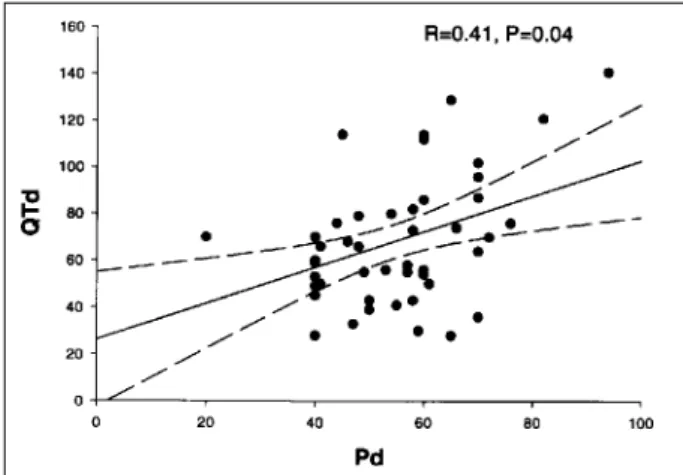
Results - The mean values of P-wave and QT disper-sions were 54±14 ms and 68±27 ms, respectively. The corre-lation between the 2 variables was R=0.41 (p=0.04). In patients with QT dispersion values > 100 ms, P-wave dis-persion was significantly greater than in those with QT dispersion values < 100 ms (58±16 vs 53±12 ms, p=0.04 ).
Conclusion - Our results suggest that, in elderly patients with heart failure, a correlation between the values of P-wave and QT dispersions exists. These fin-dings may have etiopathogenic, pathophysiologic, prog-nostic, and therapeutic implications, which should be in-vestigated in other studies.
Key words: P-wave dispersion, QT dispersion, heart failure
Arq Bras Cardiol, volume 79 (nº 5), 497-9, 2002
Cláudia Szlejf, Jairo Rays, Otávio Celso Eluf Gebara, Núbia Welerson Vieira, Humberto Pierri, Amit Nussbacher, João Batista Serro-Azul, Nelson Samesima, Carlos Alberto Pastore,
Maurício Wajngarten
São Paulo, SP - Brazil
Relation Between the Behaviors of P-Wave and QT Dispersions
in Elderly Patients with Heart Failure
Original Article
Dispersion of the QT interval (QTd) is defined as the difference between the longest and the shortest QT interval in all electrocardiographic leads, which may be possibly measured. That parameter electrocardiographically transla-tes the asynchrony of repolarization of ventricular myocar-dial rows, which is a phenomenon predisposing patients to an abnormal flow of the electric current in the heart. The in-crease in QT dispersion seems to have a prognostic mea-ning for several groups of patients and is associated with nonhomogeneous ventricular repolarization, which may favor reentry mechanisms, and, consequently, the appea-rance of ventricular arrhythmias 1,2. Batchvarov and Malik 3
have reported that QTd should be greater than 100 ms to be interpreted as a signal of abnormality in the course of ven-tricular repolarization. Some studies have shown that indivi-duals with heart failure have greater QTds, suggesting that this electrocardiographic parameter can serve as a predictor of ventricular arrhythmias 4-6.
The concept of P-wave dispersion (Pd) has been re-cently established as the difference between the longest and the shortest P-wave duration in all electrocardiogra-phic leads. An increase in Pd indicates heterogeneous atrial repolarization, favoring reentry mechanisms. Similar to that which happens with the increase in QTd, the increase in Pd can be a predictor of supraventricular arrhythmias 7-9.
Seve-ral studies have suggested that the greatest P-wave disper-sion measured in a 12-lead electrocardiogram is an indepen-dent predictor of paroxysmal atrial fibrillation 7,8,10. Other
au-thors have confirmed this hypothesis with signal-averaged electrocardiography 11-13. So far, no study using body
surfa-ce mapping has been carried out. Body surfasurfa-ce mapping is a noninvasive method that can show the spatial distribu-tion of body surface potentials, providing tridimensional details on the electrocardiogram 14,15 similar to those
ob-tained with an invasive method 16. Its 87 leads distributed all
4 9 8
Szlejf et al
Relation between P-wave and QT dispersions in elderly patients
Arq Bras Cardiol 2002; 79: 497-9.
leads distributed only on the precordium) 17. Therefore, it is
a more appropriate method for studying Pd.
Patients with heart failure have alterations in the con-duction of the stimulus in the atria and ventricles, possibly due to an increase in Pd and QTd.
Methods
We studied consecutively 47 elderly patients (age > 65 years) with heart failure, who were registered at the Heart Fai-lure Outpatient Clinic of the Cardiac Geriatric Clinical Unit of the Instituto do Coração of the Hospital das Clínicas of the Medical School of the University of São Paulo. All patients had stable heart failure and were in NYHA functional classes II or III. Their ejection fraction obtained on the echocar-diogram was lower than 50% (tab. I). All patients had under-gone extensive clinical and laboratory investigation to eli-minate any possible doubt about the diagnosis. Patients who did not have sinus rhythm at the time of the evaluation were excluded from the study. The Committee on Ethics of the hos-pital approved the study protocol, and all patients signed a written consent form before the beginning of the study.
The Mapping System HPM – 7100 from Fukuda Denshi with 87 leads was used for body surface mapping. The patients were put in the dorsal decubitus position with 59 leads on the anterior part of the thorax and 28 leads on the dorsum. The measurements were taken in a semi-auto-matic manner with 2 mobile pointers moved manually on the monitor of the device by 1 single observer. The P-wave dis-persion was calculated based on the difference between the maximum and minimum values of the interval between the beginning of the P wave and the beginning of the QRS complex on the 87 leads. The difference between the maxi-mum and minimaxi-mum values of the interval between the begin-ning of the Q wave and the end of the T wave in all leads was considered the QT dispersion.
The Pearson correlation test was used to compare QT and P-wave dispersions. In addition, the patients were se-parated into 2 subgroups according to QTd with 100 ms used as the cut-off value, as suggested by Batchvarov and Malik 3. The Student t test was used to compare Pd values
between the subgroups. P values < 0.05 were considered statistically significant.
Results
Dispersion of the QT interval was 68.2±27.1 ms, ran-ging from 28 to 141 ms. Dispersion of the P wave was 54.7±14.5 ms, ranging from 20 to 94 ms.
A significant correlation between QTd and Pd was ob-served (R=0.41 and p=0.04) (fig. 1).
The patients with QTd > 100 ms had a significantly greater Pd than those with QTd < 100 ms (tab. II).
Discussion
In the elderly patients with stable heart failure, a signi-ficant and positive correlation between Pd and QTd values
was observed. Patients with QTd > 100 ms showed a signifi-cantly greater Pd than those with QTd lower than that value. As far as we know, this is the first study assessing the relation between Pd and QTd. This relation probably indi-cates the existence of a mechanism of heterogeneous atrial and ventricular repolarization with a common substrate in patients with heart failure.
The physiopathogeny of the alterations of repolariza-tion, which lead to arrhythmias, may be considered multi-factorial with the following determinant factors: a) cardiac chamber size, b) myocardial fibrosis, and c) tonus of the au-tonomic nervous system.
Dilation and hypertrophy of the cardiac chambers due to myocardial overload in heart failure may lead to an altera-tion in the architecture of the cardiac fibers, interfering with the conduction of the electric impulses. However, Zaman et al 18 did
not find any relation between left atrial size and the duration of the P wave on signal-averaged electrocardiography of patients undergoing myocardial revascularization. A pre-vious and still unpublished study carried out at our institu-tion showed no relainstitu-tion between left atrial size and Pd in el-derly patients with heart failure.
Myocardial fibrosis, on the other hand, may cause electrical heterogeneity and anisotropic arrhythmias due to reentry 19. The presence of ischemia or the activation of the
renin-angiotensin-aldosterone system found in coronary
Table I - Case series
Age (years) 75.5 ± 6.21 (65 - 89) Males/females (n) 26/21
Ejection fraction (%) 37.3 ± 6.3 (15 - 50)
Table II - Pd and QTd greater and smaller than 100 ms
QTd > 100 ms QTd < 100 ms p
Arq Bras Cardiol 2002; 79: 497-9.
Szlejf et al Relation between P-wave and QT dispersions in elderly patients
4 9 9 artery disease could account for the remodeling of the
cardiac fibers 20. The investigation of the fibrous cardiac
area is known to depend on sophisticated imaging methods, such as magnetic nuclear resonance. Studies with speciali-zed techniques should be conducted to relate QTd and Pd to the extension of the fibrosis.
It has been reported in the literature that an increase in the sympathetic tonus in heart failure may justify the pre-sence of the prolonged QTd in these patients 4,6. Even
though differences in the atrial and ventricular autonomic tonus may be found, the sympathetic activation of both cardiac chambers due to heart failure may justify the corre-lation between Pd and QTd.
It is worth repeating that the population studied con-sisted of elderly patients with NYHA functional classes II or III for heart failure. These patients were in an advanced
stage of the disease with probable impairment of the 4 car-diac chambers. Therefore, a correlation between the disper-sion of atrial repolarization, represented by Pd, and the dis-persion of ventricular repolarization, represented by QTd, may exist. These findings cannot be extended to patients in an earlier phase of heart failure.
QTd and Pd may perhaps be considered electrocardio-graphic markers predictive of arrhythmias in patients with heart failure. However, studies with long-term follow-up should be conducted to confirm this possibility, so that these parameters can be used in clinical practice.
In conclusion, our results suggest that a relation between P-wave and QT dispersion values exists in elderly patients with heart failure. These findings may have etiopa-thogenic, physiopathologic, prognostic, and therapeutic im-plications for the appearance of arrhythmias in these patients.
References
1. Lombardi F. The QT interval and QT dispersion: the smaller, the better! Eur Heart J 1998; 19: 1279-81.
2. Zabel M, Portnoy J, Franz M. Electrocardiographic indexes of dispersion of ven-tricular repolarization: na isolated heart validation study. J Am Coll Cardiol 1995; 25: 746-52.
3. Batchvarov V, Malik M. Measurements and interpretation of QT dispersion. Pro-gress Cardiovasc Dis 2000; 42: 325-44.
4. Bonnar CE, Davie AP, Caruana L, et al. QT dispersion in patients with chronic heart failure: b blockers are associated with a reduction in QT dispersion. Heart 1999; 81: 297-302.
5. Galinier M, Vialette J-C, Fourcade P, et al. QT interval dispersion as a predictor of arrhythmic events in congestive heart failure. Importance of aetiology. Eur Heart J 1998; 19: 1054-62.
6. Peris VB, Menadas JVM, Ortuño FM, et al. Dispersión del intervalo QT em pacien-tes ingresados por insuficiencia cardíaca. Determinanpacien-tes y valor prognóstico. Rev Esp Cardiol 1999; 52: 563-9.
7. Ciaroni S, Laurence C, Bloch A. Clinical study to investigate the predictive pa-rameters for the onset of atrial fibrillation in patients with essential hypertension. Am Heart J 2000; 139: 814-19.
8. Dilaveris PE, Gialafos EJ, Andrikopoulos GK, et al. Clinical and electrocardio-graphic predictors of recurrent atrial fibrillation. PACE 2000; 23: 352-8. 9. Tükek T, Akkaya V, Demirel S, et al. Effect of Valsalva Maneuver on surface
electro-cardiographic P-wave dispersion in paroxysmal atrial fibrillation. Am J Cardiol 2000; 85: 896-8.
10. Dilaveris PE, Gialafos EJ, Sideris SK, et al. Simple electrocardographic markers for the prediction of paroxysmal idiopathic atrial fibrillation. Am Heart J 1998; 135: 733-8.
11. Kubara I, Ikeda H, Hiraki T, Yoshida T, Ohga M, Imaizumi T. Dispersion of filtered
P wave duration by P wave Signal-averaged ECG Mapping System: Its useful-ness for determining efficacy of disopyramide on paroxysmal atrial fibrillation. J Cardiovasc Electrophysiol 1999; 10: 670-9.
12. Villani GQ, Massimo P, Rosi A, Capucci A. P-wave Dispersion Index: a marker of patients with paroxysmal atrial fibrillation. Int J Cardiol 1996; 55: 169-75. 13. Yamada T, Fukunami M, Shimonagata T, et al. Dispersion of signal-averaged P
wave duration on precordial body surface in patients with paroxysmal atrial fi-brillation. Eur Heart J 1999; 20: 211-20.
14. Miller JM, Zipes DP. Management of the patient with cardiac arrhythmias. In: Braunwald E, ed. Heart Disease: A Textbook of Cardiovascular Medicine, 6th ed.
St. Louis: WB Saunders Co., 2001: 700-74.
15. Mirvis DM, Golberger AL. Electrocardiography. In: Braunwald E, ed. Heart Di-sease: A Textbook of Cardiovascular Medicine, 6th ed. St. Louis: WB Saunders
Co., 2001: 82-128.
16. Lux RL, Fuller MS, MacLeod RS, Ershler PR, Punske BB, Taccardi B. Noninva-sive indices of repolarization and its dispersion. J Electrocardiol 1999; 32(suppl): 153-7.
17. Taccardi B, Punske BB, Lux RL, et al. Useful lessons from body surface mapping. J Cardiovasc Electrophysiol1998; 9: 773-86.
18. Zaman, AG, Archbold A, Helft G, Paul EA, Curzen NP, Mills PG. Atrial fibrilla-tion after coronary artery bypass surgery: a model for preoperative risk stratifica-tion. Circulation 2000; 149: 251-66.
19. Schneider CA, Voth E, Baer FM, Horst M, Wagner R, Sechtem U. QT dispersion is determined by the extent of viable myocardium in patients with chronic Q-wave myocardial infarction. Circulation 1997; 96: 3913-20.