Effe ct o f acute and re pe ate d re straint
stre ss o n gluco se o xidatio n to CO
2
in
hippo cam pal and ce re bral co rte x slice s
Departamento de Bioquímica, Instituto de Ciências Básicas da Saúde, Universidade Federal do Rio Grande do Sul, Porto Alegre, RS, Brasil I.L.S. Torres, G.D. Gamaro,
S.N. Silveira-Cucco, M.B. Michalowski, J.B. Corrêa, M.L.S. Perry and C. Dalmaz
Abstract
It has been suggested that glucocorticoids released during stress might impair neuronal function by decreasing glucose uptake by hippocam-pal neurons. Previous work has demonstrated that glucose uptake is reduced in hippocampal and cerebral cortex slices 24 h after exposure to acute stress, while no effect was observed after repeated stress. Here, we report the effect of acute and repeated restraint stress on glucose oxidation to CO2 in hippocampal and cerebral cortex slices
and on plasma glucose and corticosterone levels. Male adult Wistar rats were exposed to restraint 1 h/day for 50 days in the chronic model. In the acute model there was a single exposure. Immediately or 24 h after stress, the animals were sacrificed and the hippocampus and cerebral cortex were dissected, sliced, and incubated with Krebs buffer, pH 7.4, containing 5 mM glucose and 0.2 µCi D-[U-14C]
glucose. CO2 production from glucose was estimated. Trunk blood
was also collected, and both corticosterone and glucose were meas-ured. The results showed that corticosterone levels after exposure to acute restraint were increased, but the increase was smaller when the animals were submitted to repeated stress. Blood glucose levels increased after both acute and repeated stress. However, glucose utilization, measured as CO2 production in hippocampal and cerebral cortex slices, was the same in stressed and control groups under conditions of both acute and chronic stress. We conclude that, al-though stress may induce a decrease in glucose uptake, this effect is not sufficient to affect the energy metabolism of these cells.
Co rre spo nde nce
I.L.S. Torres
Departamento de Bioquímica Instituto de Ciências Básicas
da Saúde, UFRGS, Lab. 32 Rua Ramiro Barcelos, 2600 (Anexo) 90035-003 Porto Alegre, RS Brasil
Fax: + 55-51-316-5540
Research supported by PRO NEX,
CNPq and FAPERGS.
Received O ctober 13, 1999 Accepted O ctober 11, 2000
Ke y wo rds
·Restraint stress ·Chronic stress ·CO2 production ·Glucose oxidation ·Hippocampus ·Cerebral cortex
The acute secretion of glucocorticoids is critical for responding to stress. On the other hand, chronic stress produces deleterious effects (1,2). A classical catabolic action of glucocorticoids in numerous peripheral tis-sues is to inhibit glucose uptake into cells (3,4). Studies have shown that in
hippocam-pal cell cultures glucocorticoids significant-ly inhibit glucose uptake and oxidation both by neurons and astrocytes (4,5).
not show the same behavior and do not experience the same consequences as ani-mals exposed to acute stress (6,7). Different neurotransmitter systems have been sug-gested to play a role in this stress desensitiza-tion process (e.g., 7,8). Probably, these neu-rotransmitters and glucocorticoids released during stress could modify glucose metabo-lism, leading to adaptation and to different glucose uptake and use.
The present study aimed to evaluate the consequences of acute or repeated exposure to restraint stress in rats on CO2 production
from glucose in slices of cerebral cortex and hippocampus at different times (0 or 24 h) after restraint stress.
Sixty-six adult male Wistar rats (60 days at the beginning of treatment) weighing 180-230 g were used. The experimentally naive animals were housed in groups of 6 to 8 rats in Plexiglas home cages (65 x 25 x 15 cm) with the floor covered with sawdust. The animals were maintained on a standard dark-light cycle (dark-lights on between 7:00 and 19:00 h), at a room temperature of 22 ± 2o
C. The rats had free access to food (standard lab rat chow) and water, except during the period when restraint stress was applied. In the experiment involving chronic stress the ani-mals were divided into three groups: stressed, handled and control. The stressed group was taken to a different room, where restraint was carried out by placing the animal in a 25 x 7-cm plastic bottle and fixing it with plas-ter tape on the outside, so that it was unable to move. A 1-cm hole was left at one end for breathing. The animals were stressed 1 h/ day, 5 days a week for 50 days. The immobi-lization procedure was performed between 10:00 and 12:00 h. The animals in the handled group were manipulated but not submitted to restraint. The animals were taken to a differ-ent room, also between 10:00 and 12:00 h. Handling consisted of gently manipulating each animal for 1 min. This group was in-cluded since handling may alter biochemical and behavioral parameters, both in young
and adult rats (9-11). Control animals were kept undisturbed in their home cages. In the experiment involving acute stress, the ani-mals were submitted to a single 1-h exposure and the control group was not stressed.
Animals were sacrificed by decapitation, immediately or 24 h after the last restraint session in the case of chronic treatment, and immediately after restraint in the case of acute stress. Immediately after decapitation, trunk blood was collected into heparinized tubes for corticosterone and glucose deter-mination. Total corticosterone concentrations were analyzed directly in plasma by 125I
radioimmunoassay (Amersham Life Science Inc., Cleveland, OH, USA). Glucose was determined in plasma by the glucose oxidase method (Bio Diagnostica, Piraquara, PA, Brazil). The brains were quickly removed and the hippocampus and cerebral cortex were dissected out on ice. After removing the cerebral cortex with a scalpel and twee-zers, it was possible to visualize the hippo-campus, which was removed with small twee-zers. The structures were weighed, and slices prepared within 5 min. Each tissue was cut into 0.3-mm slices using a McIlwain tissue chopper. About 50 mg of tissue slices were incubated in 1 ml Krebs-Ringer bicarbonate buffer, pH 7.4, containing 5 mM D-glucose and 0.2 µCi D-[U-14C] glucose (specific
ac-tivity: 3.0 mCi/mmol). Saturating glucose concentrations were used for CO2
produc-tion. The content of the flasks was gassed with 95% O2/5% CO2 for 1 min and the
flasks were sealed with rubber caps. The slices were incubated at 34o
C for 1 h in a Dubnoff metabolic shaker (60 cycles/min). Incubation was stopped by adding 0.2 ml 50% TCA through the rubber cap. Then 0.2 ml of 1 M hyamine hydroxide was injected into the central wells to trap CO2. These
were small glass wells situated inside the flasks and above the level of the incubation medium. The flasks were shaken for a fur-ther 30 min at 34o
C to trap CO2, after which
vials and assayed for CO2 radioactivity in a
liquid-scintillation counter (12).
All results are expressed considering the initial specific activity of the incubation medium, in such a way that for a given number of counts of CO2 radioactivity per
mg of tissue, the equivalent glucose con-sumed was calculated as pmol. The CO2
production rate was constant over 30, 60 and 90 min of incubation.
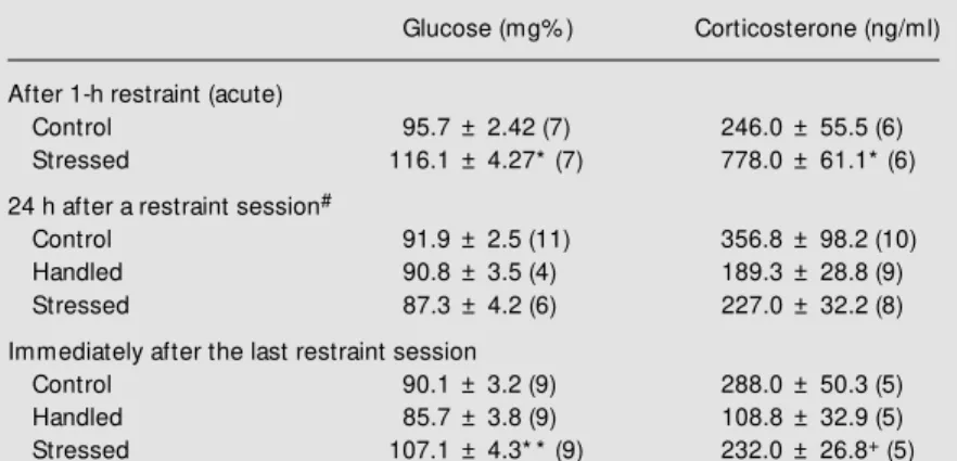
Blood glucose and corticosterone con-centrations after acute or chronic restraint stress are presented in Table 1. Corticoster-one and glucose concentrations increased in plasma after 1-h acute restraint stress (Stu-dent t-test, t = 6.45, P<0.001 for
corticoster-one and t = 4.16, P<0.001 for glucose). After exposure to repeated restraint, there was no difference in plasma corticosterone or glu-cose between groups when the hormones were measured 24 h after the last restraint session (one-way ANOVA, F = 1.790, P>0.05 for corticosterone and F = 0.535, P>0.05 for glucose), and an increase in plasma glucose again occurred after another exposure to restraint (one-way ANOVA, F = 8.784, fol-lowed by the Student-Newman-Keuls test, P<0.05); plasma corticosterone was slightly increased when compared to the handled group but not when compared to the control group (one-way ANOVA, F = 5.823, fol-lowed by the Student-Newman-Keuls test, P<0.05).
The in vitro CO2 production from
glu-cose in cerebral cortex and hippocampus slices after 1-h restraint was measured in rats sacrificed immediately or 24 h later, and the results are shown in Figure 1A and B. When CO2 production was measured immediately
or 24 h after restraint, there was no signifi-cant difference between control and stressed rats (immediately after restraint: cortex: t = 0.87, hippocampus: t = 0.084; 24 h after stress: cortex: t = 0.57, hippocampus: t = 2.17; P>0.05 in all cases). Production of CO2
from glucose was not affected by chronic restraint (Figure 1C) either in hippocampus
Figure 1 - Effect of acute and
chronic stress on CO2
produc-tion from glucose in slices of cerebral cortex and hippocam-pus. Data are reported as mean
± SEM . A, Immediately after 1-h
restraint (acute) stress (N = 4-8
per group). B, Tw ent y-f our
hours after acute stress (N =
6-7 per group). C, Animals
sub-mitted to chronic stress and sac-rificed 24 h after being submit-ted to the last restraint stress (N = 10-11 per group).
p m o l G lu c o s e m g t is s u e
-1 h -11000 800 600 400 200 0 Control Stressed
Hippocampus Cerebral cortex
p m o l G lu c o s e m g t is s u e
-1 h
-1 1200 800 400 0 p m o l G lu c o s e m g t is s u e
-1 h -1 500 400 300 200 100 0 Control Stressed
Hippocampus Cerebral cortex
Control Handled
Hippocampus Cerebral cortex
A
B
C
Table 1 - Plasma levels of glucose and corticosterone in rats submitted to acute (1 h) or repeated restraint stress.
Data are reported as mean ± SEM and the number of animals is given in parentheses.
* P<0.001 compared to control group (Student t-test); * * P<0.05 compared to control
and handled groups (Student-New man-Keuls test); +P<0.05 compared to handled
group (Student-New man-Keuls test); #no difference betw een groups (P>0.05,
one-w ay ANOVA).
Glucose (mg% ) Corticosterone (ng/ml)
After 1-h restraint (acute)
Control 95.7 ± 2.42 (7) 246.0 ± 55.5 (6)
Stressed 116.1 ± 4.27*(7) 778.0 ± 61.1*(6)
24 h after a restraint session#
Control 91.9 ± 2.5 (11) 356.8 ± 98.2 (10)
Handled 90.8 ± 3.5 (4) 189.3 ± 28.8 (9)
Stressed 87.3 ± 4.2 (6) 227.0 ± 32.2 (8)
Immediately after the last restraint session
Control 90.1 ± 3.2 (9) 288.0 ± 50.3 (5)
Handled 85.7 ± 3.8 (9) 108.8 ± 32.9 (5)
Stressed 107.1 ± 4.3* *(9) 232.0 ± 26.8+ (5)
or cerebral cortex slices, when the animals were sacrificed 24 h after exposure to stress (P>0.05, one-way ANOVA).
Glucose metabolism is essential for brain
function and structure. In the normal state, glucose is the only significant substrate for energy metabolism in the brain. The actual glucose oxidation to CO2 is not only
suffi-cient to account for total O2 consumption but
is in excess. The excess glucose is probably used to produce other intermediates of car-bohydrate metabolism, or used for synthesis of the chemical constituents of the brain rather than for energy production (13). The present experiments were conducted to evalu-ate the consequences of acute and repeevalu-ated exposure to restraint stress in rats on CO2
production from glucose in slices of cerebral cortex and hippocampus. These measure-ments are directly related to energy produc-tion from glucose.
Changes in the hippocampus after expo-sure to stress have been reported by several authors (1,2). The human hippocampus has been shown to present atrophy in different pathologic circumstances related to high cor-tisol levels (14,15). Moreover, a previous stress history was found to be related to impairment of cognitive functions in aging rats (16). These findings led to the develop-ment of the glucocorticoid hypothesis of brain aging and degeneration.
The mechanism by which glucocorticoids may exert their damaging effects on hippo-campal neurons is thought to involve disrup-tion of the energy metabolism of hippocam-pus neurons (5), and it has been suggested that a critical feature of the action of gluco-corticoids on neurons might be the inhibi-tion of glucose oxidainhibi-tion (4,5). Some reports suggest that glucocorticoids inhibit glucose uptake in the brain both in vivo (17,18) and
in cultured hippocampal neurons and glia (4,5,19). Previous results showed that when glucose uptake was measured soon after re-straint stress (1 h) there was no significant difference between control and stressed rats in glucose uptake, but when measured 24 h after the stress session there was a signifi-cant decrease in glucose uptake in both struc-tures (cortex and hippocampus) in stressed
rats (18). This effect is probably related to a corticosteroid-dependent effect, since many of the primary effects of adrenal steroids involve transcriptional regulation of a subset of genes in specific cell types that needs some time to be resumed. These results are in agreement with those obtained by other authors with cultures containing both neu-rons and glia (4,5). These authors found a time-dependent effect, with more than 4 h of steroid exposure needed for the inhibition of glucose uptake to be observed. This disrup-tive glucocorticoid effect could be mediated by a decreased expression of glucose trans-porter molecules on the cell surface. It has been shown that treatment of fibroblasts with dexamethasone for several hours reduces glucose transporters (19).
Nevertheless, there is little direct evi-dence to indicate whether brain energy me-tabolism itself is compromised after expo-sure to excess corticosterone or to stress. In the present study there was no difference in CO2 production from glucose in slices of
cerebral cortex or hippocampus from rats submitted to a restraint stress session when compared to the control group either 1 h or 24 h later. This result suggests that, although there may be decreased glucose uptake, as suggested by several investigators (4,5,17-19), the energy production is adequate. This fact suggests that the possible effects of glu-cocorticoid release on glucose transport are not sufficient to generate a physiologically important energy deficit under normal con-ditions. It is possible, however, that other metabolic routes are being affected (e.g., synthesis of lipids, glucoproteins, and glyco-gen).
acceler-ated the rate of ATP loss in neurons in response to cyanide and aglycemia.
When rats were submitted to repeated restraint and sacrificed 24 h after exposure to stress no difference was observed in CO2
production. It is important to note that the levels of corticosterone observed after acute restraint stress are higher than those ob-served after repeated restraint. In the chronic model, plasma levels of corticosterone pre-sent a modest increase immediately after restraint compared to the handled group but not when compared to the control group.
Handling is known to induce biochemi-cal and behavioral alterations, and an in-creased anxiogenic response in animals na-ive to handling has been suggested (9,10). Although corticosterone levels did not differ significantly in chronically treated animals 24 h after the last restraint session, they were
lower in the handled and stressed groups, showing that daily handling may possibly lead to an adaptation in hormonal release. Handling during the neonatal period causes alterations in the response to stress, with reduced levels of glucocorticoids, and in-creased density of glucocorticoid receptors in the hippocampus (11), but similar find-ings have not been reported in adults.
In conclusion, the present study demon-strates that although changes in glucose up-take by hippocampal and cerebral cortex cells may be part of the brains response to stress, as demonstrated by other authors, this effect does not imply a decreased energy production from glucose, at least under these conditions, when the cells were not submit-ted to any other insult. Moreover, repeasubmit-ted exposure to the same stressful event has no effect on glucose oxidation to CO2.
Re fe re nce s
1. Uno H, Tarara R, Else JG, Suleman M A & Sapolsky RM (1989). Hippocampal dam-age associated w ith prolonged and fatal
stress in primates. Journal of
Neurosci-ence, 9: 1705-1711.
2. M agarinos AM & M cEw en BS (1995). Stress-induced atrophy of apical dendrites of hippocampal CA3c neurons: involve-ment of glucocorticoid secretion and
exci-tatory amino acid receptors.
Neurosci-ence, 69: 89-98.
3. M unck A (1971). Glucocorticoid inhibition of glucose uptake by peripheral tissues: old and new evidences, molecular
mecha-nisms and physiological significance.
Per-spectives in Biology and M edicine, 14: 265-269.
4. Horner H, Packan D & Sapolsky R (1990). Glucocorticoids inhibit glucose transport in cultured hippocampal neurons and glia. Neuroendocrinology, 52: 57-63. 5. Virgin CE, Ha TP, Packan DR, Tombaugh
GC, Yang SH, Horner HC & Sapolsky RM (1991). Glucocorticoids inhibit glucose transport and glutamate uptake in hippo-campal astrocytes: implications for
gluco-corticoids neurotoxicity. Journal of
Neuro-chemistry, 57: 1422-1428.
6. Kennett GA, Dickinson SL & Curzon G (1885). Central serotoninergic responses
and behavioral adaptation to repeated immobilisation: the effect of the corticos-terone synthesis inhibitor metyrapone. European Journal of Pharmacology, 119: 143-152.
7. Lachuer J, Delton I, Buda M & Tappaz M (1994). The habituation of brainstem cate-cholaminergic groups to chronic daily re-straint stress is stress specific like that of the hypothalamo-pituitary-adrenal axis. Brain Research, 638: 196-202.
8. Cancela LM , Volosin M & M olina VA (1990). Opioid involvement in the
adap-tive change of 5-HT1a receptors induced
by chronic restraint. European Journal of
Pharmacology, 176: 313-319.
9. Boix F, Fernandez Teruel A, Escorihuela RM & Tobena A (1990). Handling-habitua-tion prevents the effects of diazepam and alprazolam on brain serotonin levels in
rats. Behavioural Brain Research, 36:
209-215.
10. File SE & Fluck E (1994). Handling alters habituation and response to stimulus
change in the holeboard. Pharmacology,
Biochemistry and Behavior, 49: 449-453. 11. Liu D, Diorio J, Tannenbaum B, Caldji C, Francis D, Feedman A, Sharma S, Pearson D, Plotsky PM & M eaney M J (1997). M a-ternal care, hippocampal glucocorticoid
receptors, and
hypothalamic-pituitary-ad-renal responses to stress. Science, 277:
1659-1662.
12. Bueno D, Azzolin IR & Perry M LS (1994). Ontogenetic study of glucose and lactate
utilization by rat cerebellum slices. M
edi-cal Science Research, 22: 631-632. 13. Clarke DD & Sokoloff L (1998). Circulation
and energy metabolism of the brain. In:
Siegel GJ (Editor), Basic Neurochemistry
-M olecular, Cellular and -M edical Aspects. 6th edn. Lippincott-Raven, Philadelphia, 637-669.
14. Bremner JD, Randall P, Scott TM , Bronen RA, Seibyl JP, Southw ick SM , Delaney RC, M cCarthy G, Charney DS & Innis RB (1995). M RI-based measurement of hip-pocampal volume in patients w ith com-bat-related posttraumatic stress disorder. American Journal of Psychiatry, 152: 973-981.
15. Starkman M N, Giordani B, Gebarski SS, Berent S, Schork M A & Schteingart DE (1999). Decrease in cortisol reverses hu-man hippocampal atrophy follow ing
treat-ment of Cushing’s disease. Biological
Psy-chiatry, 46: 1595-1602.
Fischer-344 rats. Neurobiology of Learn-ing and M emory, 66: 1-10.
17. Doyle P, Guillaum e-Gentil C, Rohner-Jeanrenaud F & Rohner-Jeanrenaud B (1994). Ef-fects of corticosterone administration on local cerebral glucose utilization of rats. Brain Research, 645: 225-230.
18. Xavier M H, Gam aro GD & Dalm az C
(1995).Efeito do estresse agudo e crônico
sobre a captação de glicose em
hipo-campo e córtex cerebral de ratos. X
An-nual M eeting of the Federação de Socie-dades de Biologia Experimental, Serra Negra, M G, Brazil, August 23-26, Abstract 1.142, 36.
19. Horner H, M unck A & Lienhard G (1987). Dexamethasone causes translocation of glucose transporters from the plasma
membrane to an intracellular site in
hu-m an fibroblasts. Journal of Biological
Chemistry, 262: 17696-17703.
20. Law rence M S & Sapolsky RM (1994). Glucocorticoids accelerate ATP loss follow -ing metabolic insults in cultured
hippo-campal neurons. Brain Research, 646: