Pathogenesis and treatment of glomerulonephritis-an update
Patogênese e tratamento da glomerulonefrite - uma atualização
A presente revisão traz os conceitos mais at-uais acerca dos fatores de risco genéticos, eventos etiológicos, respostas nefritogênicas e tratamento dos principais tipos de glomerulo-nefrite (GN) imunomediada. Tais patologias incluem GN pós-infecciosa, nefropatia por IgA, doença por anticorpo antimembrana basal glomerular (anti-MBG), vasculite asso-ciada a ANCA (VAA) e nefrite lúpica. Ape-sar da(s) etiologia(s) da maioria dos casos de GN permanecer indefinida, acredita-se que seu início se deva, em grande parte, a insul-tos ambientais, particularmente na forma de processos infecciosos que deflagram respostas de hospedeiro em indivíduos geneticamente suscetíveis, levando assim a quadros de GN. A concepção mecanicista em torno dessas pa-tologias evoluiu a partir da visão mais antiga de que a maioria seria consequência do apri-sionamento glomerular de complexos imunes pré-formados para a percepção atual de que as mesmas, em sua maioria, são doenças au-toimunes por natureza mediadas por anticor-pos e linfócitos T reativos a auto-antígenos. O tratamento da GN não tem acompanhado os progressos na compreensão de sua pa-togênese. Os papéis recentemente atribuídos a mediadores mais antigos como complemento e proteínas reguladoras do complemento lan-çam luz sobre novos alvos terapêuticos.
R
ESUMOPalavras-chave: glomerulonefrite; glomer-ulonefrite por IGA; nefrite; nefrite lúpica. This review updates current concepts
of the genetic risk factors, etiologic events, nephtitogenic responses and treatment of the major immunologically mediated types of glomerulonephritis (GN). These include post-infectious GN, IgA nephropathy, anti-glomerular basement membrane (GBM) antibody disease, ANCA-associated vasculitis (AAV) and lupus nephritis. Although the etiology(s) of most GNs remain un-defined, many are now believed to be initiated by environmental insults, par-ticularly infectious processes, that tri-gger host responses in genetically sus-ceptible individuals which lead to GN. Mechanistic concepts of these diseases have evolved from earlier views that most were consequent to glomerular trapping of preformed immune com-plexes to the current view that most of these diseases are auto-immune in na-ture mediated by both antibodies and T cells reactive with self-antigens. The-rapy of GN has lagged behind advances in understanding pathogenesis. Newly appreciated roles for older mediators like complement and complement re-gulatory proteins offer new therapeutic targets.
A
BSTRACTKeywords: glomerulonephritis; glomerulo-nephritis, IGA; lupus nephritis; nephritis.
Authors
William G Couser 1
1 University of Washington.
Submitted on: 01/20/2016. Approved on: 01/20/2016.
Correspondence to:
William G Couser. University of Washington. 16050 169th AV NE Woodinville, WA, USA.
CEP: 98072-8949 E-mail: [email protected]
DOI: 10.5935/0101-2800.20160016
I
NTRODUCTIONThe past decade has witnessed major advances in understanding the etiology and pathogenesis of glomerulonephritis (GN). Rapidly evolving molecular, genetic and data management technologies have lead to the appreciation that most of the immunologically-mediated forms of GN have an auto-immune basis and
in older male patients with significant associated co-morbidities - especially diabetes, HIV infection and malignancy.6-12
The pathogenesis of PSGN has always been assu-med to reflect the mechanisms originally defined in acute BSA-serum sickness models in rabbits that in-volve passive glomerular trapping of circulating im-mune complexes (CIC) composed of nephritogenic bacterial antigens and IgG antibody to them, activa-tion of complement (C) by IgG through the classical pathway and attraction and activation of neutro-phils that release oxidants, proteases and neutrophil extracellular traps (NETs) that inflict the resulting glomerular tissue injury.6-13 In fact, PSGN, and other IRGNs, are the only group of GNs in which exo-genous antigens, either as a component of passive-ly trapped CICs or as initiators of in situ immune deposit formation, are still regarded as essential mediators of glomerular injury whereas the mecha-nisms underlying most other forms of GN are now considered to be primarily autoimmune3 (Table 1). Autoimmune phenomena are certainly seen in PSGN as well including IgG and IgM rheumatoid factors, cryoglobulins, anti-DNA, anti-C1q, anti-endothelial cell antibody, anti-C3convertase (C3Nef), anti-ne-phritis-associated plasmin receptor and others.1,12 In fact, the only kidney elution study reported in PSGN to date found only antibody to IgG (rheumatoid fac-tor) and no antibodies to streptococcal antigens.14 However, none of these findings have been frequent or consistent enough to establish an autoimmune pa-thogenesis for PSGN.
entities.3,4 Paralleling these advances have been new approaches to therapy that include more specific, and potentially less toxic, agents, particularly biologic agents, that are now showing considerable promise in treating these diseases - indeed some have already been incorporated into current therapeutic guidelines.5
The purpose of this review is to summarize and update concepts of the etiopathogenesis and treatment of the major forms of GN (post-infectious GN, IgA nephropathy, anti-GBM nephritis, ANCA-associated GN and lupus nephritis) as they have evolved over the past decade. Diseases presenting primarily as nephrotic syndrome (minimal change/focal sclerosis spectrum, membranous nephropathy, membranoproliferative GNs and C3 nephropathies) are not covered here. In reviewing these GNs I will also emphasize newer ways of thinking about these diseases that are not always firmly established concepts today but point to new directions in which this field is moving.
P
OST-I
NFECTIOUSGN (PSGN)
PATHOGENESISOF POST-INFECTIOUS GN
Although still considered the prototype of acute GN, classic post-streptococcal GN (PSGN) has become a rare disease in developed countries.6 This has been accompanied by an increase in the incidence of non-streptococcal “post-infectious” GNs, or “infection-related” GNs (IRGN). These entities more often present with acute kidney injury (AKI) or nephrotic syndrome than with the typical acute nephritic syndrome associated with PSGN and occur primarily
TABLE 1 SERUMCOMPLEMENTPROFILESANDAUTO-IMMUNEFEATURESOFTHEMAJORFORMSOFGN. MOSTOFTHESE DISEASESARENOWBELIEVEDTOHAVEANAUTOIMMUNECOMPONENTTOTHEIRPATHOGENESIS. (ADAPTED WITHPERMISSIONFROMREFERENCE 3. COUSERWG. BASICANDTRANSLATIONALCONCEPTSOFIMMUNE-MEDIATED
GLOMERULARDISEASES. J AM SOC NEPHROL. 2012:23:381-99.)
Disease Serum C profile Auto-immune features
Post-streptococcal glomerulonephritis AP or MBL normal C1q, Low C3-C9
Anti-C1q, IgG
AECA*, anti-DNA, ANCA, protein disulide Isomerase (PDI), cardiac
myosin
IgA nephropathy Normal Anti-glycan, endothelial cell,
mesangial cell, IgG, C1q
Anti-GBM nephritis Normal Anti-GBM, ANCA (20%), anti-C1q
ANCA-positive glomerulonephritis Normal Anti-MPO, PR3, cPR3, NET, DNA,
endothelial cell, ?HLAMP2
Lupus nephritis CP, low C1q-C9
Anti-dsDNA, annexin, MPO, PR3, nucleosome, IgG, C1q, C1s, C1-INH,
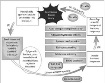
Figure 1. Schematic overview of the mechanisms linking initial exposure to an etiologic agent in a genetically susceptible individual to an autoimmune response and glomerular tissue injury. 1. Hereditable risk factors predispose certain individuals to respond to environmental factors in ways that can lead to a nephritogenic autoimmune response (Hit #1). 2. Exposure to infectious etiologic agents in the environment occurs (Hit #2), may be modiied by epigenetic factors, and activates the innate immune system through interactions with TLRs and complement. 3. Conversion of a non-antigen-speciic innate immune response to an antigen-speciic adaptive immune response directed at speciic auto-antigens can occur through several pathways that may operate simultaneously and in concert. These include defects in regulation of existing natural autoimmunity, molecular mimicry, epitope spreading, epitope conformational changes, adjuvant/ bystander efects and auto-antigen complementarity. 4. The adaptive immune response generates antigen-speciic T and B cells, usually directed at antigens that are ixed or “planted” in the glomerulus. These immune reactants, usually through inlammatory efector cells and/or complement, mediate tissue damage. (Reprinted with permission from Reference 1. Couser WG, Johnson RJ. The etiology of glomerulonephritis: roles of infection and autoimmunity. Kidney Int. 2014;86:905-14.)
However, newer serologic and immunopathologic data now suggest the pathogenesis is more complicated and implicate primarily the alternative C pathway (AP) in these diseases.1,15-17 It has long been appreciated that C3 is the dominant component seen by IF with IgG much less prominent and sometimes absent, a phenomenon not seen in traditional immune complex-mediated diseases.3,17 Some proposed nephritogenic streptococcal antigens localized in glomeruli, such as pyogenic streptococcal exotoxin B (PSEB), can activate the AP directly through the mannose-binding lectin (MBL) pathway, independently of antibody, a process that might account for the long appreciated co-localization of PSEB and C31,11,12 and the dominance of C3 in glomerular deposits.16 C activation in PSGN is also predominately via the alternative pathway (AP) whereas C activation by IgG-containing ICs usually occurs through the classical pathway.3 Indeed, both genetic and acquired deficiencies in complement
regulatory proteins like complement factor H (CFH) have now been reported in “atypical” PSGN patients who exhibit prolonged, progressive disease rather than complete recovery as is classically seen in childhood PSGN.17 This supports considering many cases of PSGN as one part of a spectrum of that includes some C3-dominant IRGNs and the recently recognized “C3 nephropathies” rather than as a traditional BSA-like immune complex (IC) disease. Thus we now recognize a spectrum of overlapping entities that include classic PSGN, IRGN, atypical PSGN, and C3 nephropathies in which infections are often precipitating events but deposition of CICs seems unlikely to be the major mechanism, and host abnormalities in C activation and regulation may play more important roles. Whether these newer mechanisms are operative only in unusual cases or play a more generalized role in PSGN awaits further study.
TREATMENT OF POST-INFECTIOUS GN
Current guidelines for treatment of PSGN, or other infection-related GNs, involve only supportive care in PSGN and treatment of on-going infection in IRGN.6-9 Although some have advocated use of pulse steroids in PSGN patients who present with AKI and crescentic glomerular lesions, there is minimal data to support the benefit of this approach, especially in patients with on-going bacterial infections. However, in response to the changing concepts of disease pathogenesis outlined above, some patients refractory to steroid therapy have been treated with Eculizumab, a humanized monoclonal anti-C5 antibody that inhibits C5 activation, and dramatic benefits have been observed.18
viral molecular patterns may become an option for genetically susceptible individuals in endemic areas in the years ahead.
Because of the favorable prognosis in PSGN, there is no data on recurrent disease in renal allografts.
IGA N
EPHROPATHY(IGAN)
PATHOGENESIS OF IGAN
Major advances in understanding the pathogenesis of IgAN, the most common form of GN in the world, have occurred over the past decade despite the fact that progress in this disease has been hindered by the lack of a good animal model caused by mechanisms similar to those defined in the human disease.20,21 New findings include appreciation that the glomerular-deposited IgA is IgA1, normally of mucosal origin, that a fraction of this IgA1 is under-glycosylated in the hinge region in both patients and disease-free relatives, and that many patients exhibit an IgG anti-underglycosylated IgA1 antibody (anti-glycan antibody) response that correlates with disease activity, outcome and recurrence.20,21 Finally, the IgG anti-glycan antibody is directed at the site of underglycosylation in the hinge region of IgA1, a site that exhibits molecular mimicry with some bacterial antigens such as TN.21
Over 100 genes associated with increased risk for IgAN have now been identified and implicate the innate immune system, likely responsible for the immediate hematuria commonly observed following upper respiratory tract or gastrointestional infections in IgAN patients, autoimmunity (HLA alleles), mucosal barrier function and the complement system.20-22 Although still poorly defined, it seems likely there is some connection between the gastrointestional system and its mucosal-associated lymphoid tissue, intestinal micro biota, the innate immune system and development of IgAN.23 The presence of predominately IgA deposits in some cases of IgA dominant post-infectious GN following infection with methicillin-resistant staph aureus24 and occasional demonstration of staph antigens in the mesangium in IgAN25 as well as the observation that IgG anti-glycan antibodies exhibit molecular mimicry with bacterial TN antigens all support a role for an infectious etiology involving the innate and mucosal immune systems. Other non-infectious causes of intestinal inflammation and barrier dysfunction such as inflammatory bowel disease, gliadin or other dietary intolerances may be etiologic as well.23
Although CICs containing galactose-deficient IgA1 and IgG, IgA or IgM antibody to it are present in the circulation, they do not correlate well with clinical or pathologic features of the disease. It remains unclear if the mesangial IgA deposits reflect primarily these preformed ICs trapped from the circulation as suggested by some authors20,21 or in situ formation of ICs due the inability of asialoglycoprotein receptors in liver and spleen to clear these high molecular weight, glycan-deficient molecules from the serum with consequent uptake in the glomerular mesangium where they serve as “planted” antigens for IC formation.3 Finally, it is increasingly clear that, although IgA is a poor activator of the classical pathway of complement compared to IgG, complement activation through the MBL or AP is ongoing in IgAN as assessed by increased glomerular deposition of short-lived C3c and increased serum levels of complement activation products that correlate with disease activity and outcomes.26-28 Sublytic C5b-9 attack on mesangial cells activates them to proliferate and over-produce oxidants, proteases, cytokines (ΤGFβ), growth factors (PDGF) and extracellular matrix material that together result in the typical focal proliferative GN with mesangial matrix expansion characteristic of IgAN (3). Thus IgAN joins most other forms of GN in occurring consequent to a genetically-determined autoimmune response to environmental, likely predominately infectious, agents.1,3
TREATMENT OF IGAN
Because of its frequency, a number of therapeutic initiatives have been well studied in IgAN. The recent development and validation of a histologic classification system, the Oxford-MEST classification, will facilitate future clinical trials and selection of patients for therapy.29 The benefit of good blood pressure control and reducing urine protein excretion to < 500 mg/day using 6 mos of conservative therapy with ACE inhibitors and/or angiotensin receptor blockers is well established and is the only therapy needed in over 75% of patients.4,30
also beneficial in reducing proteinuria and potentially slowing progression.33
Although additional immunosuppression (aza-thioprine, cyclophosphamide or MMF) has reduced rates of protein excretion in IgAN in some studies, no benefit on preservation of renal function has been shown, and these agents are not generally recom-mended except in rare patients with rapidly progres-sive, crescentic disease.30,32 Multiple studies have also failed to firmly establish a benefit for fish oil or ton-sillectomy in progressive IgAN, although fish oil in a dose of > 3.3 gm/day is recommended in some guide-lines.4,30 No credible data on the efficacy of biologic agents such as Rituximab and Eculizumab, or PDGF or ΤGFβ inhibitors, is available yet in IgAN, although several studies are in progress.5,30
IgAN recurs histologically in up to 30% of renal allografts, but usually is manifest only as mesangial deposition of IgA by IF. Recurrence has significant clinical manifestations in about 13% and has a relatively minimal impact on long term graft survival compared to other recurrent GNs.34
ANTI-GBM NEPHRITIS
PATHOGENESISOF ANTI-GBM NEPHRITIS
Despite its infrequency, anti-glomerular basement membrane antibody (aGBM) disease remains the prototype of autoimmune disease in man. The disease accounts for about 12% of US patients with rapidly progressive, crescentic GN, and over 80% of patients have crescent formation in > 50% of glomeruli.35 Much of what we understand about the immune pathogenesis of acute, inflammatory glomerular diseases in general is derived from studies in animal models of aGBM disease (“nephrotoxic nephritis”, NTN) developed by transferring heterologous anti-GBM IgG into multiple different species (rabbit, sheep, monkey, rat, mouse, guinea pig).3,36,37
The etiology of the disease remains unknown although preceding infections, both bacterial and viral, have been frequently noted clinically, and molecular mimicry between GBM and pathogen-associated molecular patterns (PAMPs) in several bacteria, especially Clostridia botulinum, has been demonstrated.1,38 Exposures to pulmonary toxins such as hydrocarbons, formaldehyde and cigarette smoke, as well as some drugs, have also been postulated to be etiologic based primarily on multiple case reports. The disease occurs with increased frequency
in association with both membranous nephropathy and anti-neutrophil cytoplasmic antibody (ANCA)-associated vasculitis (AAV), and 20-30% of patients with anti-GBM nephritis in most series have positive ANCAs as well.37,39 The molecular mechanisms accounting for these associations between anti-GBM and membranous nephropathy or AAV remain unknown.39,40
There is a strong association with HLA DRB1-1501, which is present in over 80% of patients with anti-GBM disease, and increases risk for the disease by over 8 fold, the strongest association between HLA and any autoimmune disease recognized to date. DBR1-0701 is protective.41
A role for complement activation, through both the classical and alternative pathways, in mediating the antibody-induced portion of the disease has been well established experimentally and suggested in man by glomerular deposition of components of both the classical and alternative pathways and increased serum and urinary levels of complement activation products that correlate with disease severity and outcomes.47,48
TREATMENTOF ANTI-GBM NEPHRITIS
Successful therapy of anti-GBM disease requires prompt recognition of the entity before the serum creatinine exceeds about 5.7 mg/dl, anuria develops or dialysis is required.49-51 Although the serum creatinine cut-off for successful therapy of 5.7 is arbitrary, there is no question that anti-GBM nephritis presenting as RPGN is a medical emergency, and the need for therapy is urgent. The recommended treatment regimen consists of steroids, initially given as daily “pulse” steroids, 1000 mg iv three times, oral cyclophosphamide and plasma exchange, usually carried out daily or on alternate days for 2-3 weeks until anti-GBM antibody is no longer detectable in the serum.52-54 Immunoabsorption and double filtration plasma exchange have both been shown to remove antibody somewhat more efficiently than conventional plasma exchange, although no impact of these more expensive and less available therapies on outcomes has yet been established.55,56 Seven case reports of success with B cell depletion using Rituximab have been published with some patients recovering renal function after being on dialysis,57-60 but the time required for B cell depletion and reduction in antibody levels to occur with Rituximab (2-4 weeks) in this rapidly progressive disease that demands immediate therapeutic intervention makes it less attractive as the primary induction therapy.
Transplantation is considered safe and effective if anti-GBM antibody has been undetectable for 6 months and active pulmonary disease is no longer present.34,52,61
In the absence of ANCA, anti-GBM disease rarely recurs, perhaps because of a strong rebound in regulatory T cell populations, and therefore maintenance immunosuppressive therapy is not recommended.52 However, if the patient is one of the 20-30% with dual positivity for both anti-GBM and ANCA antibodies, recurrence of vasculitic symptoms
is common and maintenance immunosuppression as described below for AAV should be implemented.37,40,52
A
NCA-A
SSOCIATEDV
ASCULITIS(AAV)
PATHOGENESIS OF AAV
Current pathologic classifications of the ANCA-associated vasculidites (AAV) that commonly involve the glomerulus include microscopic polyangitis (MPA), granulomatosis with polyangitis (GPA, formerly Wegener’s granulomatosis) and eosinophilic granulomatosis (EGA, formerly Churg-Strauss disease).62 However, recent genome wide association studies indicate that patients with anti-MPO and anti-PR3 have different genotypes that correlate better with the specificity of the ANCA antibody than with clinical manifestations of MPA or GPA suggesting that clinicians and clinical treatment trials should utilize these genetic or serologic parameters rather than MPA and GPA to define subgroups of AAV for therapeutic purposes.63 The renal manifestations of all of these small vessel vasculidites with positive ANCA antibodies often include focal necrotizing GN without significant glomerular immune deposits (“pauci-immune”), crescents in over 50% of glomeruli and often a rapidly progressive course.40,62 About 10-20% of patients with typical MPA or EGA are ANCA negative in conventional ELISA assays (see below).64-66
There is considerable evidence that infections, both bacterial and viral, are common etiologic agents in AAV, which was originally described in association with Ross River virus infection (reviewed in 1). Non-infectious environmental exposures to drugs (hydralazine, propothiouracil and recently levamisole-adulterated cocaine) have also been implicated.1,64-66. Several mechanisms by which an initial innate immune response to infection can be transformed into an antigen-specific adaptive immune response have been identified in AAV including molecular mimicry, auto-antigen complementarity and epitope conformation.1,3 The resulting autoimmune response to MPO (or PR3) includes both humoral (ANCA) and T cell components.1,3,64-66.
such as IL1 and TNF. These cytokines also increase expression of leukocyte adhesion molecules on both neutrophils and capillary endothelial cells facilitating neutrophil localization in glomerular capillaries.66 Anti-MPO IgG then binds to MPO on the neutrophil surface leading to activation of the cell and release of multiple inflammatory mediators including oxidants, proteases, MPO itself and neutrophil extracellular traps (NETs).3,65,66. NETs contain MPO (or PR3) protein and DNA in a histone/chromatin web, are thought to be the primary effectors of neutrophil-mediated injury and can circulate, present antigen to the immune system, promote hypercoagulability and activate the alternative pathway of complement.66-69
In vivo, a mouse model of ANCA-associated vasculitis has been utilized to confirm the role of both neutrophils and complement in AAV with most studies suggesting that C5a and its receptor are the key components.66,69,70
Other studies have also confirmed the ability of MPO-sensitized T cells to mediate a focal necrotizing GN with crescents in animal models with MPO localization in the capillary wall,3,71 and T cells reactive with MPO are present in increased numbers in patients with AAV.72 Finally, free MPO, which is localized in significant amounts in glomeruli in AAV,73 can react with a halide to cause halogenation of glomerular structures and severe tissue injury independent of both antibody and T cells.74 In man it is likely that all three MPO-related mechanisms of injury (antibody, T cells and direct MPO-mediated injury) are operative with antibody perhaps more important in neutrophil localization/activation and complement activation while T cells more likely contribute to focal necrosis and crescent formation.3,75
About 10-20% of patients with clinically typical MPA and EGA are ANCA-negative in conventional commercial assays.64,65 Several observations may explain this. A different ANCA antibody directed to HLAMP2, an antigen present on both neutrophils and endothelial cells, has been reported in 67% of AAV patients by one laboratory76 but not yet confirmed by others.77 This antibody recognizes the bacterial fimbrial antigen FimH strengthening the case for an infectious/molecular mimicry etiology in these patients, and it transfers disease in rodents thus supporting an anti-endothelial antibody mechanism for AAV.76 Other ANCA variants that may prove relevant include antibodies reactive with epitopes on
MPO that are blocked by circulating ceruloplasmin providing an explanation for some negative results with conventional ANCA assays.78 Finally, some 30% of patients have an anti-idiotypic antibody directed to a non-pathogenic antibody against the anti-sense strand of PR3 (complementary PR3, or cPR3) (auto-antigen complementarity).79,80 These anti-idiotypic antibodies are reactive with PR3 and also with pathogen-associated molecular patterns on several bacteria that may be etiologic in AAV.79 The roles of these several ANCA variants in mediating GN in AAV are currently under investigation.
TREATMENT OF AAV
Treatment of AAV, like treatment of lupus nephritis (see below), is divided into induction and maintenance phases.81,82 Regardless of its putative role in the pathogenesis of AAV, ANCA antibodies have generally not proven to be reliable biomarkers of disease activity.83 However, recent studies suggest that in patients with significant renal involvement conversion from negative to positive or a rise in ANCA levels can predict relapse,84,85 and it is generally true that major relapses do not occur in ANCA-negative patients. One study has suggested that patients’ subjective feelings about their disease activity more accurately predict relapse than available biomarkers.86
Corticosteroids remain a mainstay of both phases of therapy and are usually administered initially as IV “pulse” therapy (1000 mg) for 3 days followed by an oral dose of 1mg/kg for 3-4 mos and tapering to 5-10 mg/day, or on alternate days.52,64,65 Steroids are continued in low dose after remission until no signs of disease activity remain and no immunosuppressive therapy is being administered.
comparable short-term results with the iv regimen utilizing lower total doses of cyclophosphamide (CYCLOPS)90 but at the expense of a somewhat higher relapse rate.91 Recent trials (RAVE, RITUXIVAS) have compared B cell depletion with Rituximab (500-1000 mg iv every two weeks X4) to oral and IV cyclophosphamide induction and demonstrated comparable efficacy (and comparable incidence of significant adverse events).92-94 The RAVE results have been maintained out to 18 mos95 and in that subset of patients with severe renal involvement.96 In most trials, the response of MPO-ANCA patients to immunosuppression (70-80% sustained remission) has been better than that seen in PR3-ANCA patients (30% sustained remission) with lower relapse rates as well.81,82 Because of the immediate onset of action and the extensive experience with the drug, most clinicians prefer cyclophosphamide as the first choice for induction therapy in patients with severe, acute, crescentic disease.97 Currently Rituximab is the first choice for frequently relapsing patients, those with milder disease, especially if fertility or risk of malignancy are issues, and those who fail cyclophosphamide induction, reach maximal cumulative cyclophosphamide exposure (about 36 gms) or relapse following cyclophosphamide induction.98
The role of plasma exchange (PLEX) in induction therapy for AAV is uncertain.99 An initial RCT adding PLEX to conventional steroid/immunosuppressive drug therapy in patients with severe disease (serum creatinine > 5.8 mmol/L or on dialysis less than two weeks) (MEPEX trial) showed better short-term outcomes in the PLEX group at 3 and 12 mos,100 a finding confirmed by meta-analysis of all existing studies.91,93 However, longer-term follow-up confirmed a reduction in end-stage renal disease (ESRD) but no survival benefit in the PLEX group.99,101 Hopefully the on-going PEXIVAS trial involving patients with less severe disease will provide more clarity on this issue.102
The maintenance phase of therapy for AAV is designed to prevent relapses, which are clearly associated with poorer outcomes.103 Azathioprine was shown in the IMPROVE study to be more effective than mycophenolate mofetil (MMF) in maintaining remission and is usually continued for 12-18 months (with low dose steroids).103 Because of the better prognosis in MPO-ANCA patients, low
dose steroids alone may be sufficient maintenance therapy if patients are in complete remission and ANCA antibody is absent.103 However, recent studies have shown a superiority of Rituximab over AZA as maintenance therapy in AAV and reported dramatic reductions in relapses when Rituximab, usually given in induction doses every 4-6 mos, is continued as maintenance therapy.104,105 In one observational study of 172 patients, the long-term survival in AAV patients maintained on Rituximab every 4 months was indistinguishable from the general population.106 Although KDIGO guidelines recommend re-treating relapsing patients with the same regimen used for induction, the trend to increased use of B cell depletion and diminishing use of standard immunosuppressive therapy for both induction and maintenance in AAV seems likely to continue as better controlled studies, longer term follow up and newer and more effective B cell depleting or blocking agents become available in the near future. Immunosuppressive therapy for GN should not be continued for more than 4 months after a patient requires dialysis as recovery of renal function is extremely rare and the incidence of adverse events is high.52
Transplantation is considered safe and effective in AAV if signs of active disease have been absent for 12 mos or more to allow recovery from immunosuppressive therapy for the original disease.61,107 The recurrence rate is about 9% and can be renal, systemic or both but has not been documented to alter graft survival.61 No serologic parameters preclude transplantation or predict recurrence including elevated ANCA antibody levels.52
L
UPUSN
EPHRITISPATHOGENESISOF LUPUS NEPHRITIS
Etiologic factors in SLE are diverse and include viral infection, especially EBV, certain drugs and exposure to UV light.1,108,109 There is a remarkable similarity between the immune environment in SLE and that stimulated by viral infections that initiate Type I interferon (IFN-α) signalling pathways resulting in a “signature” of specific gene expression that is observed with both viral infections and SLE. 115-117 Indeed, IFN-α has been shown to be essential for
development of SLE in both spontaneous and induced animal models.117,118
Regardless of etiology, considerable data implicates defects in apoptotic cell clearing mechanisms leading to presentation of nucleosomes containing DNA in a cationic histone coating to the immune system and provoking an autoimmune response.108-110 The anti-DNA and anti-nucleosomal B cell response leads to formation of immune deposits in glomeruli containing these nucleosomal components. Studies of antibody eluted from glomeruli of patients with proliferative LN reveal it to be directed mostly against the components of NETs – nucleosomes, DNA and histones.119 Whether these deposits reflect passive trapping of preformed CICs or in situ formation of deposits probably initiated by the binding of the highly cationic histone component of nucleosomes to glomerular anionic sites to serve as “planted” antigens is not known in man. However, the degree of inflammation induced by the deposits suggests an
in situ origin.3 Mesangial and subendothelial deposits in Class II-IV proliferative disease likely have similar origins. There is also evidence that some anti-DNA antibodies can react directly with glomerular cells and other components (and that some non-DNA-specific antibodies in LN are directed to glomerular components) and can cause disease.111
The glomerular injury consequent to these deposits is believed to be primarily complement-mediated evidenced by activation of the classical, MBL and APs as judged from levels of complement components and activation products in the serum, urine and biopsy tissue.121,122 Although measurements of serum complement component levels and levels of a variety of autoantibodies, including anti-nucleosomes, anti-DNA and anti-C1q, are frequently abnormal, such studies have failed to firmly establish any of these as reliable biomarkers of disease activity in individual patients with LN. Recent reports suggest that anti-C3b levels may identify patients prone to a flare in renal disease activity.123
About 10-20% of patients with LN will have Class V, or membranous, glomerular lesions.124 These patients differ significantly from those with more proliferative lesions in pathology, clinical manifestations and probably pathogenesis. Clinically, patients with a Class V lesion are often young women who present with nephrotic syndrome but may initially not manifest serologic parameters suggestive of SLE.108,109 In contrast to primary MN in which immune deposits are exclusively subepithelial, in lupus MN IgG deposits are composed of IgG1-3 rather than the IgG4 seen in primary MN, contain other classes of immunoglobulins including IgM, IgA and IgE, can usually be found in subendothelial and mesangial locations rather than exclusively in the subepithelial space and are often accompanied by viral-like tubuloreticular structures.125,126 In addition to distinguishing these patients from ones with primary MN, it is also important to distinguish them from “lupus podocytopathy” in which lupus patients present with severe nephrotic syndrome not explainable by the paucity of immune complex deposits present in the biopsy.127 These patients are currently believed to have a variant of minimal change nephrotic syndrome (MCNS) superimposed on a relatively mild LN.127 Whether the occurrence of MCNS in some lupus patients is coincidental or pathogenetically related is unknown.
TREATMENT OF LUPUS NEPHRITIS
Like AAV discussed above, therapy of lupus nephritis is conventionally considered in terms of induction and maintenance phases.128,129 Approach to therapy is also based on the findings by renal biopsy classified according to the 2004 ISN/RPA classification,
for the management of adult and paediatric lupus nephritis134 with only minor differences between them.129
All guidelines recommend that patients with signs of renal involvement receive ACEI/ARB therapy to control blood pressure and reduce urine protein excretion to the lowest possible level, a treatment shown to significantly reduce proteinuria and lower the risk of active renal disease a decade later.52,135 Treatment of patients with hydroxychloroquine has also been shown in the LUMINA study to reduce the likelihood of developing ESRD, proteinuria or an eGFR of < 50 ml/min136 and is generally recommended for all patients.52,129,136 Recommendations for steroid therapy begin with Class II disease and proteinuria exceeding 3.0 g/day and extend to all patients with more severe disease.52,129,133,134 Steroids are usually initiated with pulse methyl prednisone, 500-1000 mg/ day on alternate days for three days followed by oral prednisone or equivalent, 1 mg/kg/day, or 2 mg/kg on alternate days, tapering slowly down to 5-10 mg/ day over about 3 months and continuing that dose until a sustained complete response is achieved and maintained and no more immunosuppression is being given.52,129 Although most patients with active LN will require additional immunosuppression, a short trial of steroids alone in some patients with early, mild disease is acceptable.
The addition of immunosuppression, primarily cy-clophosphamide, has been shown to be more effective in suppressing disease activity and preserving renal func-tion (even if initial renal funcfunc-tion is reduced) and treating “flares” than steroids alone, although 4-5 years of follow up was required initially to demonstrate significant ben-efit.137 IV cyclophosphamide therapy has been shown to be as effective as oral cyclophosphamide with lower cu-mulative doses.138 The most popular cyclophosphamide regimen currently is the “Euro-lupus” protocol which utilizes 500 mg of cyclophosphamide given bi-weekly 6 times (about 3.5 gms) before switching to a less toxic drug such as azathioprine for maintenance.138 However, for patients with severe, acute disease many clinicians still prefer to begin induction with the higher dose “NIH protocol” (500-1000 mg/m2/month for 6 mos (about 8.5 Gms) which has been better studied in such patients.128,129 Plasma exchange has not achieved a place in the treat-ment of severe LN except when a component of throm-botic microangiopathy or anti-phospholipid syndrome is present.139
In the ongoing search for drugs with equivalent or improved efficacy and less toxicity than cyclophosphamide, studies over the past decade have focused primarily on mycophenolate mofetil (MMF) and have now established MMF to be an oral drug with similar efficacy (and similar adverse event rates) to cyclophosphamide,140 but MMF is much more popular with patients who are spared the hair loss and bone marrow toxicity associated with cyclophosphamide therapy. MMF, usually used for induction in a dose of 2.5-3.0 gm/day, has equivalent efficacy to cyclophosphamide in inducing remission in the short term (50-60%),140 results confirmed in several populations including adolescents and patients with severe initial disease and eGFR < 30 ml/min.141,142 However, MMF does not have a significantly lower adverse event rate than cyclophosphamide, and it appears to be associated with a shorter time to relapse and a higher relapse rate in the longer term.143,144 Thus most clinicians still prefer to induce initial remission in severe LN (Class IV disease or > 15% crescents on biopsy, subendothelial deposits by EM, proteinuria > 2 gms/day and decreased GFR in Caucasian patients) with steroids/cyclophosphamide using the Euro-Lupus or NIH protocols.144 MMF is the preferred induction therapy in patients with milder or relapsing disease, Blacks, Asians, patients with fertility issues or patients who have failed a course of cyclophosphamide or are approaching the long-term cumulative exposure to cyclophosphamide that is associated with increased risk of malignancy (About 36 gms).109,144 With appropriate patient selection about 80% of patients with LN can achieve long-term remission.109,138,144 Recent studies have shown that Tacrolimus is equivalent to MMF in Asian patients as induction therapy in LN (although relapses and progression may be higher than with MMF) suggesting another option in patients who cannot tolerate, or do not respond to, cyclophosphamide or MMF.145
remains optimistic that a role for B cell depletion in LN will become established with larger and better designed studies.148,149 Meanwhile, some clinicians are employing Rituximab for induction, especially in younger female patients, and the drug is commonly used as rescue therapy in patients who do not respond to induction with cyclophosphamide or MMF, relapse or have contraindications to their use.141
Another possible indication of a future role for Rituximab in LN emerges from the “Rituxilup” study in which patients (40% class III-IV, 43% class V) were treated with 500 mg of steroid pulse therapy and 2 infusions of Rituximab 2 weeks apart followed by MMF maintenance and no oral steroids.151 The results (86% complete or partial remission with a 24% relapse rate at one year) are comparable to those achieved with cyclophosphamide or MMF induction in the ALMS study and better for patients with class V disease.151 More definitive RCTs of this steroid-free regimen are in progress.
About 50% of patients with LN who achieve initial remission following cyclophosphamide/ steroids or MMF/steroids induction protocols will relapse.128,129,140,144 There are no serologic parameters that accurately predict relapse better than conventional clinical parameters. Reduction of Uprot to < 1.0 gm/ day within 6 mos (or < 0.8 gms at 12 mos) predicts Scr < 1.4 at 10 years.152 Long-term follow-up of the ALMS study subjects comparing steroids/cyclophosphamide with steroids/MMF for induction included comparison of MMF and azathioprine for maintenance therapy to prevent relapses. The results showed a clear benefit of MMF over azathioprine as maintenance therapy in the entire cohort, but equivalence of the two drugs in white patients.153 There is little data on how long maintenance immunosuppressive therapy should be continued in LN, but most guidelines recommend at least one year after a complete remission and 3-4 years after a partial remission.52,129
Available data suggest that the response of patients with Class V LN to induction therapy with cyclophosphamide/steroids and MMF/steroids is about the same at 6 months but less than that achieved in the more proliferative lesions.154 Current recommendations are to treat patients with pure Class V lesions, less than 3.0 gms of proteinuria and stable renal function with supportive, antiproteinuric therapy only reserving cyclophosphamide or MMF induction for patients who do not respond to more
conservative therapy, have > 3.0 gms of proteinuria or evidence of progressive loss of GFR.128,129 The response to Rituximab in patients with membranous LN in the Rituxilup study (37% complete remission at one year) suggests a potential role for B cell depletion in these patients, but additional trial data is needed to establish that.151
Transplantation is considered safe and effective for patients with ESRD secondary to LN if signs of active disease and evidence of anti-phospholipid (APL) syndrome are absent.34,61,155 About 50% of patients will display some signs of recurrence in biopsies, but most of these are class I or II, are not associated with clinical manifestations and have a negligible effect on graft survival.61 Several risk factors for recurrent LN have been identified including young age, female sex, African-American or non-Hispanic ethnicity, living related donor and the presence of anti-phospholipid (APL) antibodies.
If these are present, or there are prominent signs of thrombosis in the biopsy, transplantation should be delayed for 6 mos and anticoagulation should be initiated and maintained.34,61,155 There are no other serologic contraindications to transplantation or predictors of recurrence in LN.
C
ONCLUSIONSare in the pipeline, promise to finally move therapy of these important renal diseases from the exclusive reliance on corticosteroids and toxic generalized immunosuppression to a new era of steroid-free, personalized renal care with agents that are safer and more effective than the drugs which have been the mainstays of therapy for the past half century.
R
EFERENCES1. Couser WG, Johnson RJ. The etiology of glomerulone-phritis: roles of infection and autoimmunity. Kidney Int 2014;86:905-14. PMID: 24621918 DOI: http://dx.doi. org/10.1038/ki.2014.49
2. Kronbichler A, Kerschbaum J, Mayer G. The Influen-ce and Role of Microbial Factors in Autoimmune Kid-ney Diseases: A Systematic Review. J Immunol Res 2015;2015:858027. PMID: 26078982 DOI:http://dx.doi. org/10.1155/2015/858027
3. Couser WG. Basic and translational concepts of im-mune-mediated glomerular diseases. J Am Soc Ne-phrol 2012;23:381-99. DOI: http://dx.doi.org/10.1681/ ASN.2011030304
4. Floege J, Amann K. Primary glomerulonephritis. Lancet, 2016 (in press)
5. Karras A, Jayne D. New biologics for glomerular disea-se on the horizon. Nephron Clin Pract 2014;128:283-91. PMID:25402272 DOI: http://dx.doi.org/10.1159/000368593 6. Kanjanabuch T, Kittikowit W, Eiam-Ong S. An
upda-te on acuupda-te postinfectious glomerulonephritis worldwi-de. Nat Rev Nephrol 2009;5:259-69. DOI: http://dx.doi. org/10.1038/nrneph.2009.44
7. Stratta P, Musetti C, Barreca A, Mazzucco G. New trends of an old disease: the acute post infectious glomerulonephritis at the beginning of the new millenium. J Nephrol 2014;27:229-39. DOI:http://dx.doi.org/10.1007/s40620-013-0018-z
8. Nasr SH, Fidler ME, Valeri AM, Cornell LD, Sethi S, Zoller A, et al. Postinfectious glomerulonephritis in the elderly. J Am Soc Nephrol 2011;22:187-95. DOI: http://dx.doi. org/10.1681/ASN.2010060611
9. Naicker S, Fabian J, Naidoo S, Wadee S, Paget G, Goetsch S. Infection and glomerulonephritis. Semin Immunopathol 2007;29:397-414. DOI: http://dx.doi.org/10.1007/s00281-007-0088-x
10. Natarajan G, Ramanathan S, Jeyachandran D, Balasu-bramaniyan T, Srinivasa Prasad ND, Thanigachalam D. Follow-up study of post-infectious glomerulonephritis in adults: analysis of predictors of poor renal outcome. Saudi J Kidney Dis Transpl 2014;25:1210-6. DOI: http://dx.doi. org/10.4103/1319-2442.144254
11. Rodríguez-Iturbe B, Batsford S. Pathogenesis of poststrep-tococcal glomerulonephritis a century after Clemens von Pirquet. Kidney Int 2007;71:1094-104. DOI: http://dx.doi. org/10.1038/sj.ki.5002169
12. Rodriguez-Iturbe B, Musser JM. The current state of poststreptococcal glomerulonephritis. J Am Soc Ne-phrol 2008;19:1855-64. DOI: http://dx.doi.org/10.1681/ ASN.2008010092
13. Dixon FJ, Feldman JD, Vazquez JJ. Experimental glome-rulonephritis. The pathogenesis of a laboratory model re-sembling the spectrum of human glomerulonephritis. J Exp Med 1961;113:899-920. PMID: 13723140 DOI:http:// dx.doi.org/10.1084/jem.113.5.899
14. Rodríguez-Iturbe B, Rabideau D, García R, Rubio L, McIntosh RM. Characterization of the glomerular an-tibody in acute poststreptococcal glomerulonephritis. Ann Intern Med 1980;92:478-81. DOI: http://dx.doi. org/10.7326/0003-4819-92-4-478
15. Eison TM, Ault BH, Jones DP, Chesney RW, Wyatt RJ. Post--streptococcal acute glomerulonephritis in children: clinical fea-tures and pathogenesis. Pediatr Nephrol 2011;26:165-80. DOI: http://dx.doi.org/10.1007/s00467-010-1554-6
16. Sorger K, Gessler U, Hübner FK, Köhler H, Schulz W, Stühlin-ger W, et al. Subtypes of acute postinfectious glomerulonephri-tis. Synopsis of clinical and pathological features. Clin Nephrol 1982;17:114-28. PMID: 7067173
17. Sethi S, Fervenza FC, Zhang Y, Zand L, Meyer NC, Borsa N, et al. Atypical postinfectious glomerulonephritis is associated with abnormalities in the alternative pathway of complement. Kidney Int 2013;83:293-9. DOI:http://dx.doi.org/10.1038/ ki.2012.384
18. Sharma A, Rosales IA, Gorstein SV, Vasilyev A, Colvin RB, Smith RN. Successful Eculizumab Treatment of Atypical Pos-tinfectious Glomerulonephritis/C3 Nephropathy. J Am Soc Ne-phrol 2015;25 Abstract Suppl:501A.
19. Hoy WE, White AV, Tipiloura B, Singh G, Sharma SK, Bloomfield H, et al. The multideterminant model of renal disease in a remote Australian Aboriginal population in the context of early life risk factors: lower birth weight, chil-dhood post-streptococcal glomerulonephritis, and current body mass index influence levels of albuminuria in young Aboriginal adults. Clin Nephrol 2015;83:75-81. DOI: http://dx.doi.org/10.5414/CNP83S075
20. Magistroni R, D'Agati VD, Appel GB, Kiryluk K. New de-velopments in the genetics, pathogenesis, and therapy of IgA nephropathy. Kidney Int 2015;88:974-89. DOI: http:// dx.doi.org/10.1038/ki.2015.252
21. Robert T, Berthelot L, Cambier A, Rondeau E, Monteiro RC. Molecular Insights into the Pathogenesis of IgA Ne-phropathy. Trends Mol Med 2015;21:762-75. DOI: http:// dx.doi.org/10.1016/j.molmed.2015.10.003
22. Kiryluk K, Li Y, Scolari F, Sanna-Cherchi S, Choi M, Verbitsky M, et al. Discovery of new risk loci for IgA nephropathy impli-cates genes involved in immunity against intestinal pathogens. Nat Genet 2014;46:1187-96. PMID:25305756 DOI: http:// dx.doi.org/10.1038/ng.3118
23. Coppo R. The intestine-renal connection in IgA nephropathy. Nephrol Dial Transplant 2015;30:360-6. DOI:http://dx.doi. org/10.1093/ndt/gfu343
24. Nasr SH, D'Agati VD. IgA-dominant postinfectious glomeru-lonephritis: a new twist on an old disease. Nephron Clin Pract 2011;119:c18-25. DOI: http://dx.doi.org/10.1159/000324180 25. Worawichawong S, Girard L, Trpkov K, Gough JC, Gregson
DB, Benediktsson H. Immunoglobulin A-dominant postin-fectious glomerulonephritis: frequent occurrence in nondia-betic patients with Staphylococcus aureus infection. Hum Pa-thol 2011;42:279-84. PMID: 21111456 DOI: http://dx.doi. org/10.1016/j.humpath.2010.07.009
26. Roos A, Rastaldi MP, Calvaresi N, Oortwijn BD, Schla-gwein N, van Gijlswijk-Janssen DJ, et al. Glomerular ac-tivation of the lectin pathway of complement in IgA ne-phropathy is associated with more severe renal disease. J Am Soc Nephrol 2006;17:1724-34. DOI: http://dx.doi. org/10.1681/ASN.2005090923
27. Maillard N, Wyatt RJ, Julian BA, Kiryluk K, Gharavi A, Fremeaux-Bacchi V, et al Current Understanding of the Role of Complement in IgA Nephropathy. J Am Soc Ne-phrol 2015;26:1503-12.
28. Daha MR, van Kooten C. Role of complement in IgA ne-phropathy. J Nephrol 2015 Nov 13. [Epub ahead of print] DOI:http://dx.doi.org/10.1007/s40620-015-0245-6 29. Barbour SJ, Espino-Hernandez G, Reich HN, Coppo R,
Roberts IS, Feehally J, et al. The MEST score provides earlier risk prediction in lgA nephropathy. Kidney Int 2016;89:167-75. PMID: 26759049 DOI:http://dx.doi. org/10.1038/ki.2015.322
31. Rauen T, Eitner F, Fitzner C, Sommerer C, Zeier M, Otte B, et al. Intensive Supportive Care plus Immunosuppression in IgA Nephropathy. N Engl J Med 2015;373:2225-36. DOI: http:// dx.doi.org/10.1056/NEJMoa1415463
32. Tesar V, Troyanov S, Bellur S, Verhave JC, Cook HT, Feehally J, et al. Corticosteroids in IgA Nephropathy: A Re-trospective Analysis from the VALIGA Study. J Am Soc Ne-phrol 2015;26:2248-58. DOI:http://dx.doi.org/10.1681/ ASN.2014070697
33. Smerud HK, Bárány P, Lindström K, Fernström A, Sandell A, Påhlsson P, et al. New treatment for IgA nephropathy: enteric budesonide targeted to the ileocecal region ameliorates protei-nuria. Nephrol Dial Transplant 2011;26:3237-42. DOI: http:// dx.doi.org/10.1093/ndt/gfr052
34. Menn-Josephy H, Beck LH Jr. Recurrent glomerular disease in the kidney allograft. Front Biosci (Elite Ed) 2015;7:135-48. 35. Jennette JC. Rapidly progressive crescentic glomerulonephritis.
Kidney Int 2003;63:1164-77. PMID: 12631105 DOI:http:// dx.doi.org/10.1046/j.1523-1755.2003.00843.x
36. Hellmark T, Segelmark M. Diagnosis and classification of Goodpasture's disease (anti-GBM). J Autoimmun 2014;48-49:108-12. PMID: 24456936 DOI: http://dx.doi.org/10.1016/j. jaut.2014.01.024
37. Cui Z, Zhao MH. Advances in human antiglomerular base-ment membrane disease. Nat Rev Nephrol 2011;7:697-705. DOI: http://dx.doi.org/10.1038/nrneph.2011.89
38. Arends J, Wu J, Borillo J, Troung L, Zhou C, Vigneswaran N, et al. T cell epitope mimicry in antiglomerular basement mem-brane disease. J Immunol 2006;176:1252-8. PMID: 16394016 DOI:http://dx.doi.org/10.4049/jimmunol.176.2.1252
39. Kambham N. Crescentic Glomerulonephritis: an upda-te on Pauci-immune and Anti-GBM diseases. Adv Anat Pathol 2012;19:111-24. DOI: http://dx.doi.org/10.1097/ PAP.0b013e318248b7a1
40. Jia XY, Hu SY, Chen JL, Qu Z, Liu G, Cui Z, et al. The cli-nical and immunological features of patients with combined anti-glomerular basement membrane disease and membranous nephropathy. Kidney Int 2014;85:945-52. DOI:http://dx.doi. org/10.1038/ki.2013.364
41. Phelps RG, Rees AJ. The HLA complex in Goodpasture's di-sease: a model for analyzing susceptibility to autoimmunity. Kidney Int 1999;56:1638-53. DOI: http://dx.doi.org/10.1046/ j.1523-1755.1999.00720.x
42. Pedchenko V, Bondar O, Fogo AB, Vanacore R, Voziyan P, Kitching AR, et al. Molecular architecture of the Goodpasture autoantigen in anti-GBM nephritis. N Engl J Med 2010;363:343-54. PMID: 20660402 DOI:http://dx.doi.org/10.1056/NEJMoa0910500 43. Wu J, Hicks J, Borillo J, Glass WF 2nd, Lou YH. CD4(+) T
cells specific to a glomerular basement membrane antigen me-diate glomerulonephritis. J Clin Invest 2002;109:517-24. DOI: http://dx.doi.org/10.1172/JCI13876
44. Ohlsson S, Herlitz H, Lundberg S, Selga D, Mölne J, Wieslan-der J, et al. Circulating anti-glomerular basement membrane antibodies with predominance of subclass IgG4 and false-ne-gative immunoassay test results in anti-glomerular basement membrane disease. Am J Kidney Dis 2014;63:289-93. PMID: 24189476 DOI:http://dx.doi.org/10.1053/j.ajkd.2013.08.032 45. Reynolds J, Preston GA, Pressler BM, Hewins P, Brown M,
Roth A, et al. Autoimmunity to the alpha 3 chain of type IV collagen in glomerulonephritis is triggered by 'autoantigen complementarity'. J Autoimmun 2015;59:8-18. DOI:http:// dx.doi.org/10.1016/j.jaut.2015.01.003
46. Cui Z, Wang HY, Zhao MH. Natural autoantibodies against glomerular basement membrane exist in normal human sera. Kidney Int 2006;69:894-9. PMID: 16518348 DOI: http:// dx.doi.org/10.1038/sj.ki.5000135
47. Ma R, Cui Z, Liao YH, Zhao MH. Complement activation contributes to the injury and outcome of kidney in human anti-glomerular basement membrane disease. J Clin Immunol 2013;33:172-8. DOI: http://dx.doi.org/10.1007/s10875-012-9772-2
48 Ma R, Cui Z, Hu SY, Jia XY, Yang R, Zheng X, et al. The alternative pathway of complement activation may be involved in the renal damage of human anti-glomerular basement mem-brane disease. PLoS One 2014;9:e91250. DOI:http://dx.doi. org/10.1371/journal.pone.0091250
49. Levy JB, Turner AN, Rees AJ, Pusey CD. Long-term outcome of anti-glomerular basement membrane antibody disease trea-ted with plasma exchange and immunosuppression. Ann Intern Med 2001;134:1033-42. PMID: 11388816DOI: http://dx.doi. org/10.7326/0003-4819-134-11-200106050-00009
50. Johnson JP, Moore J Jr, Austin HA 3rd, Balow JE, Antonovych TT, Wilson CB. Therapy of anti-glomerular basement membra-ne antibody disease: analysis of prognostic significance of cli-nical, pathologic and treatment factors. Medicine (Baltimore) 1985;64:219-27. DOI: http://dx.doi.org/10.1097/00005792-198507000-00003
51. Alchi B, Griffiths M, Sivalingam M, Jayne D, Farrington K. Predictors of renal and patient outcomes in anti-GBM disea-se: clinicopathologic analysis of a two-centre cohort. Ne-phrol Dial Transplant 2015;30(5):814-21. DOI:http://dx.doi. org/10.1093/ndt/gfu399
52. Kidney Disease: Improving Global Outcomes (KDIGO) Glome-rulonephritis Work Group. KDIGO clinical practice guideline for glomerulonephritis. Kidney Int Suppl 2012;2:139-274. 53. Radhakrishnan J, Cattran DC. The KDIGO practice guideline
on glomerulonephritis: reading between the (guide)lines-appli-cation to the individual patient. Kidney Int 2012;82:840-56. PMID: 22895519 DOI:http://dx.doi.org/10.1038/ki.2012.280 54. Greenhall GH, Salama AD. What is new in the management
of rapidly progressive glomerulonephritis? Clin Kidney J 2015;8:143-50.
55. Zhang YY, Tang Z, Chen DM, Gong DH, Ji DX, Liu ZH. Comparison of double filtration plasmapheresis with im-munoadsorption therapy in patients with anti-glomerular basement membrane nephritis. BMC Nephrol 2014;15:128. DOI: http://dx.doi.org/10.1186/1471-2369-15-128 56. Biesenbach P, Kain R, Derfler K, Perkmann T, Soleiman A,
Benharkou A, et al. Long-term outcome of anti-glomerular basement membrane antibody disease treated with im-munoadsorption. PLoS One 2014;9:e103568. DOI:http:// dx.doi.org/10.1371/journal.pone.0103568
57. Shah Y, Mohiuddin A, Sluman C, Daryanani I, Ledson T, Banerjee A, et al. Rituximab in anti-glomerular base-ment membrane disease. QJM 2012;105:195-7. PMID: 21258056 DOI: http://dx.doi.org/10.1093/qjmed/hcr001 58. Arzoo K, Sadeghi S, Liebman HA. Treatment of
refrac-tory antibody mediated autoimmune disorders with an anti-CD20 monoclonal antibody (rituximab). Ann Rheum Dis 2002;61:922-4. PMID: 12228164 DOI:http://dx.doi. org/10.1136/ard.61.10.922
59. Syeda UA, Singer NG, Magrey M. Anti-glomerular base-ment membrane antibody disease treated with rituximab: A case-based review. Semin Arthritis Rheum 2013;42:567-72. DOI: http://dx.doi.org/10.1016/j.semarthrit.2012.10.007 60. Touzot M, Poisson J, Faguer S, Ribes D, Cohen P, Geffray L,
et al. Rituximab in anti-GBM disease: A retrospective study of 8 patients. J Autoimmun 2015;60:74-9. DOI: http://dx.doi. org/10.1016/j.jaut.2015.04.003
61. Sprangers B, Kuypers DR. Recurrence of glomerulone-phritis after renal transplantation. Transplant Rev (Or-lando) 2013;27:126-34. DOI: http://dx.doi.org/10.1016/j. trre.2013.07.004
62. Jennette JC, Falk RJ, Bacon PA, Basu N, Cid MC, Ferrario F, et al. 2012 revised International Chapel Hill Consensus Conference Nomenclature of Vasculitides. Arthritis Rheum 2013;65:1-11. DOI: http://dx.doi.org/10.1002/art.37715 63. Lyons PA, Rayner TF, Trivedi S, Holle JU, Watts RA,
64. Chen M, Kallenberg CG. ANCA-associated vasculitides-advances in pathogenesis and treatment. Nat Rev Rheumatol 2010;6:653-64. DOI: http://dx.doi.org/10.1038/nrrheum.2010.158
65. Kallenberg CG. Pathogenesis and treatment of ANCA-associated vasculitides. Clin Exp Rheumatol 2015;33:S11-4.
66. Jennette JC, Falk RJ. Pathogenesis of antineutrophil cyto-plasmic autoantibody-mediated disease. Nat Rev Rheuma-tol 2014;10:463-73. DOI: http://dx.doi.org/10.1038/nr-rheum.2014.103
67. Nakazawa D, Shida H, Tomaru U, Yoshida M, Nishio S, Atsumi T, et al. Enhanced formation and disordered regulation of NETs in myeloperoxidase-ANCA-associated microscopic polyan-giitis. J Am Soc Nephrol 2014;25:990-7. DOI: http://dx.doi. org/10.1681/ASN.2013060606
68. Wang H, Wang C, Zhao MH, Chen M. Neutrophil extracellu-lar traps can activate alternative complement pathways. Clin Exp Immunol 2015:181:518-27. PMID: 25963026 DOI: http:// dx.doi.org/10.1111/cei.12654
69. Kallenberg CG, Heeringa P. Complement system activation in ANCA vasculitis: A translational success story? Mol Immunol 2015;68:53-6.
70. Xiao H, Heeringa P, Hu P, Liu Z, Zhao M, Aratani Y. Anti-neutrophil cytoplasmic autoantibodies specific for myelopero-xidase cause glomerulonephritis and vasculitis in mice. J Clin Invest 2002;110:955-63. PMID: 12370273 DOI:http://dx.doi. org/10.1172/JCI0215918
71. Ghali JR, Holdsworth SR, Kitching AR. Targeting IL-17 and IL-23 in Immune Mediated Renal Disease. Curr Med Chem 2015;22:4341-65. DOI: http://dx.doi.org/10.2174/0929867322 666151030163022
72. Nogueira E, Hamour S, Sawant D, Henderson S, Mansfield N, Chavele KM, et al. Serum IL-17 and IL-23 levels and autoanti-gen-specific Th17 cells are elevated in patients with ANCA-as-sociated vasculitis. Nephrol Dial Transplant 2010;25:2209-17. DOI: http://dx.doi.org/10.1093/ndt/gfp783
73. O'Sullivan KM, Lo CY, Summers SA, Elgass KD, McMillan PJ, Longano A, et al. Renal participation of myeloperoxidase in antineutrophil cytoplasmic antibody (ANCA)-associated glome-rulonephritis. Kidney Int 2015;88:1030-46. DOI: http://dx.doi. org/10.1038/ki.2015.202
74. Johnson RJ, Couser WG, Chi EY, Adler S, Klebanoff SJ, et al. New mechanism for glomerular injury. Myeloperoxidase-hydro-gen peroxide-halide system. J Clin Invest 1987;79:1379-87. PMID: 3033023 DOI:http://dx.doi.org/10.1172/JCI112965 75. Couser WG, Johnson RJ. What is myeloperoxidase doing in
AN-CA-associated glomerulonephritis? Kidney Int 2015;88:938-40. 76. Kain R, Exner M, Brandes R, Ziebermayr R, Cunningham D,
Al-derson CA, et al. Molecular mimicry in pauci-immune focal ne-crotizing glomerulonephritis. Nat Med 2008;14:1088-96. DOI: http://dx.doi.org/10.1038/nm.1874
77. Roth AJ, Brown MC, Smith RN, Badhwar AK, Parente O, Chung Hc, et al. Anti-LAMP-2 antibodies are not prevalent in patients with antineutrophil cytoplasmic autoantibody glomerulonephri-tis. J Am Soc Nephrol 2012;23:545-55. PMID:22021709 DOI: http://dx.doi.org/10.1681/ASN.2011030273
78. Roth AJ, Ooi JD, Hess JJ, van Timmeren MM, Berg EA, Poulton CE, et al. Epitope specificity determines pathogenici-ty and detectabilipathogenici-ty in ANCA-associated vasculitis. J Clin In-vest 2013;123:1773-83. PMID: 23549081 DOI:http://dx.doi. org/10.1172/JCI65292
79. Preston GA, Pendergraft WF 3rd, Falk RJ. New insights that link microbes with the generation of antineutrophil cytoplasmic au-toantibodies: the theory of autoantigen complementarity. Curr Opin Nephrol Hypertens 2005;14:217-22. DOI: http://dx.doi. org/10.1097/01.mnh.0000165886.93427.b1
80. Bautz DJ, Preston GA, Lionaki S, Hewins P, Wolberg AS, Yang JJ, et al. Antibodies with dual reactivity to plasminogen and complementary PR3 in PR3-ANCA vasculitis. J Am Soc Nephrol 2008;19:2421-9. DOI:http://dx.doi.org/10.1681/ ASN.2008030270
81. Zand L, Specks U, Sethi S, Fervenza FC. Treatment of ANCA--associated vasculitis: new therapies and a look at old entities. Adv Chronic Kidney Dis 2014;21:182-93. DOI: http://dx.doi. org/10.1053/j.ackd.2014.01.009
82. Pendergraft WF 3rd, Falk RJ. Understanding the role of ri-tuximab in ANCA GN: regressing toward the mean. J Am Soc Nephrol 2015;26:771-4. DOI: http://dx.doi.org/10.1681/ ASN.2014100997
83. Fussner LA, Specks U. Can antineutrophil cytoplasmic antibody levels be used to inform treatment of pauci-immune vasculitis? Curr Opin Rheumatol 2015;27:231-40.
84. Kemna MJ, Damoiseaux J, Austen J, Winkens B, Peters J, van Paassen P, et al. ANCA as a predictor of relapse: useful in pa-tients with renal involvement but not in papa-tients with nonrenal disease. J Am Soc Nephrol 2015;26:537-42. PMID: 25324502 DOI: http://dx.doi.org/10.1681/ASN.2013111233
85. Specks U. Accurate relapse prediction in ANCA-associated vascu-litis-the search for the Holy Grail. J Am Soc Nephrol 2015;26:505-7. DOI: http://dx.doi.org/10.1681/ASN.2014080817
86. Tomasson G, Davis JC, Hoffman GS, McCune WJ, Specks U, Spiera R, et al. Brief report: The value of a patient global as-sessment of disease activity in granulomatosis with polyangiitis (Wegener’s). Arthritis Rheumatol 2014;66:428-32. DOI: http:// dx.doi.org/10.1002/art.38248
87. Novack SN, Pearson CM. Cyclophosphamide therapy in Wegener’s granulomatosis. N Engl J Med 1971;284:938-42. PMID: 5551803 DOI: http://dx.doi.org/10.1056/ NEJM197104292841703
88. Fauci AS, Wolff SM, Johnson JS. Effect of cyclophosphamide upon the immune response in Wegener’s granulomatosis. N Engl J Med 1971;285:1493-6. PMID: 5127139 DOI: http://dx.doi. org/10.1056/NEJM197112302852701
89. Walsh M, Flossmann O, Berden A, Westman K, Höglund P, Ste-geman C, et al.; European Vasculitis Study Group. Risk factors for relapse of antineutrophil cytoplasmic antibody-associated vasculitis. Arthritis Rheum 2012;64:542-8. DOI: http://dx.doi. org/10.1002/art.33361
90. de Groot K, Harper L, Jayne DR, Flores Suarez LF, Gregorini G, Gross WL, et al.; EUVAS (European Vasculitis Study Group). Pulse versus daily oral cyclophosphamide for induction of re-mission in antineutrophil cytoplasmic antibody-associated vas-culitis: a randomized trial. Ann Intern Med 2009;150:670-80. DOI: http://dx.doi.org/10.7326/0003-4819-150-10-200905190-00004
91. Harper L, Morgan MD, Walsh M, Hoglund P, Westman K, Flossmann O, et al.; EUVAS investigators. Pulse versus dai-ly oral cyclophosphamide for induction of remission in AN-CA-associated vasculitis: long-term follow-up. Ann Rheum Dis 2012;71:955-60. PMID: 22128076 DOI: http://dx.doi. org/10.1136/annrheumdis-2011-200477
92. Jayne D, Rasmussen N. Twenty-five years of European Union collaboration in ANCA-associated vasculitis research. Nephrol Dial Transplant 2015;30:i1-i7.
93. Stone JH, Merkel PA, Spiera R, Seo P, Langford CA, Hoff-man GS, et al.; RAVE-ITN Research Group. Rituximab versus cyclophosphamide for ANCA-associated vasculitis. N Engl J Med 2010;363:221-32. PMID: 20647199 DOI:http://dx.doi. org/10.1056/NEJMoa0909905
94. Jones RB, Tervaert JW, Hauser T, Luqmani R, Morgan MD, Peh CA, et al.; European Vasculitis Study Group. Rituximab versus cyclophosphamide in ANCA-associated renal vasculitis. N Engl J Med 2010;363:211-20. DOI:http://dx.doi.org/10.1056/NEJ-Moa0909169
95. Specks U, Merkel PA, Seo P, Spiera R, Langford CA, Hoffman GS, et al.; RAVE-ITN Research Group. Efficacy of remission--induction regimens for ANCA-associated vasculitis. N Engl J Med 2013;369:417-27. PMID: 23902481 DOI:http://dx.doi. org/10.1056/NEJMoa1213277
for ANCA-associated vasculitis with renal involvement. J Am Soc Nephrol 2015;26:976-85. DOI: http://dx.doi.org/10.1681/ ASN.2014010046
97. Con: Kronbichler A, Jayne DR. Should all patients with anti--neutrophil cytoplasmic antibody-associated vasculitis be primarily treated with rituximab? Nephrol Dial Transplant 2015;30:1075-81.
98. Pro: Specks U Pro: Should all patients with anti-neutrophil cytoplasmic antibody-associated vasculitis be primarily trea-ted with rituximab? Nephrol Dial Transplant 2015;30:1083-7. DOI: http://dx.doi.org/10.1093/ndt/gfv217
99. Szpirt WM. Plasma exchange in antineutrophil cytoplasmic antibody-associated vasculitis-a 25-year perspective. Ne-phrol Dial Transplant 2015;30:i146-9. DOI: http://dx.doi. org/10.1093/ndt/gfv051
100. Jayne DR, Gaskin G, Rasmussen N, Abramowicz D, Ferrario F, Guillevin L, et al.; European Vasculitis Study Group. Ran-domized trial of plasma exchange or high-dosage methylpred-nisolone as adjunctive therapy for severe renal vasculitis. J Am Soc Nephrol 2007;18:2180-8. DOI: http://dx.doi.org/10.1681/ ASN.2007010090
101. Walsh M, Casian A, Flossmann O, Westman K, Höglund P, Pusey C, et al.; European Vasculitis Study Group (EUVAS). Long-term follow-up of patients with severe ANCA-associated vasculitis comparing plasma exchange to intravenous methyl-prednisolone treatment is unclear. Kidney Int 2013;84:397-402. DOI:http://dx.doi.org/10.1038/ki.2013.131
102. Walsh M, Merkel PA, Peh CA, Szpirt W, Guillevin L, Pu-sey CD, et al.; PEXIVAS Investigators. Plasma exchange and glucocorticoid dosing in the treatment of anti-neutrophil cyto-plasm antibody associated vasculitis (PEXIVAS): protocol for a randomized controlled trial. Trials 2013;14:73. DOI: http:// dx.doi.org/10.1186/1745-6215-14-73
103. Hiemstra TF, Walsh M, Mahr A, Savage CO, de Groot K, Harper L, et al.; European Vasculitis Study Group (EUVAS). Mycophenolate mofetil vs azathioprine for remission mainte-nance in antineutrophil cytoplasmic antibody-associated vas-culitis: a randomized controlled trial. JAMA 2010;304:2381-8. DOI: http://dx.doi.org/10.1001/jama.2010.1658
104. Guillevin L, Pagnoux C, Karras A, Khouatra C, Aumaître O, Cohen P, et al.; French Vasculitis Study Group. Rituximab versus azathioprine for maintenance in ANCA-associated vas-culitis. N Engl J Med 2014;371:1771-80. DOI:http://dx.doi. org/10.1056/NEJMoa1404231
105. Kattah AG, Fervenza FC, Roccatello D. Rituximab-based novel strategies for the treatment of immune-mediated glome-rular diseases.. Autoimmun Rev 2013;12:854-9. DOI: http:// dx.doi.org/10.1016/j.autrev.2012.09.002
106. Pendergraft WF 3rd, Cortazar FB, Wenger J, Murphy AP, Rhee EP, Laliberte KA, et al. Long-term maintenance therapy using rituximab-induced continuous B-cell depletion in patients with ANCA vasculitis. Clin J Am Soc Nephrol 2014;9:736-44. DOI: http://dx.doi.org/10.2215/CJN.07340713
107 Moroni G, Torri A, Gallelli B, Quaglini S, Pozzi C, Banfi G, et al. The long-term prognosis of renal transplant in patients with systemic vasculitis. Am J Transplant 2007;7:21333-9. DOI: http://dx.doi.org/10.1111/j.1600-6143.2007.01904.x
108. Liu Y, Anders HJ. Lupus nephritis: from pathogene-sis to targets for biologic treatment. Nephron Clin Pract 2014;128:224-31. PMID: 25401461 DOI: http://dx.doi. org/10.1159/000368581
109. Schwartz N, Goilav B, Putterman C. The pathogene-sis, diagnosis and treatment of lupus nephritis. Curr Opin Rheumatol 2014;26:502-9. DOI: http://dx.doi.org/10.1097/ BOR.0000000000000089
110. van der Vlag J, Berden JH. Lupus nephritis: role of antinu-cleosome autoantibodies. Semin Nephrol 2011;31:376-89. DOI: http://dx.doi.org/10.1016/j.semnephrol.2011.06.009 111. Munroe ME, James JA. Genetics of Lupus Nephritis: Clinical
Implications. Semin Nephrol 2015;35:396-409. DOI:http:// dx.doi.org/10.1016/j.semnephrol.2015.08.002
112. Yang W, Lau YL. Solving the genetic puzzle of systemic lupus erythematosus. Pediatr Nephrol 2015;30:1735-48. DOI: http:// dx.doi.org/10.1007/s00467-014-2947-8
113. Chung SA, Brown EE, Williams AH, Ramos PS, Berthier CC, Bhangale T, et al.; International Consortium for Systemic Lu-pus Erythematosus Genetics. LuLu-pus nephritis susceptibility loci in women with systemic lupus erythematosus. J Am Soc Nephrol 2014;25:2859-70. DOI: http://dx.doi.org/10.1681/ ASN.2013050446
114. Caster DJ, Korte EA, Nanda SK, McLeish KR, Oliver RK, et al. ABIN1 dysfunction as a genetic basis for lupus nephritis. J Am Soc Nephrol 2013;24:1743-54. DOI: http://dx.doi.org/10.1681/ ASN.2013020148
115. Gatto M, Zen M, Ghirardello A, Bettio S, Bassi N, Iaccari-no L, et al. Emerging and critical issues in the pathogenesis of lupus. Autoimmun Rev 2013;12:523-36. DOI: http://dx.doi. org/10.1016/j.autrev.2012.09.003
116. Seredkina N, Van Der Vlag J, Berden J, Mortensen E, Rekvig OP. Lupus nephritis: enigmas, conflicting models and an emer-ging concept. Mol Med 2013;19:161-9. DOI: http://dx.doi. org/10.2119/molmed.2013.00010
117. Kulkarni OP, Anders HJ. Lupus nephritis. How latest insi-ghts into its pathogenesis promote novel therapies. Curr Opin Rheumatol 2012;24:457-65. DOI: http://dx.doi.org/10.1097/ BOR.0b013e328354c877
118. Boackle SA. Advances in lupus genetics. Curr Opin Rheu-matol 2013;25:561-8. DOI:http://dx.doi.org/10.1097/ BOR.0b013e328363eb4e
119. Bruschi M, Galetti M, Sinico RA, Bonanni A, Radice A, Tin-cani A, et al. Glomerular Autoimmune Multicomponents of Human Lupus Nephritis In Vivo (2): Planted Antigens. J Am Soc Nephrol 2015;26:1905-24. DOI:http://dx.doi.org/10.1681/ ASN.2014050493
120. Yung S, Chan TM. Mechanisms of Kidney Injury in Lu-pus Nephritis - the Role of Anti-dsDNA Antibodies. Front Immunol 2015;6:475. DOI: http://dx.doi.org/10.3389/fim-mu.2015.00475
121. Giles BM, Boackle SA. Linking complement and anti-dsDNA antibodies in the pathogenesis of systemic lupus erythematosus. Immunol Res 2013;55:10-21. DOI: http://dx.doi.org/10.1007/ s12026-012-8345-z
122. Birmingham DJ, Hebert LA. The Complement System in Lupus Nephritis. Semin Nephrol 2015;35:444-54. DOI:http://dx.doi. org/10.1016/j.semnephrol.2015.08.006
123. Birmingham DJ, Bitter JE, Ndukwe EG, Dials S, Gullo TR, Con-roy S, et al. Relationship of Circulating Anti-C3b and Anti-C1q IgG to Lupus Nephritis and Its Flare. Clin J Am Soc Nephrol 2016;11:47-53. DOI:http://dx.doi.org/10.2215/CJN.03990415 124. Moroni G, Quaggin S, Gravellone L, Gallelli B, Leoni A,
Mes-sa P, et al. Membranous nephropathy in systemic lupus erythe-matosus: long-term outcome and prognostic factors of 103 patients. Semin Arthritis Rheum 2012;41:642-51. DOI:http:// dx.doi.org/10.1016/j.semarthrit.2011.08.002
125. Song YS, Min KW, Kim JH, Kim GH, Park MH. Differential diagnosis of lupus and primary membranous nephropathies by IgG subclass analysis. Clin J Am Soc Nephrol 2012;7:1947-55. DOI:http://dx.doi.org/10.2215/CJN.04800511
126. Sam R, Joshi A, James S, Jen KY, Amana F, Hart P, et al. Lupus--like membranous nephropathy: Is it lupus or not? Clin Exp Ne-phrol 2015;19:395-402.
127. Rezende GM, Viana VS, Malheiros DM, Borba EF, Silva NA, Silva C, et al. Podocyte injury in pure membranous and prolife-rative lupus nephritis: distinct underlying mechanisms of protei-nuria? Lupus 2014;23:255-62.
128. Chan TM. Treatment of severe lupus nephritis: the new ho-rizon. Nat Rev Nephrol 2015;11:46-61. DOI:http://dx.doi. org/10.1038/nrneph.2014.215