238
Case RepoRtSolitary plexiform neurofibroma determining
pyloric obstruction: a case report
Neurofibroma plexiforme solitário determinando obstrução pilórica: relato de caso
Eduardo Cambruzzi1; Karla Lais Pêgas2; Andreza Mariane de Azeredo3; Isadora Bombassaro3
First submission on 13/01/14; last submission on 23/02/14; accepted for publication on 24/02/14; published on 20/06/14
1. Postdoctoral student in Cardiovascular Pathology, Instituto de Cardiologia at Fundação Universitária de Cardiologia (IC-FUC); pathologist; pathology professor at Universidade Federal do Rio Grande do Sul (UFRGS), and Universidade Luterana do Brasil (ULBRA).
2. MSc in Pathology at Universidade Federal de Ciências da Saúde de Porto Alegre (UFCSPA); pathologist at Santa Casa de Porto Alegre (ISCMPA). 3. Medical student at ULBRA.
abstRaCt
Solitary gastric plexiform neuroibroma (PN) is a very rare tumor that originates from the peripheral nerves. PN is a rare cause of pyloric obstruction. A 58 year-old man, reported epigastric discomfort, nausea, and vomiting for two months. Upper digestive endoscopy showed a moderate/accentuated pyloric stenosis. Computed tomography (CT) and echoendoscopy revealed a pyloric nodule. The patient underwent to distal gastrectomy. Macroscopically, a gray nodule measuring 1.1 × 1.0 × 1.0 cm was identiied. Using microscopy, a benign tumor composed of enlarged tortuous nerve fascicles showing a neuroibromatous proliferation with mild atypia and myxoid matrix was found. The lesion showed positive immunoexpression for S100, Leu7, and epithelial membrane antigen (EMA), and was negative for CD117, DOG-1, desmin, and smooth muscle actin. The diagnosis of PN was then determined.
Key words: neuroibroma; pylorus; gastric cancer; gastrointestinal tract; pathology.
J Bras Patol Med Lab, v. 50, n. 3, p. 238-241, junho 2014
IntRoduCtIon
Primary gastric neurogenic tumors are rare. They are found into two major groups: those arising from the peripheral nerve sheath origin (schwannomas, neuroibromas, ganglioneuromas, neuromas, and perineuromas), and those of sympathetic or chromafin system (neuroblastomas, ganglioneuromas, and paragangliomas). Gastric neuroibromas develop either as sporadic isolated lesions or as a more diffuse involvement in neuroibromatosis type 1 (NF1) patients. Isolated gastric neuroibromas are uncommon lesions related to bleeding, melena, pain, and obstruction. Plexiform variant is a subtype of neuroibroma that usually compromises soft tissue and rarely affects gastric wall(1, 3, 4, 6, 13, 16, 18). Herein, the authors report an uncommon case of solitary gastric plexiform neuroibroma (PN), and discuss pathologic and clinical indings of this tumor.
Case report
A 58-year-old man was admitted to hospital service referring epigastric discomfort, episodes of nausea, and vomiting for
two months. Physical examination revealed discrete epigastric pain on abdominal palpation. Other organs and systems have not showed clinical alterations, as there was no previous history of relevant disease. Upper gastrointestinal endoscopy showed areas of enanthematous gastritis at the antrum and a moderate to accentuated pyloric stenosis. Biopsy specimen
contained moderate chronic gastritis. Helicobacter pylori
were not found. Computed tomography (CT) scans revealed thickening area of pylorus. Upper digestive echoendoscopy revealed a wall pyloric nodule measuring 1.0 cm in diameter. Chest and central nervous system CT/magnetic resonance imaging (MRI) scans did not show abnormalities. The patient underwent distal gastrectomy. Macroscopically, a gray oval nodule measuring 1.1 × 1.0 × 1.0 cm was identified; it was affecting the muscular layer of the pylorus and perigastric tissue. Using microscopy, a benign soft tissue tumor was found. The process was composed of enlarged tortuous nerve
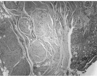
fascicles (Figure 1), showing neurofibromatous proliferation
with elongated, mildly atypical cells with oval to elongate
nuclei (Figure 2). Neoplastic cells were loosely dispersed
in a moderately myxoid matrix. Some collagen fibers were
239
seen. The lesion showed positive immunoexpression for S100 (Figure 3), Leu7, and epithelial membrane antigen (EMA) (rare peripheral cells), and was negative for CD117, DOG-1, desmin, and smooth muscle actin. The diagnosis of PN was then determined. No other clinical evidences of NF1 were found.
dIsCussIon
Neurofibromas are well-differentiated, benign peripheral sheath tumors consisted of Schwann, perineurial-like, and fibroblasts cells, and cells with intermediate features. Residual myelinated or unmyelinated axons are often present. Many of these lesions are associated with a recognizable nerve. In general population, neurofibromas are much more sporadic than associated with neurofibromatosis. Neurofibromas occur in a variety of architectural types, including cutaneous (localized and diffuse), intraneural (localized or plexiform type), massive soft tissue tumors (composed of both diffuse and plexiform elements), and visceral. The clinical presentation and gross appearance differ considerably between the different forms. As isolated lesions, both plexiform and massive soft tissue tumors are almost pathognomonic of NF1, although there are cases in which the syndrome is not identified, at least at the time of surgery. PN are related to alterations in the NF1 gene, including secondary, somatic mutations. Visceral neurofibroma consists of solitary or multiple, sporadic or NF1-associated neurofibromas of localized or plexiform type. Viscera affected include the small bowel, mesentery, large bowel, stomach, liver, and the genitourinary tract. Laryngeal or cardiac lesions are rare(1-3, 5, 9, 10, 12, 14, 17).
PN is deined as a neuroibromatous involvement of multiple fascicles of a nerve, and often of its branches. PN most often presents in children of either sex, and less frequently occurs in young adults. The tumor arises mostly in large nerves related to cervical, brachial, or lumbosacral plexuses. Most visceral and mesenteric neuroibromas are of the plexiform type. PN of major nerves are considered a precursor lesion to the majority of malignant peripheral nerve sheath tumors. Malignant transformation occurs in 5% of sizable plexiform tumors, but is a rare event in diffuse cutaneous and massive soft tissue neuroibromas. PN associated to NF1 essentially always develop during early childhood, often
before the cutaneous neuroibromas have fully developed(1, 2, 5, 12,
14, 15, 19-21). In the present case, the authors describe a PN originated
in the pylorus which determined gastric obstruction. The Table
shows some cases of gastrointestinal PN found in the international literature compared with our case study.
Eduardo Cambruzzi; Karla Lais Pêgas; Andreza Mariane de Azeredo; Isadora Bombassaro
FIguRe 1 – Plexiform neurofibroma: tortuous expansion of multiple nerve
fascicles caused by neurofibroma cells, affecting gastric wall, HE, 40× HE: hematoxylin and eosin stain.
FIguRe 2 – Plexiform neurofibroma: a benign soft tissue tumor composed of
cells which have oval to elongate normochromatic nuclei, expanding the nerve fascicle, HE, 200×
HE: hematoxylin and eosin stain.
FIguRe 3 – Plexiform neurofibroma: positive immunoexpression for S100
240
Solitary plexiform neuroibroma determining pyloric obstruction: a case report
PN more commonly consists of grossly expanded nerves or nerve ibers which are largely replaced by neuroibromatous tissues. These expanded nerves form thick, convoluted cords and nodules macroscopically. PN have smooth glistening external surface. The cut surface of PN is uniformly light tan or gray, glistening, semitranslucent, irm, and without hemorrhage or necrosis. In the gastrointestinal tract, PN usually determines a nodular lesion affecting muscular layer and perivisceral adipose tissue. Less commonly, PN extend from the submucosa across the muscularis mucosae into the mucosa where they expand the gland and distort the crypts(1, 2, 5, 6, 8, 13, 17-19, 21, 22). At microscope, the tumor is composed by a tortuous mass of expanded nerve branches, which are better seen in various planes of section. PN is composed of a growth of cells with oval to elongate normochromatic nuclei, which are loosely dispersed in a variably myxoid matrix intermingled with collagen ibers. Nuclei of neuroibroma cells are about one-third the size of schwannoma cell nuclei and their cell processes are indistinguishable from collagen ibers. The cells grow alongside nerve ibers of fascicle origin. The fascicle is expanded by the tumor but maintenance of original contour. Each nodule of PN is outlined by an evident perineurium. PN can show nuclear atypia and areas of heightened cellularity. Uncommon histologic indings in PN are pseudomeissnerian bodies, densely aggregated hyperchormatic nuclei, melanin pigmentation, discrete formation of neoplastic Schwann cells, and true epithelial cell differentiation. Electron microscopy of PN demonstrates that Schwann cell is the predominant cell type, and it is surrounded by basal lamina. A signiicant number of ibroblasts are also present. PN shows immunopositivity for vimentin and Leu7, and only a few cells can be highlighted for S100 protein. Positive immunostaining for glial ibrillary acidic protein (GFAP) and EMA can be found(1, 2, 5, 6, 8, 13,17-19, 21, 22).
Differential diagnosis includes schwannomas, plexiform schwannomas (PS), plexiform ibrohistiocytic tumors (PFT), and gastrointestinal stromal tumors (GIST). Schwannomas occur at all ages but are more common in persons between the ages of 20 and 50 years. Schwannomas affect more commonly the nerve roots of the head, neck, and lexor surfaces of upper and lower extremities. Most schwannomas are uninodular masses surrounded by ibrous capsule consisting of epineurium and residual nerve ibers. The tumor shows some areas composed by compact spindle cells with twisted nuclei arranged in short bundles (Antoni A areas, including nuclear palisading), and less cellular zones showing spindle or oval cells arranged haphazardly in a loosely textured matrix (Antoni B areas). PS are composed of uniform Schwann cells, can show Verocay bodies, lack of a diffuse extraneural component, exhibit large cells when compared to PN, and uniform S100 protein immunopositivity. PFT show female predilection. They are small and irm, do not demonstrate large expanded nerves, lack of an underlying nerve association, composed of myoibroblasts, epithelioid, and giant cells, and show anti-muscle speciic actin (HHF35) immunoreactivity rather than S100 protein positivity. The stomach is the most common site of localization for GISTs, which are generally benign lesions, with well-deined borders. Gastric GIST usually exhibits two histologic patterns. One is a cellular spindle stromal tumor characterized by fascicles of spindle cells exhibiting monotonous and uniform nuclei. The epithelioid GIST contains round epithelioid cells with prominent clear cytoplasm and cytoplasmic perinuclear vacuolization, arranged in sheets or packets. Gastric GIST shows variable positive immunoexpression for CD117, CD34, smooth muscle actin, heavy caldesmon. GISTs strongly express the DOG-1 gene, and infrequently exhibit immunopositivity for desmin and S100(1, 2, 8, 13, 19, 20).
TABLE – Summary of some published cases of gastrointestinal PS
Authors Age/gender Clinical findings Topography Treatment modality Outcome Beck et al.(4) 14/F Hematemesis Stomach Gastrectomy Unavailable Ganeshan et al.(7) 67/M Dysphagia Esophagus Esophago-gastrectomy Unavailable Leslie et al.(11) 76/F Abdominal pain Small bowel Partial enterectomy Died due lung adenocarcinoma
Park(15) 11/M Abdominal discomfort Ileum Partial tumor resection No clinical evidence of tumor growth after 10 months
Rezende et al.(18) 42/M Abdominal discomfort, black
stools, and anemia Antrum Distal gastrectomy
No evidence of recurrence after 36 months of follow-up
Present report 58/M Epigastric discomfort, nausea,
and vomiting Pylorus Distal gastrectomy
No evidence of recurrence after 03 months of follow-up
241
ReFeRenCes
1. AGAIMY, A. et al. Peripheral nerve sheath tumors of the gastrointestinal tract: a multicenter study of 58 patients including NF1-associated gastric schwannoma and unusual morphologic variants. Virchows Arch, v. 456, n. 4, p. 411-22, 2010.
2. ANTONESCU, C. R.; WOODRUFF, J. M. Neurofibroma. In: MCLENDON, R. E.; ROSENBLUM, M. K.; BIGNER, D. D. Russell & Rubinstein’s pathology of tumors of the nervous system. 7. ed. New York: Hodder Arnold, 2006. p. 803-14. 3. BAKKER, J. R. et al. Gastrointestinal neuroibromatosis: an unusual case of gastric outlet obstruction. Am Surg, v. 71, n. 2, p. 100-5, 2005. 4. BECK, D. E. et al. Gastric neuroibroma in an adolescent. South Med J, v. 79, p. 359-61, 1986.
5. COLMAN, D. S. et al. Benign neuroibromas in type I neuroibromatosis (NF1) show somatic deletions of the NF1 gene. Nat Gene, v. 11, p. 90-2, 1995. 6. FULLER, C. E.; WILLIAMS, G. T. Gastrointestinal manifestations of type I neuroibromatosis (von Recklinghausen’s disease). Histopathology, v. 19, p. 1-19, 1991.
7. GANESHAN, A. et al. Plexiform neuroibroma of the oesophagus: a mimicker of malignancy. Br J Radiol, v. 78, p. 1095-7, 2005.
8. JOHNSON, M. D. et al. Immunohistochemical evaluation of Leu-7, myelin basic-protein, glial-ibrillary-acidic-protien, and LN3 immunoreactivity in nerve sheath tumors and sarcomas. Arch Pathol Lab Med, v. 112, p. 155-60, 1988.
9. KASAPOGLU, F. et al. Laryngeal plexiform neuroibroma in a child. Ear Nose Throat J, v. 92, n. 6, p. E31, 2013.
10. KIM, S. et al. Early gastric cancer with neuroibroma mimicking a metastatic node: a case report. J Gastric Cancer, v. 13, n. 3, p. 185-7, 2013. 11. LESLIE, A. et al. Plexiform neuroibroma of the small bowel iniltrated with metastatic adenocarcinoma. Br J Radiol, v. 72, p. 604-6, 1999. 12. LISTERNICK, R. et al. Segmental neuroibromatosis in childhood. Am J Med Genet A, v. 121A, n. 2, p. 131-5, 2003.
13. MACARRON, K. F.; GOLDBLUM, J. R. Plexiform neuroibroma with and without associated malignant peripheral nerve sheath tumor: a
clinicopathologic and immunohistochemical analysis of 54 cases. Mod Pathol, v. 11, p. 612-7, 1998.
14. MATSUKI, K. et al. Mesenteric plexiform neuroibroma associated with Recklinghausen’s disease. Pediatr Radiol, v. 27, p. 255-6, 1997. 15. PARK, J. Mesenteric plexiform neuroibroma in an 11-year-old boy with von Recklinghausen disease. J Pediatr Surg, v. 42, p. E15-8, 2007. 16. OGASAWARA, N. et al. Gastric schwannoma with adjacent external progression harbored aberrant NF2 gene. Dig Endosc, v. 21, n. 3, p. 192-5, 2009.
17. RÁLIS, Z.; EMERY, J. L. Congenital plexiform neuroibroma of the vagus with cardiac, pulmonary and visceral involvement. J Pathol, v. 107, n. 1, p. 55-7, 1972.
18. REZENDE, N. A. et al. Plexiform neuroibroma: an unusual cause of GI bleeding and intestinal obstruction. Gastrointest Endosc, v. 70, n. 2, p. 396-8, 2009.
19. SCHEITHAUER, B. W. et al. Neurofibroma. In: LOUIS, D. N.; OHGAKI, H.; WIESTLER, O. D.; CAVENEE, W. K. WHO Classiication of tumors of the central nervous system. Lyon: IARC Press, 2007. p. 156-7. 20. SIDERAS, P. A. et al. Simultaneous presentation in the neck and abdomen of malignant peripheral nerve sheath tumors involving two different tracts. J Clin Neurosci, v. 20, n. 4, p. 602-4, 2013.
21. SOUZA, F. H. et al. Bilateral plexiform neuroibromas of the brachial and lumbosacral plexuses. Arq Neuropsiquiatr, v. 71, n. 2, p. 128-30, 2013. 22. WATANABE, A. et al. An individual with gastric shwannoma with pathologically malignant potential surviving two years after laparoscopy-assisted partial gastrectomy. Case Rep Gastroenterol, v. 5, p. 502-7, 2011.
MaIlIng addRess
Eduardo Cambruzzi
Hospital Conceição de Porto Alegre; Av. Francisco Trein, 596, 2º andar; Laboratório de Patologia; Cristo Redentor; CEP: 91350-200; Porto Alegre-RS, Brazil; Phone/fax: +55 (51) 3357-2164; e-mail: [email protected].
ResuMo
Neurofibroma plexiforme (NP) gástrico solitário é um tumor muito raro originado a partir dos nervos periféricos. É uma causa rara de obstrução pilórica. Paciente masculino, 58 anos, relatava desconforto epigástrico, náuseas e vômitos durante dois meses. A endoscopia digestiva superior mostrou estenose moderada/acentuada do piloro. Tomografia computadorizada (TC)/ecoendoscopia revelaram nódulo no piloro. O paciente foi submetido a gastrectomia distal. À macroscopia, identificou-se nódulo cinzento medindo 1.1 × 1 × 1 cm. À microscopia, encontrou-se tumor benigno composto por fascículos nervosos dilatados/tortuosos, exibindo proliferação neurofibromatosa com atipias leves e matriz mixoide. A lesão exibiu imunoexpressão positiva para S100, Leu7 e antígeno da membrana epitelial (EMA), e negatividade para CD117, DOG-1, desmina e actina de músculo liso. O diagnóstico de PN foi, então, determinado.
Unitermos: neurofibroma; piloro; neoplasias gástricas; trato gastrointestinal; patologia.
Eduardo Cambruzzi; Karla Lais Pêgas; Andreza Mariane de Azeredo; Isadora Bombassaro