109
REV. HOSP. CLÍN. FAC. MED. S. PAULO 58(2):109-112, 2003
From the Department of Gastroenterology, Division of Gastrosurgery and Coloprocto-logy, Hospital das Clínicas, Faculty of Medicine, University of São Paulo.
Received for publication on May 13, 2002.
BLUE RUBBER BLEB NEVUS SYNDROME: CASE
REPORT
Magaly Gemio Teixeira, Marcos Vinicius Perini, Carlos Frederico S. Marques, Angelita Habr-Gama, Desidério Kiss and Joaquim J. Gama-Rodrigues
TEIXEIRA MG et al. - Blue rubber bleb nevus syndrome: case report. Rev. Hosp. Clín. Fac. Med. S. Paulo 58(2):109-112, 2003.
The case of a patient with blue rubber bleb nevus syndrome who is infected by acquired immunodeficiency syndrome virus due to multiple blood transfusions is presented. This case shows that although it is a rare systemic disorder, blue rubber bleb nevus syndrome has to be considered in the differential diagnosis of chronic anemia or gastrointestinal bleeding. Patients should be investigated by endoscopy, which is the most reliable method for detecting these lesions. The patient underwent gastroscopy and enteroscopy via enterotomy with identification of all lesions. Minimal resection of the larger lesions and string-purse suture of the smaller ones involving all the layers of the intestine were performed. The string-purse suture of the lesions detected by enteroscopy proved to be an effective technique for handling these lesions, avoiding extensive intestinal resection and stopping the bleeding. Effective management of these patients demands aggressive treatment and should be initiated as soon as possible to avoid risks involved in blood transfusions, as occurred in this case.
DESCRIPTORS: Blue rubber bleb nevus syndrome. Hemorrhage. Surgery.
Blue rubber bleb nevus syndrome is an uncommon systemic disorder characterized by cutaneous and gastrointestinal hemangiomas with an approximate incidence of 1:14,0001,2.
It was first described by Bean3 in 1958,
who named the syndrome because of the bluish color of the nevi and their rubbery consistency at palpation. The typical finding in this disease is ve-nous malformations, which are present at birth or may appear in childhood and increase in size and number with age. Early diagnosis and management are important because of the potential for serious or fatal bleeding4. We report
a case of late successful treatment of a patient that due to several blood infu-sions was infected with acquired im-munodeficiency syndrome virus (AIDS).
CASE REPORT
A 19-year-old woman was admit-ted with a 10-year history of sporadic episodes of melena and fatigue. She had been admitted several times at the emergency service because of anemia, receiving blood transfusions, with temporary improvement of her condi-tion. She underwent an operation to excise a head tumor at the age of 3. The admission blood cell count re-vealed Hb = 4.9 and Ht = 16% and positive tests for AIDS. Other
labora-tory tests were normal. The patient un-derwent the following examinations:
· Upper gastrointestinal endos-copy revealing 3 wine-colored lesions of about 4 to 6 mm in the stomach and 1 in the duodenum;
• Colonoscopy revealing polypoid lesions in rectum, sigmoid, and transverse colon;
• Enteroscopy with visualization of 20 cm from the Treitz angle show-ing a bluish lesion;
sta-110
REV. HOSP. CLÍN. FAC. MED. S. PAULO 58(2):109-112, 2003 Blue rubber bleb nevus syndrome: case report
Teixeira MG et al.
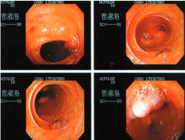
ples through gastrostomy and duo-denectomy. We performed an enter-otomy between the angle of Treitz and the ileocecal valve. An enteroscopy was performed through this opening proximally and distally. Several bluish vascular lesions were detected (Fig. 1). Each one was treated with a purse-string suture involving all the intesti-nal layers (Fig. 2). Only one was resected and sent for histopathological examination (Fig. 3). The study and treatment were completed with a colonoscopy that detected 3 lesions; 1 was resected, and 2 were treated with a purse-string suture. The postopera-tive recovery was uneventful.
The pathological study of the sur-gical specimens revealed vascular mal-formation with hamartomatous vascu-lar ectasias in the mucosa and submu-cosa, with interstitial hemorrhage in the stomach; similar lesions in the duodenum were associated with calci-fication. The same pattern was ob-served in the small bowel and colon.
The patient remains without bleed-ing or anemia 1 year after the surgical procedure.
DISCUSSION
Blue rubber bleb nevus syndrome is characterized by cutaneous vascular malformations associated with gastro-intestinal hemorrhage due to similar lesions in the gastrointestinal tract. The cutaneous malformations are blu-ish lesions that range from a few millimeters to several centimeters in diameter and occur preferentially in the trunk and upper extremities5.
Al-though it is a rare syndrome, blue rub-ber bleb nevus syndrome must be con-sidered in the differential diagnosis of anemia.
The intestinal lesions are more fre-quent in the small bowel, as occurred in the present case, although any ana-tomical site may be affected in the
gastrointestinal tract5-7. In the colon,
they are usually distal4.
Intussuscep-tion, volvulus, and infarction may also occur7-10. Lesions outside the
gastro-intestinal tract rarely bleed.
Diagnosis can be established by upper endoscopy and colonoscopy. Soft tissue masses not specific for hemangiomas can be revealed by CT scans. Selective arteriography and
Figure 3 - Resection of lesion of the small bowel.
111
REV. HOSP. CLÍN. FAC. MED. S. PAULO 58(2):109-112, 2003 Blue rubber bleb nevus syndrome: case report Teixeira MG et al.
scintillography are useful in cases of active bleeding.
In our case, the massive bleeding indicated surgical treatment. Enteros-copy performed during laparotomy al-lowed us to detect all lesions and to treat them by resection (larger ones) or by purse-string suture. We believe that all lesions must be treated in order to stop or prevent bleeding. In this case, the 1-year followup indicates that the
procedure, although time-consuming, was worthwhile. We think that the de-tection of lesions by intraoperative en-doscopy and treatment by purse-string suture is a desirable approach, since it avoids the risks of extensive intestinal resections as reported by Place11.
Additional lesions will probably develop subsequently12. For a long
pe-riod of time, the only therapeutic meas-ure used in this case consisted of
blood transfusions to replace blood loss when bleeding occurred; this strat-egy unfortunately led to infection by HIV virus. Patients with iron defi-ciency anemia should be investigated for the possibility of blue rubber bleb nevus syndrome. Endoscopy is the most reliable method for detecting these lesions and can act therapeuti-cally through sclerosis13.
RESUMO
TEIXEIRA MG e col. - Síndrome do blue rubber bleb nevus: relato de caso. Rev. Hosp. Clín. Fac.
Med. S. Paulo 58(2):109-112,
2003.
É descrito um caso da síndrome do blue rubber bleb nevus associada a in-fecção pelo vírus da imunodeficiência adquirida em conseqüência de múlti-plas transfusões de sangue. Este caso demonstra que, embora rara, esta sín-drome deva ser considerada no
diag-nóstico diferencial da anemia crônica ou sangramento gastrointestinal. O melhor método diagnóstico é a endos-copia. A doente foi operada e subme-tida a esofagogastroduodenoscopia e endoscopia através de enterotomia com identificação de todas as lesões que foram tratadas, as maiores, por ressecção mínima e as demais por su-tura em bolsa ao redor do nevo inte-ressando toda a parede intestinal. A su-tura em bolsa das lesões detectadas por enteroscopia demonstrou ser uma
téc-nica efetiva no tratamento destas le-sões, evitando assim ressecções intes-tinais extensas e parando o sangra-mento. O manuseio destes doentes de-manda tratamento agressivo e deve ser iniciado precocemente para evitar ris-cos associados a transfusões sangüí-neas como ocorreu neste caso.
112
REV. HOSP. CLÍN. FAC. MED. S. PAULO 58(2):109-112, 2003 Blue rubber bleb nevus syndrome: case report
Teixeira MG et al.
REFERENCES
1 . GENTRY R, DOCKERTY MB, CLAGETT OT - Vascular malformations and vascular tumours of the gastrointestinal tract. Int Abst Surg 1949; 88:281-323.
2 . CAMILLERI M, CHADWICK VS, HODGSON JF - Vascular anomalies of the gastrointestinal tract. Hepato Gastroenterol 1984; 31:149-153.
3 . BEAN WB - Blue rubber bleb nevi of the skin and gastrointestinal tract. In CHARLES C THOMAS - Vascular spiders and related lesions of the skin. Springfield, Illinois, 1958. p. 178-185. 4 . ÖKSUZOGLU BC, OKSUZOGLU G, ULKEM C et al. - Blue
rubber bled nevus syndrome Am J Gastroenterol 1996; 91: 780-782.
5 . RODRIGUES D, BOURROUL MLM, FERRER APC et al. - Blue rubber bleb nevus syndrome. Rev Hosp Clin Fac Med S Paulo 2000; 55:29-34.
6 . MACANNEL CA, HOENIG J, UMLAS J et al. - Orbital lesions in the blue rubber bleb syndrome. Ophtalmology 1996; 103 :933-936.
7 . RADKE M, WALDSCHMIDT J, STOLPE HJ, et al. - Blue rubber bleb nevus syndrome with predominate urinary bladder hemangiomatosis. Eur J Pediatr Surg 1993; 3:313-316.
8 . MOODLEY M, RANDIAL P - Blue rubber bleb nevus syndrome: Case report and review of the literature. Pediatrics 1993: 92; 160-162.
9 . WONG SH, LAU WY - Blue rubber bleb nevus syndrome. Dis Colon Rectum 1982; 25:371-374.
10. BROWNE AF, KATZ S, MISER J, BOLES T - Blue rubber bleb nevi as a cause of intussusception. J Ped Surg 1983; 18:7-9. 11. PLACE RJ - Blue rubber bleb nevus syndrome: a case report with
long-term follow-up. Mil Med 2001; 166:728-730. 12. SANDHU KS, COHEN H, RADIN R, et al. - Blue rubber bleb
nevus syndrome presenting with recurrences. Dis Dis Sci 1987; 32:214-219.