1190
RADIOLOGY PAGE
Vol. 43 (6): 1190-1191, November - December, 2017doi: 10.1590/S1677-5538.IBJU.2016.0555
Gas surrounding the urinary bladder in emphysematous
cystitis
_______________________________________________
Zhenyu Yang
1, Chang Sheng
11 Department of Urology, Pudong New Area People’s Hospital, Shanghai, China
ABSTRACT
ARTICLE
INFO
______________________________________________________________ ______________________
We report a rare case of emphysematous cystitis in a 66-year-old woman with a history of diabetes mellitus. The predisposition of diabetes mellitus and infection of gas-forming bacteria is considered to precede the manifestation of emphysematous cystitis. The present recommended diagnosis test is computed tomography, which have definite value in the evaluation of gas accumulation in bladder wall, or an air-fluid level in bladder.
Keywords:
Cystitis; Tomography, X-Ray Computed; Diabetes Mellitus
Int Braz J Urol. 2017; 43: 1190-1
_____________________
Submitted for publication: October 09, 2016
_____________________
Accepted after revision: December 27, 2016
_____________________
Published as Ahead of Print: May 15, 2017
INTRODUCTION
A 66-year-old woman with a 10-year his-tory of diabetes mellitus presented to the emer-gency department for painful urination and gross hematuria. Similar episodes had occurred several times in the 6 months preceding presentation, along with episodes of acute urinary retention and bladder catheterization. The physical examination was unremarkable. Laboratory investigations re-vealed mild anemia (Hb:9.2g/dL) and elevated blood glucose (BG:171mg/dL). Urinalysis findin-gs indicated urinary tract infection. The culture of voided midstream urine showed evidence of Escherichia coli. Urinary system ultrasonography
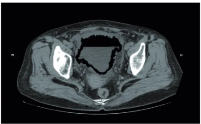
revealed an irregular thickened bladder wall with post-void residual volume of 140mL. Computed tomography (CT) of the abdomen and pelvis wi-thout administration of contrast material revea-led diffuse gas within the bladder wall (Figure-1) and a prominent air-fluid level (Figure-2). This pattern of gas surrounding the urinary bladder on computed tomography is a typical manifestation of emphysematous cystitis (EC) in which natural fermentation of glucose for gas-forming bacteria infected mostly in diabetic women (1, 2).
IBJU| RADIOLOGY PAGE
1191
being diabetic patients (1). Diabetes mellitus and female gender are the highest risks for developing EC. The typical presentation spectrum of EC inclu-des lower abdominal pain, bacteremia, and dysu-ria. Urinalysis often indicates bacteriuria, pyuria and hematuria. CT is the most sensitive diagnos-tic protocol for EC (3). Current concepts about the pathogenesis of gas formation in the bladder is postulated that bacteria such as Escherichia coli. ferment the glucose in the urine of diabetic pa-tients but in non-diabetic papa-tients remains still Figure 1 - Gas surround the bladder wall on computed tomography.
Figure 2 - A prominent air-fluid level within the bladder on computed tomography.
unknown. EC is often successfully managed with drainage and appropriate antibiotics. About 10% of cases require surgery and estimated mortality rate is 7% (4, 5). Our patient was treated with le-vofloxacin 500mg for 5 days and was discharged in stable condition.
CONFLICT OF INTEREST
None declared.
REFERENCES
1. Grupper M, Kravtsov A, Potasman I. Emphysematous cystitis: illustrative case report and review of the literature. Medicine (Baltimore). 2007;86:47-53.
2. Mokabberi R, Ravakhah K. Emphysematous urinary tract infections: diagnosis, treatment and survival (case review series). Am J Med Sci. 2007;333:111-6.
3. Huang JJ, Tseng CC. Emphysematous pyelonephritis: clinicoradiological classification, management, prognosis, and pathogenesis. Arch Intern Med. 2000;160:797-805. 4. Quint HJ, Drach GW, Rappaport WD, Hoffmann CJ.
Emphysematous cystitis: a review of the spectrum of disease. J Urol. 1992;147:134-7.
5. Thomas AA, Lane BR, Thomas AZ, Remer EM, Campbell SC, Shoskes DA. Emphysematous cystitis: a review of 135 cases. BJU Int. 2007;100:17-20.